Real-time force feedback during
flexion-distraction procedure for low back pain:
A pilot study
Maruti Ram Gudavalli,
PhD*James M. Cox,
DC, DACBR*** Associate Professor
Palmer Center for Chiropractic Research 741 Brady Street
Tel. (563) 884-5260 Fax (563) 884-5227
Email: [email protected] ** Cox Chiropractic Medicine Inc
Fort Wayne, IN 46805
Conflicts of Interest: Dr. Cox is the developer of this technique, he teaches these procedures to practicing chiropractors, and is a paid consultant to Haven Innovations Inc.
Acknowledgements: This investigation was conducted in a facility constructed with support from research facilities improvement program grant # C06 RR15443-01 from the National Center for Research Resources, National Institutes of Health. The authors also acknowledge the donations received from practicing Doctors of Chiropractic.
©JCCA 2014
A form of chiropractic procedure known as Cox flexion-distraction is used by chiropractors to treat low back pain. Patient lies face down on a specially designed table having a stationery thoracic support and a moveable caudal support for the legs. The Doctor of Chiropractic (DC) holds a manual contact applying forces over the posterior lumbar spine and press down on the moving leg support to create traction effects in the lumbar spine. This paper reports on the development of real-time feedback on the applied forces during the application of the flexion-distraction procedure. In this pilot study we measured the forces applied by experienced DCs as well as novice DCs in using this procedure. After a brief training with real-time feedback
Real-time force feedback during flexion-distraction procedure for low back pain: A pilot study
Introduction
Musculoskeletal conditions are common causes of pain and disability with low back pain representing a prevalent complaint and costly societal burden.1-7 Doctors of chiro-practic (DCs) treat low back pain patients to relieve dis-comfort and improve function. DCs may deliver several types of chiropractic adjustments or spinal manipulation therapy (SMT) to the spine for the treatment of muscu-loskeletal (MSK) conditions. SMT includes manual high velocity low amplitude spinal manipulative (HVLA-SM) procedures, handheld instrument assisted techniques, low-velocity distraction procedures, drop piece high-vel-ocity techniques.8
Chiropractic students traditionally learn the technique of delivering SMT procedures by observing someone skilled in a procedure. The expert teacher demonstrates a technique and the student then practices its delivery on other students or volunteer patients. The teacher observes the student performing a manual procedure and provide hands-over-hands guidance, and provide verbal feedback as the student develops proficiency. Experienced DCs provide training in a similar manner with student interns in clinical situations. Triano et al. have reviewed on the training methods used in the literature.9
Chiropractic techniques are measurable biomechanical events involving the application of forces to specific re-gions of interest, causing vertebral movements.10-13 Sev-eral investigators have measured the forces delivered by DCs during manipulations of the lumbar, thoracic and cervical spine.14-20 HVLA-SM is characterized by clinical
force delivery, loading durations, loading rates, coordina-tion index, and transmitted loads to the spine.
Over the past decade, educators have incorporated in-novative bioengineering technologies into the training of chiropractic students and licensed doctors to give feed-back on the forces, durations, loading rates, and coordina-tion indexes. Mechanical instruments, mannequins, and human volunteers were used for training. Subsequently, researchers have demonstrated quantified force-time pro-file characteristics.16,21-25 Most of these studies focused on HVLA-SM, with the majority evaluating the thoracic and lumbar spine.16;21-24 Few studies have measured the biomechanical characteristics of HVLA-SM delivery to the cervical spine24,25, and few studies on these parameters with mobilization procedures26-29.
James Cox, DC developed manual distraction, or the flexion distraction procedure, to treat patients with spin-al problems.30,31 Several case reports, case series, and a randomized clinical trial have been published for treat-ing neck and low back pain problems ustreat-ing this proced-ure.32-38 During Cox Flexion-Distraction procedure, the patient lies face down on a specially designed chiropractic table. The DC gently moves the caudal section of the table while holding a broad manual contact over the posterior part of the low back with a vertebral level selected, with an intent to create traction effects in the lumbar spine. This paper reports on the development of real-time force feedback at the Palmer Center for Chiropractic Research, which provides clinicians with real time vis-ual graphical feedback on the magnitude of forces at the
novice DCs have improved on the magnitude of the applied forces. This real-time feedback technology is promising to do systematic studies in training DCs during the application of this procedure.
(JCCA 2014; 58(2):193-200)
k e y w o r d s: Cox, flexion-distraction, technique, real-time, chiropractic
Après une brève formation avec rétroaction en temps réel, les DC débutants s’étaient améliorés relativement à la magnitude des forces appliquées. Cette technologie de rétroaction en temps réel est prometteuse pour la réalisation d’études systématiques sur la formation des DC durant l’application de cette procédure.
(JCCA 2014; 58(2):193-200)
contact hand of the DC on the participant’s lumbar spine. This novel training tool was used to collect pilot data while Cox Flexion-Distraction was applied to simulated asymptomatic volunteers by experienced DCs as well as novice DCs.
Methods
The Palmer College of Chiropractic (PCC) institutional review board approved this study. Human simulated pa-tient volunteers and the doctors of chiropractic volun-teers signed written informed consent to participate in the study.
Recruitment
Four asymptomatic volunteers (2 male and 2 female age range 22-52 years old) served as simulated patients, re-cruited from the doctors attending a Cox certification course. DCs screened volunteers for any contraindica-tions and safety consideracontraindica-tions relative to receiving the Cox flexion-distraction procedure before study inclusion. Five experienced (>15 years experience in using flexion distraction procedure) DCs and 5 Novice DCs (<1 year experience in using flexion-distraction procedure) partici-pated in the measurement of force delivery.
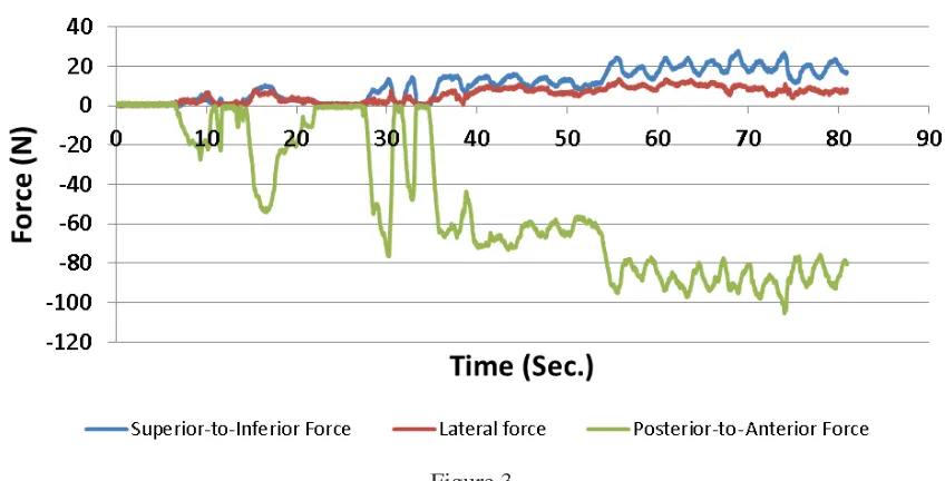
Force Transducer and Force Feedback Software During the Cox Flexion-distraction procedure the DC contacts the posterior aspect of the lumbar spine using one hand and applies downward motion of the caudal section of the table where the ankles are cuffed to the table. DCs apply posterior-to-anterior forces (PAF) as well as inferior-to-superior forces (ISF) at the stabilizing hand contact on the posterior aspect of the lumbar spine. Figure 1 shows the table, the patient in a prone position, and the hand contacts. A three dimensional force trans-ducer (Model # Mini-45, ATI-Industrial Automation, Apex, NC) was used to measure the three dimensional forces applied by the DC at the lumbar spine contact. Figure 2 shows the force transducer and the negative Fz axis is directed in the posterior-to-anterior direction of the patient, positive Fx axis is directed along the in-ferior-to-superior-direction of the volunteer participant. A rubber padding is placed between the patient and the transducer. The measurement of forces is achieved with the help of a three-dimensional force transducer, ampli-fiers, analog-to-digital converters, laptop portable com-puter, and custom written Labview software. A custom written software provides the graphical visual feedback in real time as a function of time during the delivery of Figure 1.
Cox Flexion-Distraction Table with hand contacts for treating low back.
Figure 2.
Real-time force feedback during flexion-distraction procedure for low back pain: A pilot study
the treatment. Figure 3 shows force-time graph with the possibility to change the applied force while delivering the treatment (visual real-time graphical feedback). The software was written in Labview (Version 7, National Instruments, Austin, TX). The data is collected at a sam-pling rate of 100Hz. Magnitude of forces in the inferior-to-superior direction and posterior-to-anterior direction at the hand contact can be simultaneously incorporated into the training.
We have independently tested the force transducer measures (Model: Mini45, ATI industrial Automation, Apex, NC) against a 3-D force plate (Model 4060NC, Bertec Corporation, Columxbus, OH) (20) in both normal and shear directions and found good agreement (less than 3% difference). During Cox flexion-distraction procedure for treating low back, forces are delivered in a gentle slow manner at a rate of approximately 0.5 Hz. Cox flexion distraction for low back pain is a form of low velocity variable amplitude spinal manipulation (LVVA SM). The procedure is performed with a participant lying prone on a specially designed table with a fixed section of the table under the trunk, and a moveable caudal section that al-lows guided flexion and traction movement in the lumbar spine. The clinician gently grasps the posterior aspect of the participant’s back with a thenar contact (contact hand)
at a specific vertebral level. With the opposite hand, the clinician grasps the control handle of the moving piece near the ankles. Using the contact hand, the clinician ex-hibits traction while attempting to maintain a contact at a single vertebral level and ensuring a gentle movement of the caudal section via contact with the control handle. The goal is to create a slow rhythmic distractive movement. Figure 1 shows a manual contact used by DCs while performing the low back pain procedure. Because low back stiffness and lumbar spine anatomy differ between patients, force-feedback training provides clinicians an opportunity to perceive and gauge force magnitudes on different body types.
We have collected the data from five experienced clinicians with 15, 17, 21, 26, and 35 years experience in using the flexion-distraction technique and five nov-ice clinicians (less than one year experience) in using this technique. Novice clinicians were given a brief training approximately 5 minutes while practicing using this force transducer and the real-time visual graphical feedback. After brief training we measured the forces of the five novice clinicians while delivering on asymptomatic vol-unteers.
The data is exported into an excel file and then to a custom written MathCad software program (version12, Figure 3.
Parametric Technologies, Natick, MA). The magnitudes of forces corresponding to the preload and peak force are extracted and averaged for each doctor over five cycles. The averages for the experienced and the novice doctors are averaged and compared descriptively.
Results
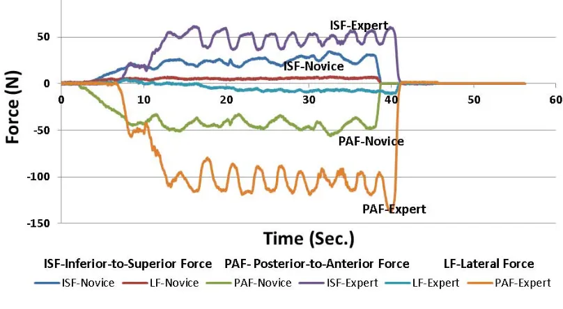
Participants who received the lumbar Cox flexion-distrac-tion procedure consisted of 2 males and 2 females (total of 4 participants). The mean age was 45 years old (SD: 12). The mean height of participants was 172.8cm (SD: 7.7cm) and mean weight was 79.6kg (SD: 22.0kg). Five experienced field clinician DCs (2 males and 3 females) with a wide range of clinical experience (15-35 years experience using flexion-distraction procedure) performed the lumbar flexion-distraction on all the four participants. This provided a reference data for compari-son. Five recently graduated DCs with less than one year experience using flexion-distraction procedure (3 male and 2 female) participated in training. Figure 4 shows the graphical data on the forces used by a typical experienced DC as well as a novice DC. Table 1 provides the forces comparing the experienced and novice doctors. Table 2
provides the data on the novice doctors before and after a brief training using the software developed for training.
Discussion
To the best of these authors’ knowledge, this is the first investigation in developing real-time force feedback and visual graphical display to deliver Cox flexion-distraction for lumbar spine. This real-time force feedback provides a foundation to monitor clinician force delivery and train clinicians to alter the delivery of force ranges. This real-time force feedback developed in this study is portable and could be easily implemented in classrooms, teaching clinics, and field settings.
This is a pilot study in collecting data on experienced and novice DCs using flexion-distraction procedure. Forces applied by experienced DCs are higher compared to the novice DCs. After a brief training of 5 minutes the force magnitudes have improved in preload as well as peak forces for the novice doctors. This improvement was observed for both posterior-to-anterior forces as well as inferior-to-superior forces.
Traditional approaches to technique training for Cox-flexion distraction have included observation and feed-Figure 4.
Real-time force feedback during flexion-distraction procedure for low back pain: A pilot study
back by an instructor/mentor. This method is based pri-marily on the subjective evaluation of distraction tech-nique as a complex psychomotor skill rather than measur-ing the biomechanical event. The real-time visual graph-ical feedback of forces developed in this project extends this subjective evaluation process by providing real-time quantitative force data. As seen in Figure 3 one can notice the improvement of the application of forces during train-ing. Initially the novice DC was applying light forces with no pre-load, gradually improving on the magnitude of the pre-load as well as peak forces by using the real-time vis-ual graphical feedback on the computer monitor. This al-lows clinicians and students the opportunity to hone in their ability to deliver specific biomechanical forces. Peer and participant feedback/debriefing, delivered verbally, remained an essential component of clinician training. Other investigators have used training instruments and instrumented mannequins to obtain visual feedback on forces and force-time profiles16-22 during HVLA-SM, comparing force-time characteristics of students and clin-icians. Our study is different from these studies in two ways: a) our study is based on real time graphical feed-back while delivering treatment on human volunteers and b) for delivering a low velocity procedure such as flexion distraction and the DC can vary the treatment forces dur-ing the delivery with visual graphical feedback similar to the study reported on posterior to anterior mobilization forces on cervical spine29.
Manual therapists apply forces to the spine for sever-al reasons including improving joint mobility, reducing
muscular hypertonicity, stimulating proprioceptive activ-ity, and to relieve pain.26 Force-magnitude related thera-peutic effects have not been studied, but this technology will also allow to train clinicians to deliver treatment within specified force values. Applying treatment within specific force ranges can be a first step toward developing clinical studies designed to investigate optimum force-dosage in clinical settings. This will also allow clinical/ physiological outcomes evaluation of patients as a func-tion of different force ranges as an intervenfunc-tion.
Limitations
This study with a small sample size is not designed to test the differences between experienced DCs and nov-ice DCs. Neither the study is designed to test the training process using a control group. This study is designed to provide time visual graphical feedback. This real-time force feedback could be used to design and conduct control studies to evaluate training and proficiency of novice DCs, and chiropractic students. The improvement in the delivery of the forces could be related to immediate learning effect. Considering this possibility, future stud-ies should be undertaken to quantify the retention of this training procedure.
Conclusions
Real-time visual graphical feedback was developed and used to train novice DCs to change the force magnitudes applied during flexion-distraction procedure. This tech-nology has the potential to design and undertake well
de-Table 2.
Descriptive comparison of forces of novice Doctors of Chiropractic before and after training
Variable Before Training (N=5)
Mean (SD)
After Training (N=5) Mean (SD) Inferior-to-Superior Forces
Pre-load (N) 19 ( 6) 31 (12)
Peak Force (N) 41 (12) 52 (12)
Posterior-to-Anterior Forces
Pre-load (N) 46 (27) 69 (30)
Peak Force (N) 86 (45) 102 (43)
N-Newtons
Table 1.
Descriptive values of Forces by experienced and novice Doctors of Chiropractic
Variable Novice DCs (N=5)
Mean (SD)
Experienced DCs (N=5)
Mean (SD) Inferior-to-Superior Forces
Pre-load (N) 19 ( 6) 44 (16)
Peak Force (N) 41 (12) 65 (10)
Posterior-to-Anterior Forces
Pre-load (N) 46 (27) 95 (34)
Peak Force (N) 86 (45) 140 (43)
signed studies in training and assessing the delivery of forces during flexion-distraction procedure. The system developed in this study is portable with a laptop computer and can be easily implemented in any field clinician’s of-fice.
References
1 Murray CJ, Vos T, Lozano R, Naghavi M, Flaxman AD, Michaud C, et al. Disability-adjusted life years (DALYs) for 291 diseases and injuries in 21 regions, 1990-2010: a systematic analysis for the Global Burden of Disease Study 2010. Lancet. 2012 Dec 15;380(9859):2197-223. 2 Vos T, Flaxman AD, Naghavi M, Lozano R, Michaud C,
Ezzati M, et al. Years lived with disability (YLDs) for 1160 sequelae of 289 diseases and injuries 1990-2010: a systematic analysis for the Global Burden of Disease Study 2010. Lancet. 2012 Dec 15;380(9859):2163-96. 3 Buchbinder R, Blyth FM, March LM, Brooks P, Woolf
AD, Hoy DG. Placing the global burden of low back pain in context. Best Pract Res Clin Rheumatol. 2013 Oct;27(5):575-89.
4 Deyo RA, Ching A. Surgical trials for pain relief: in search of better answers. Pain. 2012 Nov;153(11):2155-6.
5 Hayden JA, Dunn KM, van der Windt DA, Shaw WS. What is the prognosis of back pain? Best Pract Res Clin Rheumatol. 2010 Apr;24(2):167-79.
6 Hayden JA, Chou R, Hogg-Johnson S, Bombardier C. Systematic reviews of low back pain prognosis had variable methods and results: guidance for future prognosis reviews. J Clin Epidemiol. 2009 Aug;62(8):781-96.
7 Tiira AH, Paananen MV, Taimela SP, Zitting PJ, Jarvelin MR, Karppinen JI. Determinants of adolescent health care use for low back pain. Eur J Pain. 2012 Nov;16(10):1467-76.
8 Christensen MG, Mark G., Kollash MW, and Hyland JK. Practice Analysis of Chiropractic 2010: A project report, survey analysis, and summary of chiropractic practice in the United States. Greeley, Colorado: National Board of Chiropractic Examiners; 2010.
9 Triano JJ, Descarreaux M, Dugas C. Biomechanics – review of approaches for performance training in spinal manipulation. J Electromyogr Kinesiol. 2012 Oct;22(5):732-9.
10 Hessell BW, Herzog W, Conway PJ, McEwen MC. Experimental measurement of the force exerted during spinal manipulation using the Thompson technique. J Manipulative Physiol Ther. 1990 Oct;13(8):448-53. 11 Forand D, Drover J, Suleman Z, Symons B, Herzog W.
The forces applied by female and male chiropractors during thoracic spinal manipulation. J Manipulative Physiol Ther. 2004 Jan;27(1):49-56.
12 Herzog W, Conway PJ, Kawchuk GN, Zhang Y, Hasler
EM. Forces exerted during spinal manipulative therapy. Spine (Phila Pa 1976 ). 1993 Jul;18(9):1206-12. 13 Herzog W, Kats M, Symons B. The effective forces
transmitted by high-speed, low-amplitude thoracic manipulation. Spine (Phila Pa 1976 ). 2001 Oct 1;26(19):2105-10.
14 Herzog W, Zhang YT, Conway PJ, Kawchuk GN.
Cavitation sounds during spinal manipulative treatments. J Manipulative Physiol Ther. 1993 Oct;16(8):523-6.
15 Symons B, Wuest S, Leonard T, Herzog W. Biomechanical characterization of cervical spinal manipulation in living subjects and cadavers. J Electromyogr Kinesiol. 2012 Mar 6.
16 Cohen E, Triano JJ, McGregor M, Papakyriakou M. Biomechanical performance of spinal manipulation therapy by newly trained vs. practicing providers: does experience transfer to unfamiliar procedures? J Manipulative Physiol Ther. 1995 Jul;18(6):347-52. 17 Triano J, Schultz AB. Loads transmitted during
lumbosacral spinal manipulative therapy. Spine (Phila Pa 1976 ). 1997 Sep 1;22(17):1955-64.
18 Stemper BD, Hallman JJ, Peterson BM. An experimental study of chest compression during chiropractic manipulation of the thoracic spine using an anthropomorphic test device. J Manipulative Physiol Ther. 2011 Jun;34(5):290-6.
19 Descarreaux M, Dugas C. Learning spinal manipulation skills: assessment of biomechanical parameters in a 5-year longitudinal study. J Manipulative Physiol Ther. 2010 Mar;33(3):226-30.
20 Gudavalli MR, DeVocht J, Tayh A, Xia T. Effect of sampling rates on the quantification of forces, durations, and rates of loading of simulated side posture high-velocity, low-amplitude lumbar spine manipulation. J Manipulative Physiol Ther. 2013 Jun;36(5):261-6. 21 Descarreaux M, Dugas C, Lalanne K, Vincelette M,
Normand MC. Learning spinal manipulation: the importance of augmented feedback relating to various kinetic parameters. Spine J. 2006 Mar;6(2):138-45. 22 Descarreaux M, Dugas C, Raymond J, Normand MC.
Kinetic analysis of expertise in spinal manipulative therapy using an instrumented manikin. J Chiropr Med. 2005;4(2):53-60.
23 Triano JJ, Rogers CM, Combs S, Potts D, Sorrels K. Developing skilled performance of lumbar spine manipulation. J Manipulative Physiol Ther. 2002 Jul;25(6):353-61.
24 Triano JJ, Rogers CM, Combs S, Potts D, Sorrels K. Quantitative feedback versus standard training for cervical and thoracic manipulation. J Manipulative Physiol Ther. 2003 Mar;26(3):131-8.
Real-time force feedback during flexion-distraction procedure for low back pain: A pilot study
26 Chiradejnant A, Latimer J, Maher CG. Forces applied during manual therapy to patients with low back pain. J Manipulative Physiol Ther. 2002 Jul;25(6):362-9. 27 Sheaves EG, Snodgrass SJ, Rivett DA. Learning lumbar
spine mobilization: the effects of frequency and self-control of feedback. J Orthop Sports Phys Ther. 2012 Feb;42(2):114-24.
28 Snodgrass SJ, Rivett DA, Robertson VJ, Stojanovski E. Cervical spine mobilisation forces applied by physiotherapy students. Physiotherapy. 2010 Jun;96(2):120-9.
29 Snodgrass SJ, Rivett DA, Robertson VJ, Stojanovski E. Real-time feedback improves accuracy of manually applied forces during cervical spine mobilisation. Man Ther. 2010 Feb;15(1):19-25.
30 Cox JM. Low back pain: Mechanism, diagnosis and treatment. Baltimore: Williams and Wilkins; 2011. 31 Cox JM. Neck, Shoulder, Arm Pain: Mechanism,
Diagnosis, Treatment, 3rd Edition. 3rd ed. 2004.
32 Cox JM, Aspegren DD. Degenerative spondylolisthesis of C7 and L4 in same patient. J Manipulative Physiol Ther. 1988 Jun;11(3):195-205.
33 Gudavalli S, Kruse RA. Foraminal stenosis with
radiculopathy from a cervical disc herniation in a
33-year-old man treated with flexion distraction decompression manipulation. J Manipulative Physiol Ther. 2008 Jun;31(5):376-80.
34 Kruse RA, Cambron JA. Large C4/5 spondylotic disc bulge resulting in spinal stenosis and myelomalacia in a Klippel-Feil patient. J Altern Complement Med. 2012 Jan;18(1):96-9.
35 Kruse RA, Cambron J. Chiropractic management of postsurgical lumbar spine pain: a retrospective study of 32 cases. J Manipulative Physiol Ther. 2011 Jul;34(6):408-12. 36 Kruse RA, Imbarlina F, De Bono VF. Treatment of cervical
radiculopathy with flexion distraction. J Manipulative Physiol Ther. 2001 Mar;24(3):206-9.
37 Cambron JA, Gudavalli MR, Hedeker D, McGregor M, Jedlicka J, Keenum M, et al. One-year follow-up of a randomized clinical trial comparing flexion distraction with an exercise program for chronic low-back pain. J Altern Complemen Med. 2006 Sep 1;12(7):659-68. 38 Gudavalli MR, Cambron JA, McGregor M, Jedlicka J,