Chronic recurrent multifocal osteomyelitis in a
13 year old female athlete: a case report
Brad Ferguson,
BSc, DCaDavid Gryfe,
BSc, DC, FRCCSS(C)bWilliam Hsu,
BSc, DC, DACBR, FCCR(C)ca Division of Graduate Studies, Sports Sciences, Canadian Memorial Chiropractic College, 6100 Leslie Street, Toronto, Canada b Private Practice Toronto, Ontario
c Clinical Radiologist, Associate Professor, Diagnostic Imaging Department, Canadian Memorial Chiropractic College, 6100 Leslie Street,
Toronto, Canada
Corresponding author: Dr. Brad Ferguson [email protected]
T: (416) 482-2340 ext. 315 F: (416) 482-2560| Disclaimers: None
Patient consent was obtained for the use of clinical information and imaging with respect to this case report. Sources of financial support: none
©JCCA 2013
Chronic recurrent mutlifocal osteomyelitis (CRMO) is an extremely rare skeletal disorder in the younger population. It presents with multifocal bony lesions that often mimic more sinister diagnoses such as infection or neoplasm. The cause of this condition remains unknown and there is limited evidence on effective treatments. In this case, a 13-year-old female athlete presented to a sports chiropractic clinic with non-traumatic onset of right ankle pain. After failed conservative management, radiographs and MRI were obtained exhibiting a bony lesion of the distal tibia resembling osteomyelitis. The patient was non-responsive to antibiotics, which lead to the diagnosis of CRMO. CRMO should be considered as a differential diagnosis for chronic bone pain with affinity for the long bones of the lower extremity in children and adolescents. The role of the primary clinician in cases of CRMO is primarily that of recognition and referral for further diagnostic investigations.
(JCCA 2013; 57(4):334-340)
k e y w o r d s: osteomyelitis, bone lesion, athlete, leg
pain, adolescent
L’ostéomyélite multifocale chronique récurrente (OMCR) est une maladie du squelette extrêmement rare qui touche les jeunes. Elle présente des lésions osseuses multifocales qui imitent souvent des diagnostics plus sinistres tels qu’une infection ou une tumeur. La cause de cette maladie reste inconnue et il y a peu de preuves sur les traitements efficaces. Dans ce cas, une athlète de 13 ans s’est présentée à une clinique chiropratique sportive avec l’apparition non traumatique de douleurs à la cheville droite. Après l’échec des traitements conservateurs, les radiographies et l’IRM ont montré une lésion osseuse du tibia distal ressemblant à une ostéomyélite. Comme les antibiotiques n’agissaient pas sur la patiente, on en conclut qu’elle souffrait d’une OMCR. L’OMCR doit être considérée comme un diagnostic différentiel des douleurs osseuses chroniques, surtout des os longs des membres inférieurs chez les enfants et les adolescents. Le rôle du médecin traitant en cas d’OMCR est de la reconnaître surtout et de renvoyer le patient pour des tests diagnostics avancés.
(JCCA 2013; 57(4):334-340)
m o t s c l é s : ostéomyélite, lésion osseuse, athlète,
Introduction
Chronic Recurrent Multifocal Osteomyelitis (CRMO) is an extremely rare skeletal disorder most commonly found in the long bones of children and adolescents. CRMO was first described in the literature in 1972 by Giedon et al.1 who described the lesion as “an unusual form of
multifocal bone lesions with subacute and chronic sym-metrical osteomyelitis”. Since 1972 approximately 200-300 cases have been reported in literature2, and none in
the chiropractic literature. Its estimated prevalence is 1-2 per 1 million people and accounts for only 2-5% of osteomyelitis diagnoses.3 But this statistic may be
mis-representative of the true prevalence, as this condition is thought to be under reported or misdiagnosed. As more research becomes available and awareness for CRMO increases, the reported number of cases may increase ac-cordingly.
The name CRMO was derived from its radiographic appearance which is similar to osteomyelitis. It generally presents as lytic destruction with sclerotic borders within the metaphysis, mimicking an infectious or neoplastic process. Despite the radiographic similarities, CRMO is a misnomer. The condition although chronic and recur-rent, is not an infectious process. Hence, CRMO is com-monly used interchangeably with Chronic Non-Bacterial Osteomyelitis (CNO). Also, it need not be multi-focal as cases have shown uni-focal presentations as well. CRMO is widely believed to be a pediatric equivalent of SAPHO syndrome.4 SAPHO syndrome (synovitis, acne,
pustu-losis, hyperostosis and osteitis) is an inflammatory bone disorder that commonly presents with skin manifesta-tions.
This case report focuses on CRMO of the distal tibia and highlights the importance of considering this diag-nosis for chronic recurrent, lower extremity bone pain in adolescents. This case report outlines the pertinent aspects of epidemiology, clinical presentation, diagnosis and management of this condition.
Case
A 13 year old female patient presented to a sports chiro-practic clinic with right ankle pain of 2 weeks duration. She did not recall a precipitating event and related the pain to her active lifestyle. She localized the pain to the anterior region of the right medial malleolus and described the pain as sharp and painful to touch. The ankle pain
was aggravated during her dance and volleyball practices when she was jumping, lunging or running. Although rest improved her symptoms, the pain remained constant and rated between a 6-8/10 in intensity.
She had no history of lower extremity injuries but re-ported a history of bilateral Achilles tightness, sore feet and right ankle stiffness attributed to her training. She reported a history of chronic musculoskeletal complaints including bilateral knee pain, back pain, shoulder pain and headaches. She additionally noted that she suffered from dry, sensitive eczematous skin. She reported no symptoms of fever, lethargy or weakness.
Her family history revealed that her father suffers from chronic spinal pain particularly in the thoracic region with notable hyperkyphosis of the thoracic spine, bilateral wrist pain with weakness and aquagenic pruritis.
On examination the patient presented with no notice-able swelling, bruising or deformity of the right ankle. On standing observation there was moderate pes planus bi-laterally, but otherwise foot and ankle alignment was un-remarkable. The patient reported mild pain with walking and displayed a mild (<100) out-toeing on the right. On
palpation the patient reported tenderness over the right medial malleolus and distal tibia, and muscular tender-ness along extensor hallicus longus, tibialis anterior and tibilias posterior on the right. Active range of motion re-vealed a 25% decrease in dorsiflexion of the right ankle in comparison to the left. Resisted testing of anterior, lateral and posterior compartments of the lower leg revealed full strength. Anterior drawer test, Kleiger’s test and Syndes-motic testing was negative. Reflexes, sensory and motor testing of L4-S1 was within normal limits bilaterally. The clinical impression was chronic repetitive strain of the anterior and deep posterior compartments. The patient was educated on the possibility of a stress fracture and was advised to visit her family physician regarding an x-ray.
The patient underwent a course of treatment including myofascial release of lower leg muscles, mobilization/ manipulation of the ankle mortise joint and Tanda laser therapy (3-660 nm red SLDs; 33-870nm infrared SLDs; delivering 5 Joules/cm2 over 3 minutes). The patient was
seen weekly for 4 weeks. The patient was also recom-mended to take a two week rest period from active partici-pation in dance and volleyball.
re-sumed her normal activities and had considerable relief of symptoms. She was recommended to continue with her normal activities and follow up in two weeks.
Two weeks later, the pain began to intensify again and plain film radiographs were ordered. (Figures 1 and 2). A diagnosis of osteomyelitis was rendered and the patient was started on a course of oral antibiotics for 4 weeks. During the course of antibiotic treatment, the patient reported a worsening of symptoms. She was subsequent-ly recommended for an MRI to assist in ruling out any other causes of her symptoms. MRI of both ankles was obtained with gadolinium contrast injection (Figures 3-4). The patient was referred to a rheumatologist and was pre-scribed a course of celebrex and symptomatic relief was achieved. Based off the MRI findings, clinical presenta-tion, ineffective response to antibiotic treatment and ef-fective NSAID treatment the diagnosis of Chronic Recur-rent Multifocal Osteomyelitis was made.
Once symptomatic relief was achieved the patient was removed from the course of celebrex and has since been symptom free. The patient currently takes Aleve on an as needed basis. The patient has returned to full activities of
daily living and sport with no recurrence of symptoms to date.
Epidemiology
CRMO is a rare pediatric bone disorder that has unknown etiology. Although exact cause of CRMO is unknown, theories include inflammatory disorder, genetic disor-der, autoimmune disordisor-der, juvenile seronegative spon-dyloarthropathy (SNSA) or a low virulence infectious organism.5
CRMO is most commonly found in the metaphysis of long bones of the lower extremity in children and ado-lescents. Common locations for CRMO include the tibia, pelvis and femur, but some cases have been shown in the calcaneus, spine, clavicle, mandible, sternum among other locations.6 The mean age of onset is 10 years of age
(range 4-17) and CRMO appears more commonly (up to 67-85%) in females.7
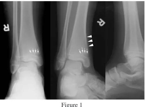
The clinical course of CRMO is prolonged, recurrent and self-limiting. Prognosis of CRMO is unpredictable in nature and can last for upwards of 25+ years and is thought to resolve spontaneously, regardless of inter-Figure 1
Plain films of AP, medial oblique and lateral views of right ankle. Radiographic images of the right ankle show
a geographic lucent lesion with a hazy band of sclerosis at the medial aspect of the distal tibial metaphysis abutting the growth plate spanning 1/3 of the tibial girth
(white arrows).
Figure 2
AP comparison view of both ankles. A comparison view the ankles allows for better appreciation of the lucent
metaphyseal lesion. A subtle thin layer of periosteal reaction (arrow heads) is observed at the medial metadiaphysis of the distal tibia on the anteroposterior view and better appreciated on the medial oblique view
Figure 3
Medial parasagittal images of the right ankle in STIR, T1 and T1 with gadolinium contrast sequences with comparison left parasagittal STIR image in the same plane show marrow edema in the distal tibial metaphysis and epiphysis and surrounding soft tissue edema in the right distal tibia.
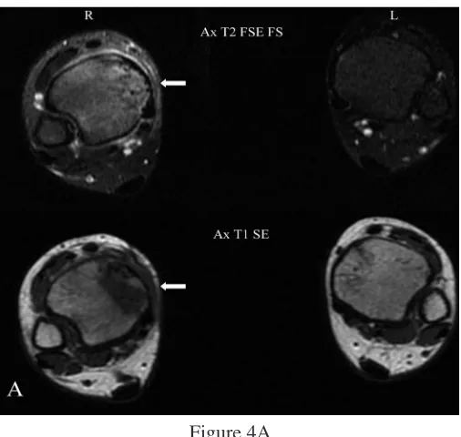
Figure 4A
Axial T2 FSE FS and T1 SE images at the level of distal tibial metaphysis above the physis show marrow edema (hyperintense T2 and hypointense T1)
at the medial third of the tibial metaphysis when compared to the normal left distal tibia.
Figure 4B
Axial T2 FSE FS and T1 SE images at the level of distal tibial metadiaphysis show marrow edema,
surrounding soft tissue edema as well as a layer of periosteal reaction (white arrows) at the
vention. Retrospective studies looking at the course of CRMO have documented a prevalence of 25-59% after a median of 10 years follow up.7
Clinical Presentation
The clinical presentation of CRMO is variable, dependent upon the location of the lesion and stage of the disorder. CRMO is a chronic disorder that can cycle in and out of painful bouts of localized bony pain and tenderness for months to years. Many cases may have been mistaken as growing pain and thus delay the correct diagnosis. The pain is insidious in onset and can present with associated swelling. The lesions are commonly multi-focal and sym-metrical.8 The average number of lesions is 3-4, but has
ranged from 1-13. Symptoms can recur repeatedly at the same location, or new areas can be affected with subse-quent flare ups. The flare ups occur insidiously and pain is often worse at night. Patients may experience low grade fever or general malaise associated with flare ups.9 This
has led to the incorrect diagnosis of osteomyelitis in many cases of CRMO. CRMO does not respond to antibiotic therapy as shown in this case. Similar to SAPHO, patients with CRMO may also present with an accompanied skin disorder such as pustulosis palmoplantaris or acne. In fact, current estimates suggest that up to 25% of CRMO patients have some sort of accompanying inflammatory disorder.10 Also, it is estimated that nearly 50% of first
degree and second degree relatives have an inflammatory disorder, suggesting a significant genetic component.
Diagnosis
An adolescent presenting with bony pain of the lower extremity generates a large list of differentials. CRMO should be included as one of these differential diagnoses. However, CRMO is a diagnosis of exclusion relying upon combination of clinical findings, diagnostic imaging and negative histological and microbiological examinations. Since there is no gold standard test to diagnose this condi-tion, the path to diagnosis relies upon ruling out all other possibilities.11 Due to the complex presentation, there is a
large and variable list of differential diagnoses to consider (Figure 5). The mean time from symptom onset to diag-nosis is estimated at 18 months (ranging from few weeks to several years), highlighting the difficulty of arriving at this diagnosis.7
Iyer et al.12, have formulated systematic approach to
assist in making the diagnosis of CRMO by exclusion using the following criteria:
1. Lack of causative organism
2. No abscess, fistula or sequestra formation 3. Radiographic appearance of sub-acute or
chron-ic osteomyelitis
4. Atypical location compared to infectious osteo-myelitis
5. Non-specific histopathologic and laboratory findings compatible with sub-acute or chronic osteomyelitis or other known disease process 6. Prolonged (> 6 months) and recurrent painful
symptoms
7. Accompanying pustulosis palmoplantaris or acne
The laboratory findings suggest an inflammatory pro-cess with evidence of elevated ESR, C-reactive protein and alkaline phosphatase in approximately two-thirds of cases.3 A tissue biopsy is commonly required in order to
rule out more sinister diagnoses such as tumour or
infec-DIFFERENTIAL DIAGNOSES FOR CRMO
• Bacterial Osteomyelitis
• Bone Bruise
• Fracture
• Osteonecrosis
• Juvenile Idiopathic Arthritis
• Osteosarcoma
• Ewing’s Sarcoma
• Osteoblastoma
• Osteoid Osteoma
• Leukemia
• Lymphoma
• Neuroblastoma
Figure 5.
Differential diagnoses for
tion.13 Cultures of blood and bone, along with microbial
laboratory assays are negative for infectious processes.10 Imaging
Plain film imaging should be the initial assessment for children and adolescents with musculoskeletal symptoms that do not resolve within the natural history of soft tissue injury. Due to the variability of growth centres and subtle-ness of CRMO in the initial development, a comparison view of the contralateral side is crucial for proper imaging assessment of CRMO.
Diagnosing CRMO is challenging as it may manifest radiographically as normal, lytic lesion, sclerotic lesion or mixed lesion.15 The varying manifestations are in part
related to the age of the lesion and severity of the disease. Initially, CRMO manifests as a juxta-physeal lucent le-sion in the metaphysis of long bones. Other sites include medial clavicle, vertebral bodies, mandible, pelvis and ribs. Due to its inflammatory nature, reactive bone forma-tion surrounding the lucent lesion as well as periosteal re-action eventually develop. Radiographically, this state of CRMO can be confused with chronic physeal injuries in adolescent athletes.16 With some CRMO cases where the
course of the disease is recurrent and progressive, hyper-ostosis, medullary sclerosis and rare bony deformity may develop. In majority of CRMO cases, the disease is mild and the affected bones eventually remodel and normalize prior to skeletal maturity.
Use of bone scan, particularly the whole-body bone scan, is supported by many authors for the detection of multiple lesions, some of which may be asymptomatic at presentation. Increased uptake at the metaphysis of long bone in soft-tissue and delayed whole-body scintigraphy confirms abnormal bony activity at the metaphysis. Bi-lateral and multifocal presentation lends support to the diagnosis of CRMO which should be correlated and con-firmed with plain film study. If plain film study is non-contributory, advanced imaging such as magnetic reson-ance imaging (MRI) is the imaging modality of choice. Clinicians should choose MRI over CT scan in the im-aging of young patients suspected of CRMO, particularly as it has no radiation hazard and it is very sensitive in detecting early subclinical lesions. Inflammatory process of CRMO manifests as marrow edema which shows up as hypointense on T1 and hyperintense T2 signals in the affected metaphysis, the adjacent epiphysis as well as the
surrounding soft tissue.17 As the disease progresses,
hy-pointense T1 and T2 signal in the medullary space and cortex represents medullary sclerosis and cortical thick-ening observed on plain films as well as CT scan. Gado-linium contrast will enhance the bony lesion of CRMO and show up as hyperintense area.
Management
Due to the rarity of CRMO, there has been no randomized control trials regarding treatment of this condition and treatment has yet to be standardized. Treatment recom-mendations rely on expert opinion, and relatively small retrospective or prospective case series. No treatment to date has shown any promise for treating the disease itself. However, the most commonly used and accepted first line therapy is NSAIDs.14 NSAIDs anecdotally have
been shown to decrease symptoms in upwards of 80% of the population. NSAIDs can be used during painful tacks or can be used as maintenance therapy to prevent at-tacks. The goal of the treatment is to eliminate symptoms and minimize bone destruction until the disease resolves on its own. If NSAIDs do not provide pain relief then TNF-alpha antagonists, glucocorticoids, sulfasalazine, colchicine and bisphosphonates are considered for more severe cases. Although originally thought to be an infec-tious process because of its radiographic presentation and symptomatology, anti-microbial therapies have no effect on disease symptoms or progression and should therefore not be administered for CRMO.5
CRMO was first thought to lead to no long lasting de-formity or disability in the majority of cases.11 However,
recent data suggests that residual physical impairments may persist in up to 50% of patients with CRMO.2 These
physical impairments include chronic pain, bone deform-ities, leg length inequalities and early growth plate clos-ures. Due to nature of the pathological process in combin-ation with some of the mediccombin-ations prescribed for CRMO, the integrity of the bone may be compromised. Patients should be advised of this, as pathological fractures have occurred in this condition.10 The patient should be placed
on modified activities until the integrity of the bones is more thoroughly assessed with imaging.
Discussion
infection. This case demonstrates a typical case of CRMO in an adolescent female. CRMO should be considered as a differential diagnosis for chronic, bone pain with affin-ity for the long bones of the lower extremaffin-ity in children and adolescents. The key to diagnosing CRMO relies on ruling out a long list of potential alternative causes. The role of the primary clinician in cases of CRMO is pri-marily that of recognition. The first step of investigations for CRMO is radiographs. Due to the subtle radiographic findings of CRMO in early state of the disease and vari-ability of adolescent bones, a radiograph of the unaffected side should be included as comparison. As articulated in this case, CRMO can resemble bacterial osteomyelitis which results in treatment with anti-microbial therapy. When a patient with suspected osteomyelitis does not re-spond to anti-microbial therapy then CRMO should rise higher on your list of differential diagnoses. Other fac-tors that should raise your clinical suspicion for CRMO would be a personal or family history of autoimmune or inflammatory conditions, multiple lesions and prolonged, fluctuating nature of symptoms. It often takes months to years to arrive at diagnosis of CRMO.
This case illustrates a relatively quick arrival at diag-nosis as the health care providers ordered radiographs and subsequent MRI when the patient was not responding to care. When diagnosed with CRMO, NSAIDs appear to be the first line of therapy and a trial should be administered. Clinicians should inform patients on the self-limiting, re-current and prolonged nature of CRMO. They should also be aware that the nature of this condition, along with some of the medications for it can lead to decreased integrity of the bone. There have been reported pathological fractures with CRMO and this rare, but severe consequence should be articulated. Although CRMO is an extremely rare con-dition, the symptoms it manifests can lead to a patient seeking chiropractic care. A child or adolescent with in-sidious, recurrent bony pain will put any clinician on high alert. As a primary care clinician it is important to keep the diagnosis of CRMO in your mind under the circumstances stated in this case report, as this condition can commonly go undetected, misdiagnosed or mistreated.
References
1. Giedion A, Holthusen W, Masel LF, et al. Subacute and chronic “symmetrical” osteomyelitis. Ann Radiol. 1972; 15:329-42.
2. Huber AM, Lam PY, Duffy CM, et al. Chronic recurrent
multifocal osteomyelitis: clinical outcomes after more than five years of follow up. J Pediatr. 2002; 141:198-203. 3. Wipff J, Adamsbaum C, Kahan A, Job-Deslandre C.
Chronic recurrent multifocal osteomyelitis. Joint Bone Spine. 2011; 78: 555-560.
4. Chamot AM, Benhamou CL, Kahn MF, et al. Acne-pustulosis-hyperostosisosteitis syndrome. Results of a national survey. 85 cases. Rev Rhum Mal Osteoartic. 1987; 54:187–96.
5. Boutin RD, Resnick D. The SAPHO syndrome: an evolving concept for unifying several idiopathic disorders of bone and skin. AJR Am J Roentgenol. 1998 Mar;170(3):585-91.
6. Jurik AG, Moller SH, Mosekilde L. Chronic sclerosing osteomyelitis of the iliac bone. Etiological possibilities. Skeletal Radiol. 1988;17(2):114-8.
7. Catalano-Pons C, Comte A, Wipff J, et al. Clinical outcome in children with chronic recurrent multifocal osteomyelitis. Rheumatology (Oxford). 2008;47:1397–9. 8. Fritz J, Tzaribatschev N, Claussen CD, Carrino JA,
Horger MS. Chronic recurrent multifocal osteomyelitis: comparison of whole-body MR imaging with radiography and correlation with clinical and laboratory data.
Radiology. 2009; 252:842–851.
9. Carr JA, Cole WG, Robertston DM, Chow CW. Chronic multifocal osteomyelitis. J Bone Joint Surg Br. 1993; 75:582–591.
10. Ferguson P, Sandu M. Current understanding of the pathogenesis and management of chronic recurrent multifocal osteomyelitis. Curr Rheumatol Rep. 2012; 14: 130-141.
11. Girschick H, Zimmer C, Klaus G, Darge K, Dick A, Morbach H. Chronic recurrent multifocal osteomyelitis: What is it and how should it be treated? Rheumatology. 2007; 3(12): 733-738.
12. Iyer R, Thapa M, Chew F. Chronic recurrent multifocal osteomyelitis: Review. Am J Roentgenology. 2011; 196: 87-91.
13. Bracamonte J, Roberts C. Chronic recurrent mutlifocal osteomyelitis mimicking osteosarcoma. Radiology Case Reports. 2006; 1(2): 42-46.
14. Schultz C, Holterhus PM, Seidel A, et al. Chronic recurrent multifocal osteomyelitis in children. Pediatr Infect Dis J. 1999; 18:1008–1013.
15. Falip C, Alison M, Boutry N, Job-Deslandre C, Cotton A, Azoulay R. Adamsbaum C. Chronic recurrent Multifocal osteomyelitis (CRMO): a longitudinal case series review. Pediatr Radiol. 2013 Mar;43(3):355-75.
16. Caine D, DiFiori , Maffulli N. Physeal injuries in children’s and youth sports: reasons for concern? Br J Sports Med. 2006; 40:749-760.