1
2019-nCoV: A possible progenitor for SARS-CoV with bat origin?
Yongchang Xu1,2,4#, Leyi Wang3#, Xu Jia2, Youjun Feng1,2,4*
1 Department of Pathogen Biology & Microbiology, and Department of General Intensive Care Unit of the Second Affiliated Hospital, Zhejiang University School of Medicine, Hangzhou, Zhejiang 310058, China
2 Non-coding RNA and Drug Discovery Key Laboratory of Sichuan Province, Chengdu Medical College, Chengdu, Sichuan 610500, China
3 Department of Veterinary Clinical Medicine and the Veterinary Diagnostic
Laboratory, College of Veterinary Medicine, University of Illinois, Urbana, IL, 61802, USA
4 College of Animal Sciences, Zhejiang University, Hangzhou, Zhejiang 310058, China
# these authors contribute equally to this work.
*Correspondence: Youjun Feng ([email protected])
2
Abstract
A novel coronavirus (2019-nCoV) that is initially found to trigger human severe respiratory illness in Wuhan City of China, 2019, has been recognized as a public health emergency of international concern. In the past two months, this deadly agent has caused 77,785 cases with 2,666 deaths via rapid person-to-person transmission and reached at least 25 countries. However, its evolutionary origin is poorly
understood. Here we show integrative evidence that 2019-nCoV is a possible progenitor for SARS-CoV with bat origin. Our finding underscores the importance of tracing origin in the efficient monitoring, and effectively preventing the interspecies transmission of such emerging/re-emerging coronaviruses.
Key words: Bat SARS-like CoV, SARS-CoV, 2019-nCoV, Phylogeny, Spike protein, Viral and host fusion
3
Introduction
Emerging and re-emerging infectious diseases (like Severe Acute Respiratory Syndrome (SARS) 1, 2 and Ebola Virus Disease (EVD) 3) are threatening global
public health 4, 5. As such zoonotic pathogens, coronaviruses (CoVs) result in a
number of notorious infections (ranging from respiratory, enteric, to neurologic diseases) 4, 6. Essentially, these causative agents constitute an expanding family Coronaviridae of enveloped viruses that harbor a single-stranded, positive-sense RNA genome (26 to 32kb) 3. In general, four genera in family Coronaviridae are
classified 7, namely α-CoV (such as HCoV-229E & HCoV-NL63), β-CoV exemplified
with SARS-CoV and Middle Eastern Respiratory Syndrome CoV (MERS-CoV), γ-CoV (such as infectious bronchitis virus), and δ-γ-CoV (such as porcine δ-γ-CoV).
Particularly, natural reservoirs of α-CoV and β-CoV are bats and rodents 8-11. Prior to
the outbreak of 2019 acute respiratory diseases (formerly called 2019-nCoV
pneumonia in Wuhan), six known CoV species cause human diseases. In general, the four species of HCoVs (229E, NL63, OC43 and HKU1) are mild causatives of common cold-like syndrome, in that they preferentially cause self-limiting upper respiratory tract diseases. However, SARS-CoV and MERS-CoV are pandemic agents that are responsible for two large-scaled outbreaks of fatal human respiratory diseases: i) SARS originated from South China, 2002-2003 (774 deaths of 8000 cases, ~10% mortality) 2, 12, and ii) MERS occurred in Middle East, 2012 (804 deaths
4
It seems likely that the novel CoV initially found in Wuhan City of Hubei Province, China, 2019 (initially called 2019-nCoV, and then formally named COVID-19 by WHO) represents the 7th human CoV pathogen 15, 16. The clinical feature of
those patients infected with 2019-nCoV refers to atypical viral pneumonia with
abnormal findings on chest computed tomography (CT), implicating acute respiratory distress syndrome 17, 18. Epidemiological studies reveal that certain inpatients with
unknown pneumonia consistently display the history of exposure to Huanan seafood wholesale wet market in Wuhan City, China 15, 16, 19, implying the possibility of its wild
animal-to-human transmission at the initial stage. Subsequently, a large-scale retrospective study of 425-confirmed human cases including familial clusters of pneumonia 18, 20, underlines that 2019-nCoV also acquires an ability of
person-to-person transmission 15. As of preparing this draft, not only has 2019-nCoV almost
disseminated into the whole China, but also spread abroad to no less than 25 countries (or regions), including United States and Vietnam
(https://www.cdc.gov/coronavirus/2019-ncov/locations-confirmed-cases.html).
2019-5
nCoV epidemic is 2.68 (95%, CtI. 2.47-2.86), suggesting a possibility of appropriate 75, 815 individuals with atypical viral pneumonia (95%, CrI 37,304–130,330) in Wuhan City alone, as of Jan 25, 2020. Therefore, this unexpected outbreak of acute respiratory illness is due to the emergence of a novel CoV, 2019-nCoV of global health concern 16, 19, 22. To prevent its further spread worldwide, this requires close
monitoring, effective interference strategies, and development of promising vaccines (and/or anti-viral therapies) against this ongoing, and quickly-devastating challenge with 2019-nCoV 19, 22.
Despite that clinical and genomic aspects of 2019-nCoV outbreaks are
6
Materials and Methods
Phylogenetic Analysis and Bioinformatics
All the genome sequences of 2019-nCoVs are sampled from GISAID
(https://www.gisaid.org/epiflu-applications/next-betacov-app/). Phylogenetic tree of 2019-nCoV isolates at the whole genome level was constructed with MEGA software in which the method of Maximum likelihood is used. The program of SimPlot
(https://www.mybiosoftware.com/simplot-3-5-1-sequence-similarity-plotting.html) was applied in the measurement of viral genomic similarity. As for both S and N protein, sequence alignments were conducted with Clustal Omega
(https://www.ebi.ac.uk/Tools/msa/clustalo/), and its resultant output of phylogeny was given with the program TreeView (https://treeview-x.en.softonic.com/). The final output of RBD (and/or ACE2) alignment was produced using the program of ESPript 3.0 (http://espript.ibcp.fr/ESPript/cgi-bin/ESPript.cgi), along with ClustalOmega (https://www.ebi.ac.uk/Tools/msa/clustalo/).
Structural Modeling and Analysis
In addition to cellular ACE2 protein 23, viral proteins of SARS-CoV with known
structures used in this study, included S protein 23, fusion core 24, and 3CL 25, 26. The
7
accessed via the accession numbers of 1Z1I and 6LU7, respectively. Structural modeling was performed
8
Results and Discussion
Genomic Insights into 2019-nCoV
The availability of viral genomes renders it possible to determine origin,
evolution, and pathogenesis of 2019-nCoV, this deadly agent of atypical pneumonia. On January 28, 2020, a Chinese scientist, Yong-Zhen Zhang completed a first genome sequence of 2019-nCoV (29,903bp, Acc. no.: NC_045512), which refers to Wuhan-Hu-1, a patient isolate of Wuhan seafood market collected in December, 2019 27. Distinct from the two human pathogens (hCoVs 229E and NL63) of α-CoV
(Fig. S1), genomic context of 2019-nCoV demonstrates that it exclusively locates within lineage B of β-CoVs (Fig. S1). The paradigm member of lineage B denotes SARS-CoV, rather than MERS-CoV within Lineage C (Fig.S1). In addition to a large polypeptide ORF1a/1b, three structural proteins are localized, namely spike (S) glycoprotein, envelope (E) protein, and matrix (M) protein (Fig. 1A). Among them, S protein of 2019-nCoV is a paradigmatic fusion protein responsible for viral entry into host cells (Fig. S2).
SimPlot-based analysis of similarity unveils that Wuhan-Hu-1, the isolate of 2019-nCoV at that time matches two bat SARS-like CoVs we have ever reported at highest level of genomic coverage (Fig. 1B)10. Namely, they give 87.8% similarity to
CoVZC45 (29,802bp, Acc. no.: MG772933) and 87.6% similarity to bat-SL-CoVZXC21 (29,732bp, Acc. no.: MG772934)]. In contrast, it only exhibits 79.4% similarity to the paradigm strain of SARS-CoV Tor2 (29,753bp, Acc. no.:
9
this supports the placement of 2019-nCoV as a new CoV, and hints its bat origin. So far, more than 114 viral isolates with full genome sequences are been accumulated, which include two confirmed US cases exported from Wuhan City [2019-nCoV/USA-IL1/2020 (29,882bp, Acc. no.: MN988713) and 2019-nCoV/USA-WA1/2020
(29,882bp, Acc. no.: MN985325)] and one Shenzhen isolate from the familial cluster of pneumonia, returned from Wuhan City (2019-nCoV_HKU-SZ-002a_2020
(29,838bp, Acc. no.: MN938384) 18. Not only does extensive genomic analysis
reveals that all the 2019-nCoV isolates are almost identical with little change of nucleotide acids (Fig. 1B), but also updates the most-homologous virus (96.0% similarity to 2019-nCoV) from the two bat SARS-like CoVs (SL-CoVZC45 & bat-SL-CoVZXC21) 10 to another newly-released bat CoV
(β-CoV/bat/Yunnan/RaTG13/2013, Acc. no.: EPI_ISL_402131) collected from Yunnan Province, 2013 (Fig. 1B) 29. Taken together, this provides genomic evidence that
these newly-emerging viral pathogens originate from bats 27, 30.
Phylogeny of 2019-nCoV
10
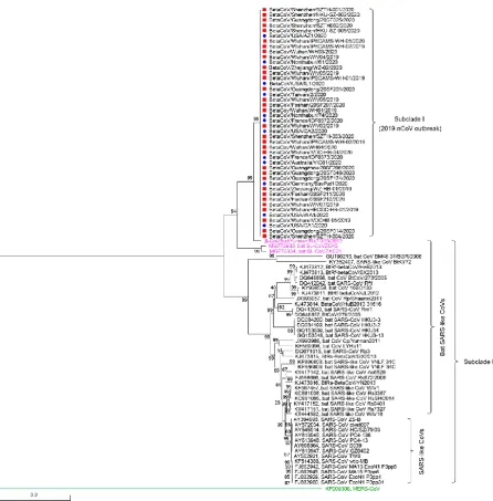
almost identical, and clustered into the same subclade, termed Subclade I (Fig. 2); ii) three bat SARS-like CoVs are closely-related cousins of Subclade I, namely
β-CoV/bat/Yunnan/RaTG13/2013, bat-SL-CoVZC45, and bat-SL-CoVZXC21 (Fig. 2); and iii) Subclade II, a neighboring cluster of Subclade I, is majorly consisted of human/civet SARS-CoVs and bat SARS-like CoVs (Fig. 2). Intriguingly, the afore-mentioned 3 bat SARS-like versions bridge Subclade I and Subclade II. This raises a possibility that Subclade I, is phylogenetically placed in the position of an
ancestor/progenitor for Subclade II, rather than in paralleled evolution. To further consolidate this hypothesis, we also performed phylogenetic analyses with two evolutionarily conserved proteins (S protein and nucleoprotein (N)). As expected, similar scenarios are also seen in phylogeny of both S protein (Fig. S3) and N protein (Fig. S4). Not only is 2019-nCoV closely adjacent to the two bat-SARS-like CoVs (bat SL-CoVZC45, and bat-SL-CoVZXC21), but also it is evolutionarily placed as an ancestor/progenitor for SARS-CoV-forming subclade (Figs S3-S4). Although it leads to the outbreak of 2019(20) atypical pneumonia, in parallels to SARS pandemic in 2002-2003, we favor believing 2009-nCoV as an ancestry version, rather than a domesticated version of SARS-CoV. This is because the latter’s survival presumably requires its already being adapted gradually to human hosts. As a result, it evolves to only give a mild illness of cold-like syndrome to some extent. On the contrary, the fact that the onset of severe respiratory
11
Entry of 2019-nCoV by S Protein
The surface S glycoprotein of 2019-nCoV plays critical roles in successful entry into the infected host cells. This is dependent on efficient binding of 2019-nCoV S to angiotensin converting enzyme 2 (ACE2) receptor, followed by the formation of membrane fusion between CoV and host (Fig. S5). In general, the S protein of 2019-nCoV consists of two distinct domains (S1 and S2, Fig. S2). As predicted, a putative receptor-binding domain (RBD, 331-583aa) is also detected in C-terminus of S1 protein (its counterpart of SARS-CoV locates at the position 318-569aa).
Furthermore, overall architecture of 2019-nCoV RBD (Fig. S5C) is in appreciable similarity to the counterparts of SARS-CoV (Fig. S5B) and other 3 bat SARS-like CoVs (Figs S5D-F). In spite that functional motifs’ organization of S protein is almost identical, receptor-binding motifs (RBM) is highly variable (Fig. S5A). When
compared with those of bat-SL-CoVZC45 and bat-SL-CoVZXC21, two discontinuous sequence inserts consistently appear in 2019-nCoV, SARS-CoV, and
β-CoV/bat/Yunnan/RaTG13/2013 (Fig. S5A). In brief, the two inserts correspond to the two regions (i.e., 434-438aa and 473-486aa for 2019-nCoV, equivalent to
431-435aa, 460-472aa in SARS-CoV, in Fig. S5A). Very recently, a modeling study by Wan et al. 31 indicates that a putative RBM of 2019-nCoV possesses five distinct
12
important amino acids (K31, E35, D38, and K353) of human ACE2 receptor (Fig. S6)
32. This might suggest the possible diversity of CoV/ACE2 receptor.
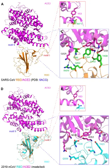
Re-analysis of cryo-EM structure of SARS-CoV RBD and its ACE2 receptor (PDB: 6ACG) allows us to illustrate a binding interface comprising two motifs (motif I & II, Fig. 3A). Obviously, binding residues of SARS-CoV RBM we propose here (Figs 3A-C) are quite different from the prediction by Wan et al. 31 on the basis of SARS-CoV RBD domain (Acc. no.: 2AJF). Indeed, N473 of RBM interacts with Q24 and Y83 of ACE2 in motif I (Fig. 3B); motif II involves at least eight pairs of hydrogen bonds [i.e., the six residues (Y408, R426, G482, Y484, T486 & Q492) provided by SARS-CoV RBM cross-talk with the six residues (D38, Y41, Q42, Q325, N330, &
K353) donated by human ACE2, Fig. 3C]. Structural modeling of 2019-nCoV
RBD/ACE2 complex also gives two unique binding motifs (Fig. 3D): i) N487 of RBM interacts with Q24 and Y83 of ACE2 in motif I (Fig. 3E); ii) though that the contact residues of ACE2 remains intact, the residues of RBM are changed into Y421, G496, T500 & Q506 (Fig. 3F). Of note, the two binding-residues (D38 and K353) of ACE2 are consistently revealed in our analysis along with other reports 31, 32. The variable
snapshots on RBM/ACE2 are in part (if not all) due to the different structural
templates we utilized 23, 31, 32. However, it might constitute a relatively comprehensive
picture on receptor recognition by 2019-nCoV S protein.
13
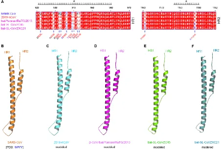
bat SARS-like CoVs (Figs 4A and S2). The two HR motifs of CoVs participate in formation of membrane fusion core with coiled-coil structure (Figs 4B-F). Not surprisingly, fusion core of 2019-nCoV (Fig. 4C) is structurally similar to those of SARS-CoV (Fig. 4B) and the other bat SARS-like CoV (Figs 4D-F). Thus, S protein of 2019-nCoV is a paradigmatic type I viral fusion protein, assuring its efficient entry into host cells.
Structural Similarity of SARS-CoV and 2019-nCoV 3CL
The chymotrypsin-like protease (3CLpro), also called main protease (Mpro), is a
prevalent nonstructural protein that mediates the proteolytic processing of viral replicase polypeptides, pp1a (486 kDa) and pp1ab (790 kDa) across different species of CoV. Unlike that papain-like protease 2 (PL2pro) cleaves three sites 33-37,
the paradigm 3CL (~34kDa) of SARS-CoV recognizes 11 cleavage sites, giving numbers of functional enzymes, like RNA-dependent RNA polymerase 25, 26. The
maturation of 3CL is dependent on its auto-processing at both N-terminal and C-terminal sites 25. Like that of SARS-CoV (Fig. 5A), a 3CLPro homologue is predicted
within the polypeptide ORF1a/b of 2019-nCoV (Fig. S2), and supposed to be excised by its own auto-cleavage ability 38. Sequence alignment indicates that the
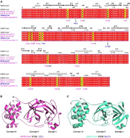
3CL enzymes have 98%~99% similarity each other, with the limited substitution of only ~10 residues (Fig. 5A). In relative to the prototypical version (H41 and C145) of SARS-CoV 26, an evolutionarily-conserved catalytic dyad (H41 and C145) is also
14
to define that SARS-CoV 3CL is composed of three unique domains 24. Among them,
the former two domains (Domain I & II) are structurally featuring with anti-parallel β-barrels and represent the reminiscent of trypsin-like serine proteases (Fig. 5B). By contrast, the last one (Domain III) that is linked to Domain II, seems to display a globular structure comprising five α-helices (Fig. 5B). A similar scenario is also seen in the overall structure of 2019-nCoV 3CL (PDB: 6LU7). More importantly, the fusion inhibitors targeting HR1 domain is a promising therapy against the 2019-nCoV infections 39. Such almost-identical structural snapshots of 3CL proteases validate
15
Conclusion
As of preparing this manuscript, WHO(https://www.who.int/) renamed the severe respiratory illness caused by 2019-nCoV epidemic, as Corona Virus Infectious Disease (COVID-19). Meanwhile, the International Committee on Taxonomy of Viruses has replaced 2019-nCoV with SARS-CoV-2
(https://talk.ictvonline.org/) on February 11, 2020. Obviously, such declaration aims to formally recognize it as a cousin of SARS-CoV. Given that the clinical
consequence of 2019-nCoV is paralogous to that of SARS, we agree with this announcement of renaming it to some extents. It rules out the possibility that 2019-nCoV is regarded a variant of SARS-CoV domesticated in an ongoing evolution. However, our phylogeny of 2019-nCoV combined with series of structural analysis also proposes an alternative explanation for the emergence of 2019-nCoV. This denotes that 2019-nCoV can be a progenitor for SARS-CoV with bat origin.
16
Acknowledgements
This work was financially supported by National Natural Science Foundation of China (31830001, YF).
Declaration of interest statement
The authors declare that they have no competing interests.
Author contributions
17
References
1. Lee, N. et al. A major outbreak of severe acute respiratory syndrome in Hong Kong. N Engl J Med 348, 1986-1994 (2003).
2. Ksiazek, T.G. et al. A novel coronavirus associated with severe acute respiratory syndrome. N Engl J Med 348, 1953-1966 (2003).
3. Holmes, E.C., Dudas, G., Rambaut, A. & Andersen, K.G. The evolution of Ebola virus: Insights from the 2013-2016 epidemic. Nature 538, 193-200 (2016).
4. Cui, J., Li, F. & Shi, Z.L. Origin and evolution of pathogenic coronaviruses. Nat Rev Microbiol 17, 181-192 (2019).
5. Gao, G.F. From "A"IV to "Z"IKV: Attacks from Emerging and Re-emerging Pathogens. Cell 172, 1157-1159 (2018).
6. Perlman, S. & Netland, J. Coronaviruses post-SARS: update on replication and pathogenesis. Nat Rev Microbiol 7, 439-450 (2009).
7. Zumla, A., Chan, J.F., Azhar, E.I., Hui, D.S. & Yuen, K.Y. Coronaviruses - drug discovery and therapeutic options. Nat Rev Drug Discov 15, 327-347 (2016).
8. Poon, L.L. et al. Identification of a novel coronavirus in bats. J Virol 79, 2001-2009 (2005).
9. Lau, S.K. et al. Severe acute respiratory syndrome coronavirus-like virus in Chinese horseshoe bats. Proc Natl Acad Sci U S A 102, 14040-14045 (2005).
10. Hu, D. et al. Genomic characterization and infectivity of a novel SARS-like coronavirus in Chinese bats. Emerg Microbes Infect 7, 154 (2018).
11. Feng, Y. & Gao, G.F. Towards our understanding of SARS-CoV, an emerging and devastating but quickly conquered virus. Comp Immunol Microbiol Infect Dis 30, 309-327 (2007).
12. Rota, P.A. et al. Characterization of a novel coronavirus associated with severe acute respiratory syndrome. Science 300, 1394-1399 (2003).
13. Assiri, A. et al. Epidemiological, demographic, and clinical characteristics of 47 cases of Middle East respiratory syndrome coronavirus disease from Saudi Arabia: a descriptive study. Lancet Infect Dis 13, 752-761 (2013).
14. de Groot, R.J. et al. Middle East respiratory syndrome coronavirus (MERS-CoV): announcement of the Coronavirus Study Group. J Virol 87, 7790-7792 (2013).
15. Li, Q. et al. Early Transmission Dynamics in Wuhan, China, of Novel Coronavirus-Infected Pneumonia. N Engl J Med (2020).
16. Zhu, N. et al. A Novel Coronavirus from Patients with Pneumonia in China, 2019. N Engl J Med (2020).
17. Huang, C. et al. Clinical features of patients infected with 2019 novel coronavirus in Wuhan, China. Lancet (2020).
18. Chan, J.F. et al. A familial cluster of pneumonia associated with the 2019 novel coronavirus indicating person-to-person transmission: a study of a family cluster. Lancet (2020).
19. Wang, C., Horby, P.W., Hayden, F.G. & Gao, G.F. A novel coronavirus outbreak of global health concern. Lancet (2020).
20. Phan, L.T. et al. Importation and Human-to-Human Transmission of a Novel Coronavirus in Vietnam. N Engl J Med (2020).
18
22. The, L. Emerging understandings of 2019-nCoV. Lancet (2020).
23. Song, W., Gui, M., Wang, X. & Xiang, Y. Cryo-EM structure of the SARS coronavirus spike glycoprotein in complex with its host cell receptor ACE2. PLoS Pathog 14, e1007236 (2018). 24. Xu, Y. et al. Crystal structure of severe acute respiratory syndrome coronavirus spike protein fusion
core. J Biol Chem 279, 49414-49419 (2004).
25. Muramatsu, T. et al. SARS-CoV 3CL protease cleaves its C-terminal autoprocessing site by novel subsite cooperativity. Proc Natl Acad Sci U S A 113, 12997-13002 (2016).
26. Yang, H. et al. The crystal structures of severe acute respiratory syndrome virus main protease and its complex with an inhibitor. Proc Natl Acad Sci U S A 100, 13190-13195 (2003).
27. Wu, F. et al. A new coronavirus associated with human respiratory disease in China. Nature (2020). 28. Marra, M.A. et al. The Genome sequence of the SARS-associated coronavirus. Science 300,
1399-1404 (2003).
29. Zhou, P. et al. A pneumonia outbreak associated with a new coronavirus of probable bat origin. Nature (2020).
30. Lu, R. et al. Genomic characterisation and epidemiology of 2019 novel coronavirus: implications for virus origins and receptor binding. Lancet (2020).
31. Wan, Y., Shang, J., Graham, R., Baric, R.S. & Li, F. Receptor recognition by novel coronavirus from Wuhan: An analysis based on decade-long structural studies of SARS. J Virol (2020).
32. Li, F., Li, W., Farzan, M. & Harrison, S.C. Structure of SARS coronavirus spike receptor-binding domain complexed with receptor. Science 309, 1864-1868 (2005).
33. Sulea, T., Lindner, H.A., Purisima, E.O. & Menard, R. Deubiquitination, a new function of the severe acute respiratory syndrome coronavirus papain-like protease? J Virol 79, 4550-4551 (2005). 34. Lindner, H.A. et al. The papain-like protease from the severe acute respiratory syndrome
coronavirus is a deubiquitinating enzyme. J Virol 79, 15199-15208 (2005).
35. Harcourt, B.H. et al. Identification of severe acute respiratory syndrome coronavirus replicase products and characterization of papain-like protease activity. J Virol 78, 13600-13612 (2004). 36. Gao, F., Ou, H.Y., Chen, L.L., Zheng, W.X. & Zhang, C.T. Prediction of proteinase cleavage sites in
polyproteins of coronaviruses and its applications in analyzing SARS-CoV genomes. FEBS Lett 553, 451-456 (2003).
37. Barretto, N. et al. The papain-like protease of severe acute respiratory syndrome coronavirus has deubiquitinating activity. J Virol 79, 15189-15198 (2005).
38. Muramatsu, T. et al. Autoprocessing mechanism of severe acute respiratory syndrome coronavirus 3C-like protease (SARS-CoV 3CLpro) from its polyproteins. FEBS J 280, 2002-2013 (2013). 39. Xia, S. et al. Fusion mechanism of 2019-nCoV and fusion inhibitors targeting HR1 domain in spike
19
Figure legends
Figure 1 Genomic organization and similarity analysis of 2019-nCoV
A. Scheme for genomic organization of 2019-nCoV
B. Genome-wide similarity analyses of 2019-nCoV with five closely-related coronaviruses
20
Figure 2 Phylogenetic analysis of 2019-nCoV in the context of β-coronaviruses Maximum likelihood-based phylogeny of full genomes of 2019-nCoV isolates was generated with the MEGA software. MERS-CoV acts as an outgroup here. It infers that i) all the 2019-nCoV isolates form a unique subclade, termed Subclade I; ii) the representative isolates of SARS-CoV and bat SARS-like CoV can be organized in a neighboring subclade, named Subclade II; and three bat SARS-like CoVs
21
22
23
A. Complex structure of SARS-CoV RBD and its receptor ACE2
RBD of SARS-CoV spike protein is in orange, and ACE2 is given in magenta. Two critical binding motifs (motif I and motif II) are squared with dashed lines.
Enlarged views of the interface between SARS-CoV RBD and ACE2, consisting of motif I (B) and motif II (C)
The binding of SARS-CoV spike protein its cellular receptor ACE2 is revealed by Song et al. 23 with Cryo-EM structure deposited into NCBI under accession number:
6ACG.
D. Modeled structure of 2019-nCoV RBD complexed with its putative receptor ACE2 Unlike that ACE2 is in purple, RBD of 2019-nCoV S protein is in cyan. The two motifs (motif I and motif II) essential for RBD/ACE2 binding are squared with dashed lines. Structural snapshots of the binding of 2019-nCoV RBD to ACE2, which are featuring with motif I (E) and motif II (F)
The important residues from ACE2 are labeled with dark letters, and those of SARS-CoV RBD are indicated with green letters. In contrast, the residues of 2019-nSARS-CoV RBD implicated into its contact with ACE2 are highlighted in cyan.
24
Figure 4 Sequence and structural analyses of membrane protein fusion cores of 2019-nCoV
A. Sequence comparison of two heptad regions (HR), HR1 and HR2, from 2019-nCoV and its related four coronaviruses
The detailed position of HR1 (HR2) in spike proteins are depicted in Fig. S2.
B. Ribbon presentation of SARS-CoV membrane protein fusion core, a heterodimer bundle of HR1 and HR2
It is derived from the S protein structure of SARS-CoV (PDB: 1WYY).
C. Modeled structure of 2019-nCoV membrane protein fusion core Ribbon structures of membrane protein fusion cores of
25
26
Figure 5 Sequence and structural similarity of SARS-CoV and 2019-nCoV 3CL enzymes
A. Sequence alignment of 3CL proteases
The catalytic dyad (H41 and C145) is highlighted with triangle. The residue substitution is indicated.
27
2019-nCoV 3CL protease. However, only three residues alteration is present in 3CL protease of 2019-nCoV when compared with CoVZXC21 (and/or bat-SL-CoVZC45). Two residues of 3CL enzyme are different between 2019-nCoV and β-CoV/bat/Yunan/RaTG/2013.
B. Crystal structure of SARS-CoV 3CL protease in monomeric form (PDB: 1Z1I)
C. Ribbon structure of 2019-nCoV 3CL enzyme (PDB: 6LU7)
28
Supplementary Materials for
Figure S1 Genomic context of 2019-nCoV and other viral relatives
Figure S2 Linear scheme for distinct domains/motifs in spike protein of different coronaviruses
Figure S3 Phylogeny of 2019-nCoV surface spike protein
Multiple sequence alignment of spike protein was conducted with Clustal Omega (https://www.ebi.ac.uk/Tools/msa/clustalo/), and the phylogenic analysis was given with TreeView.
Figure S4 Phylogeny of 2019-nCoV nucleoprotein
Phylogenetic tree of nucleoproteins was generated with TreeView, which follows the multiple sequence alignment with Clustal Omega
(https://www.ebi.ac.uk/Tools/msa/clustalo/).
Figure S5 Structural analyses of RBD from 2019-nCoV and four closely-related CoVs
29
B. Ribbon structure of RBD from SARS-CoV
C. Modeled structure of 2019-nCoV RBD
Structural architectures of RBD from β-CoV/bat/Yunnan/RaTG/2013 (D), bat-SL-CoVZC45 (E), and bat-SL-CoVZC45 (F)
Figure S6 Sequence analysis of the ACE2 receptor