Page 1 of 44
Manuscript
(Review Article)
A comprehensive review on inherited Sensorineural Hearing Loss and their
syndromes
Authors
Muhammad Noman 1, Shazia Anwer Bukhari 1, Muhammad Tahir 2, & Shehbaz Ali 3*
Author’s information
1 Department of Biochemistry, Molecular Biology laboratory, Government College, University,
Faisalabad, 38000, Pakistan.
2 Department of Oncology, Allied Teaching Hospital Faisalabad, Faisalabad Medical University,
Faisalabad, 38000, Pakistan.
3 Department of Biosciences and Technology, Khwaja Fareed University of Engineering and
information technology, Rahim Yar Khan, Punjab, Pakistan.
*Correspondence:
Shehbaz Ali: shehbaz205@gmail.com
Tel: +92-333-7477407
Abstract
Hearing impairment is an immensely diagnosed genetic cause, 5% of the total world population
effects with different kind of congenital hearing loss (HL). In third-world countries or countries
where consanguineous marriages are more common the frequency rate of genetic disorders are at
its zenith. Approximately, the incidence of hearing afflictions is ostensibly 7-8:1000 individuals
whereas it is estimated that about 466 million peoples suffer with significant HL, and of theses
deaf cases 34 million are children’s up to March, 2020. Several genes and colossal numbers of
pathogenic variants cause hearing impairment, which aided in next-generation with recessive,
dominant or X-linked inheritance traits. This review highlights on syndromic and non-syndromic
HL (SHL and NSHL), and categorized as conductive, sensorineural and mixed HL, which having
autosomal dominant and recessive, and X-linked or mitochondrial mode of inheritance. Many
hundred genes involved in HL are reported, and their mutation spectrum becomes very wide.
Page 2 of 44
Mapping of pathogenic genes in consanguinity family is facilitated to understand the disease
history. Review presents the bases of HL and also focused on various genetic factors that cause
deafness like the basics of genetic inheritance, and classic and well-characterized inherited factors
of it. It also overviews the application of linkage analysis, SNPs genotyping and whole exome
sequencing methods, in mapping and identification of new locus, causative genes and their variants
in families inherited with HL. Conclusively, this review supports researchers in understanding the
location of chromosome, the causative genes and specific locus which causing deafness in humans.
Keywords: Hereditary HL, genetics of syndromic and non-syndromic HL, methods for diseased
locus/gene identification
Introduction
Deafness or hearing failures are seen as an extenuate form [1], in human it is one of the high
prevailing neurosensory deficits that harshly negotiate the life value of individuals and can cause
their social separation [2]. Both genetics and environmental factors cause hearing failure [3], and
the genetic factors contributes about 50% of all hearing loss (HL) patients [4]. The worldwide
estimated results report as of March, 2020 defines rounded 466 million peoples suffer with
significant HL, and of theses deaf cases 34 million are children’s (under 6 years old) (WHO
MARCH 2020). Furthermore, before maturity 3/1000 children become deaf [5]. According to
WHO prediction, hearing disability will affect ~900 million (or 1:10 peoples) by 2050 [6].
Basically, HL was categorized in two main groups non-syndromic sensorineural HL (NSHL) and
syndromic sensorineural HL (SSHL). Genetic subsidizing factors to NSHL are remarkably diverse
coverings, over autosomal (recessive and dominant), to X-linked (recessive and dominant), to
mitochondrial patterns of inheritance [7]. The SSHL, hearing disability appeared with multiple
physiological anomalies (diseases), and it is limited only to the inner ear [8].
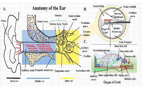
Many hundreds or even thousands of genes are involved in hearing process and helps in proper
functioning of inner ear, which is the most sensitive part of the ear in human body (figure 2B).
Several genes and their expressed protein families like (Myosin family, Gap-junction family and
solute carrier proteins etc.) in inner ear function as, in control of adhesion of hair cell, in
neurotransmitter release, intercellular transport, maintenance of ionic homeostasis and protection
Page 3 of 44
During the period of last 10-12 years, the identification rate of causative genes associated with
hearing loss becomes very high. Several hundred genes associated with hearing loss are reported,
and their mutation spectrum becomes very wide, so that identification of disease-causing mutation
is still more difficult. Linkage studies and auto zygosity methods used for mapping and
identification of pathogenic genes in consanguineous families and with the advancement in
technologies, Next-generation Sequencing, target-enrichment method and sanger sequencing
makes it easy to identifying the novel gene and their variant in inherited heterogeneous disorders
[11-14]. Whole Exome Sequencing (WES), used as stream-line approach now a days, for
identifying the disease causing (causative) gene variants (mutation), which results specific
phenotypic disorder [10, 15]. This review presents an overview and description of the currently
known genes related to hereditary HL. It reviews the basics of genetic inheritance, and also
focusing on the classic and well-characterized, inherited factors that cause deafness. Brief
overview of this review study shown in figure 1.
Figure 1: Overview and schematic illustration of complete review of literature.
Page 4 of 44
Auditory system of mammals is highly sensitive, integrated and the most complicated structure,
which is planned to achieve both functions of interpreting the sound waves in an organized manner
to nerve impulse and also to sustain the balance of the body. The vestibular systems of the human
ear specific to sustain the balance of the body are composed of two parts: the membranous
labyrinth and the bony labyrinth [16]. The function of the human ear is to collect sound waves
from the sounding and interpret of these sound waves of different sound frequencies of range 20
to 20,000 Hz [17, 18]. Ear can be defined as a microcomputer or an analytic microphone, that
conducts sound waves towards the brain in type of nerve impulse, and it is divided into three
structural partitions, which works like a unit; the outer ear; pinna, the auditory canal, and the
tympanic membrane, middle ear; tympanic cavity, Ossicles bones (incus, malleus, and stapes),
middle ear muscles and Eustachian tube, and the inner ear that perform two functions, transduction
of sound waves into neurochemical signals which completed in cochlea, a main functioning organ
of the ear and to maintains the optic fixation and support to sustain sanding body posture that takes
place in the vestibular system during the process of movement (figure 2A) [19-21].
Figure 2: A. Outer ear the pinna and auditory canal separated from the middle ear by tympanic
Page 5 of 44
to the Eustachian tube at the back of nose. The inner ear holds Cochlea and vestibular structures
specific to generate nerve impulse and sustain balance of the body. B. Cochlea; the boney tube,
filled with perilymph, in which membranous labyrinth floats filled with endolymphatic fluid.
Perilymph separates the Scala media to Scala tympani. C. A cross-section of single piece Cochlea
display comprehensive picture of the membranous labyrinth, and the Basilar membrane keeps the
epithelial cells of hearing –the organ of Corti. The organ of Corti holds; inner hair cells (IHC’s),
three outer hair cells (OHC’s), Hensen cells (HC’s), Deitters cells (DC’s), Pillar cells (PC’s) and
the Inner Supporting cells (ISC’s) respectively. The auditory nerve linked to inner hair cell at their
tip-link.
Organ of corti and hair cells
The power of identifying and separating the variable frequencies sounds of human cochlea mainly
based on the portion of sensory epithelia named as organ of Corte; at the sensorineural end, the
organ necessary for listening a sound (figure 2C) [22]. It contains placodal origin (membranous
labyrinth) polarized epithelial cells (supporting and hair cells), the basil membrane (specialized
basement membrane having layer of matrix), tectorial membrane and nerve endings [19, 23, 24].
In mammals, two types of hair cells are present, the inner hair cells (IHCs) and outer hair cells
(OHCs). The “IHCs” actually the type of true sensory cell, transmits impulses through the auditory
nerve, and OHCs are obliging in to increase the working capacity of the cochlea, quantitatively
(increased sensitivity) and qualitatively (increased selectivity) [25, 26]. The name "hair" cell was
derived from the tuft of stereocilia that protrude from the apical domain of every cell [21, 27, 28].
Mechanism and types of hearing failure
Hearing failure may be partial or complete and it developed either in response of a damage, injury,
physiological causes or congenital diseases which specify as conductive HL [29].Whereas when
any injury or damage occurs in the inner ear, brain or vestibular nerve caused sensorineural hearing
loss (SNHL), and mixed hearing damage caused both conductive and SNHL. The SNHL mostly
occurred due to genetic variations in genes that regulate the intracellular transport, the adhesion of
hair cells, ionic homeostasis, neurotransmitter release and structure of hair cells results to damage
of the cochlea and the inner ear [30]. In the current century, with new inventions of genetic variants
in congenital hearing loss, new treatment opportunity and genetic counseling have appeared and
Page 6 of 44
Detection of hearing
Hearing level of suspects was evaluated through behavioral testing and pure tone audiometry.
Behavioral testing includes visual reinforcement audiometry (VRA) and behavioral observation
audiometry (BOA) [32]. VRA is used for testing hearing level of child between age from six
months to 2 ½ years and can provide reasonable complete information for audiogram while the
BOA is used for evaluating the hearing level of infants from birth to six month age and this kind
of testing is highly dependents on the skills of the testing persons, and is subject to error [33, 34].
Pure tone audiometry means to identify the minimum frequency on which a person "hear" a pure
tone, whereas the "bone conduction audiometry, depends on the sound waves reach the ear through
a vibrator consists on the forehead mastoid bone, now the thresholds depend on the condition of
the inner ear, by bypassing the outer and middle ear and the calculated/obtained values are plotted
on a graph paper [35-38], for sample of audiogram (figure 3).
Figure 3: The audiogram sample, defining various types of hearing loss constructed on the
basis of types of ear defect. Horizontal axis represents frequencies in "Hz" while the vertical axis
Page 7 of 44
audiogram, the right ear is denoted with the symbol "O" and the left ear is denoted with symbol
"X".
Molecular genetics of HL
Hearing failure is the most common sensory impairment. It shows highly heterogeneous behavior.
The early 1990s, the identification and localization of genes causing deafness/HL is started, but
till 1994 only a few gene loci have been mapped/identified on human genome; causing hearing
loss/deafness either NSHL/SHL [27, 39]. Inherited HL consists 50–60% of all HL cases. The
inherited form of HL is further classified to different categories [40-42]. Recent advances in
genetics and genomics have led us to identification of over 300 SHLs and more than 100
chromosomal loci and more than 40 genes responsible for NSHL [30, 43, 44]. Better understanding
of impaired genes and their structure and function will open a new window for screening as well
as the genetic approach to treatment of HL. In contrast, the identification of the single causative
gene of linking in NSHL becomes very difficult in a single family, because it needs positional
cloning; the linkage analysis and WES make it feasible.
Non-syndromic sensorineural Hearing Loss (NSHL)
Hereditary HL (HHL) is an immensely studied neurosensory disorder in worldwide. It is highly
heterogeneous genetic disorder, and most often autosomal recessive and non-syndromic is
approximately 80% of congenital HL [11, 45-48]. Most of the studies on NSHL predominantly focused
on three main aspects; the kind of hearing defect, its degree of severity, and the configuration or
inheritance pattern. Almost 60% cases of the congenital HL are on the account of genetic factors [49].
In humans hearing failure is a sensory disability that ambits from mild, moderate, severe and profound.
Approximately, the profound HL is comprised of 20-25%, while a higher ratio of individuals is
damaged with moderate to severe HL [50].
Moreover, the NSHL are sorted in consonance with their inheritance patterns; as autosomal (dominant
or recessive) or linked. Autosomal inheritance patterns of NSHL found ubiquitous, while the
X-linked inheritance pattern found tremendously rare [51]. In pre-lingual HL; inherited X-X-linked trait
(1%-3%), autosomal recessive trait (70%-80%), while autosomal dominant trait (12%-24%), and
mitochondrial (2%-3%) are observed [52]. In the NSHL, either an autosomal dominant or recessive
inheritance pattern characterizes thrilling genetic heterogeneity, as more than hundreds specific
Page 8 of 44
causative-genes, most of them were reported from Pakistan according to hereditary hearing loss
homepage [53].
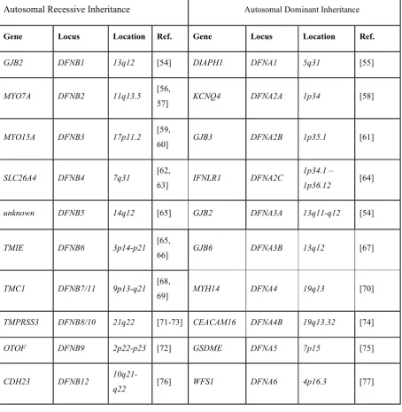
Table 1: Pathogenic genes, locus and their positions on chromosomes, causing non-syndromic
hearing loss in Humans
MUTATED GENES OF HEREDITARY NON-SYNDROMIC HEARING LOSS
Autosomal Recessive Inheritance Autosomal Dominant Inheritance
Gene Locus Location Ref. Gene Locus Location Ref.
GJB2 DFNB1 13q12 [54] DIAPH1 DFNA1 5q31 [55]
MYO7A DFNB2 11q13.5 [56,
57] KCNQ4 DFNA2A 1p34 [58]
MYO15A DFNB3 17p11.2 [59,
60] GJB3 DFNA2B 1p35.1 [61]
SLC26A4 DFNB4 7q31 [62,
63] IFNLR1 DFNA2C
1p34.1 –
1p36.12 [64]
unknown DFNB5 14q12 [65] GJB2 DFNA3A 13q11-q12 [54]
TMIE DFNB6 3p14-p21 [65,
66] GJB6 DFNA3B 13q12 [67]
TMC1 DFNB7/11 9p13-q21 [68,
69] MYH14 DFNA4 19q13 [70]
TMPRSS3 DFNB8/10 21q22 [71-73] CEACAM16 DFNA4B 19q13.32 [74]
OTOF DFNB9 2p22-p23 [72] GSDME DFNA5 7p15 [75]
CDH23 DFNB12
Page 9 of 44
unknown DFNB1
3 7q34-36 [78] LMX1A DFNA7 1q21-q23 [79, 80]
unknown DFNB14 7q31 [78] TECTA DFNA8 11q22-24 [81]
GIPC3 DFNB15 3q21-q25 [82,
83] COCH DFNA9 14q12-q13 [84]
STRC DFNB16
15q21-q22 [85] EYA4 DFNA10 6q22-q23 [86]
unknown DFNB17 7q31 [87,
88] MYO7A DFNA11 11q12.3-q21 [89]
USH1C DFNB18
11p14-15.1 [90] TECTA DFNA12 11q22-24 [81]
unknown DFNB19 18p11 [91] COL11A2 DFNA13 6p21 [92]
unknown DFNB20
11q25-qter [93] WFS1 DFNA14 4p16.3 [94]
TECTA DFNB21 11q [95] POU4F3 DFNA15 5q31 [96]
OTOA DFNB22 16p12.2 [97] unknown DFNA16 2q24 [98]
PCDH15 DFNB23
10p11.2-q21 [99] MYH9 DFNA17 22q [100]
RDX DFNB24 11q23 [101] unknown DFNA18 3q22 [102]
GRXCR1 DFNB25 4p13 [103] ACTG1 DFNA20 17q25 [104,
105]
unknown DFNB26 4p31 [106] unknown DFNA21 6p21 [107]
unknown DFNB27 2q23-q31 [108,
109] unknown DFNA22 6q13 [110]
Page 10 of 44
CLDN14 DFNB29 21q22 [113] unknown DFNA24 4q [114]
MYO3A DFNB30 10p11.1 [115] unknown DFNA25 12q21-24 [116]
WHRN DFNB31 9q32-q34 [117] ACTG1 DFNA26 17q25 [118]
CDC14A DFNB32/10
5
1p13.3-22.1 [119] unknown DFNA27 4q12 [120]
unknown DFNB33 9q34.3 [121] GRHL2 DFNA28 8q22 [122]
ESRRB DFNB35
14q24.1-24.3 [123] unknown DFNA30 15q25-26 [124]
ESPN DFNB36 1p36.3 [125] unknown DFNA31 6p21.3 [126]
MYO6 DFNB37 6q13 [127] unknown DFNA33 13q34-qter [128]
unknown DFNB38 6q26-q27 [129] NLRP3 DFNA34 1q44 [130]
HGF DFNB39 7q21.1 [131] DFNA36 DFNA36 9q13-q21 [69]
unknown DFNB40 22q [132] WFS1 DFNA6 4p16.3 [77, 94]
ILDR1 DFNB42
3q13.31-q22.3 [133] DSPP DFNA39 4q21.3 [134]
ADCY1 DFNB44
7p14.1-q11.22 [135] P2RX2 DFNA41 12q24-qter [136]
unknown DFNB45 1q43-q44 [137] unknown DFNA42 5q31.1-q32 [138]
unknown DFNB46 18p11.32
-p11.31 [139] unknown DFNA43 2p12 [140]
unknown DFNB47
2p25.1-p24.3 [141] CCDC50 DFNA44 3q28-29 [142]
CIB2 DFNB48
15q23-q25.1
[143,
Page 11 of 44 MARVELD2
/ BDP1 DENB49
5q12.3-q14.1 [146] MYO1A DFNA48 12q13-q14 [145]
unknown DENB51
11p13-p12 [147] MIRN96 DFNA50 7q32.2 [148]
COL11A2 DENB53 6p21.3 [149] TJP2 DFNA51 9q21 [150]
unknown DENB55
4q12-q13.2 [151] unknown DFNA52 4q28 [138]
PJVK DENB59
2q31.1-q31.3 [152] unknown DFNA53 14q11.2-q12 [153]
SLC22A4 DENB60
5q23.2-q31.1 [154] unknown DFNA54 5q31 [155]
SLC26A5 DENB61 7q22.1 [156] TNC DFNA56 9q31.3-q34.3 [157]
unknown DENB62
12p13.2-p11.23
[158,
159] unknown DFNA57 19p13.2 [160]
LRTOMT/
COMT2 DENB63
11q13.2-q13.4 [161] unknown DFNA58 2p12-p21 [162]
unknown DENB65
20q13.2-q13.32 [163] unknown DFNA59
11p14.2-q12.3 [164]
DCDC2 DENB66 6p21.2—
22.3
[165,
166]
SMAC/DIAB
LO DFNA64
12q24.31-q24.32 [167]
LHFPL5 DENB66/67 6p21.31 [168] TBC1D24 DFNA65 16p13.3 [169]
S1PR2 DENB68 19p13.2 [170] CD164 DFNA66 6q15-21 [171]
BSND DENB73 1p32.3 [172] OSBPL2 DFNA67 20q13.33 [173]
MSRB3 DENB74
12q14.2-q15
[174,
Page 12 of 44
SYNE4 DENB76 19q13.12 [177] MCM2 DFNA70 3q21.3 [178]
LOXHD1 DENB77
18q12-q21 [179] KITLG
Unknown
12q21.32-q23.1 [180]
TPRN DENB79 9q34.3 [181] PTPRQ DFNA73 12q21.31
Unknown DENB80
2p16.1-p21 [182] DMXL2 Unknown 15q21.2 [183]
Unknown DENB81 19p [83] MYO3A Unknown 10p12.1 [184]
Unknown DFNB83
2p25.1-p24.3 [185] REST DFNA27 4q12 [120]
PTPRQ/
OTOGL DENB84 12q21.2 [186] COL11A1 DFNA37 1p21 [187]
Unknown DENB85
17p12-q11.2 [188] PDE1C Unknown 7p14.3 [189]
TBC1D24 DENB86 16p13.3 [190] TRRAP Unknown 7q22.1 [191]
ELMOD3 DENB88
2p12-p11.2 [192] PLS1 Unknown 3q23 [193]
KARS DENB89
16q21-q23.2 [194] SCDS Unknown 4q21.22 [195]
Unknown DENB90
7p22.1-p15.3 [196] SLC12A Unknown 5q23.3 [197]
SERPINB6 DENB91 6p25 [198]
SEX-LINKED INHERETANCE
CABP2 DENB93
11q12.3-11q13.2 [199]
FAM65B DENB104 6p22.3 [200] Gene Locus Locatio
n
Referenc
Page 13 of 44
CDC14A DFNB32/10
5
1p13.3-22.1 [201] PRPS1 DFNX1 Xq22 [202]
GIPC3 DENB95 19p13 [203] POU3F4 DFNX2 Xq21.1 [204]
Unknown DENB96
1p36.31-p36.13 [205] Unknown DFNX3 Xp21.2 [206, 207]
MET DENB97
7q31.2-q31.31 [208] SMPX DFNX4 Xp22 [209]
TSPEAR DENB98
21q22.3-qter
[203,
210] AIFM1 DFNX5 Xq26.1
[211]
TMEM132E DENB99 17q12 [205,
212] COL4A6 DFNX6 Xp22.3
[51]
PPIP5K2 DENB100
5q13.2-q23.2 [213] Unknown DFNY1 Y [214]
GRXCR2 DENB101 5q32 [210,
215]
EPS8 DENB102 12p12.3 [212]
WBP2 Unknown 17q25.1 [216]
ESRP1 Unknown 1p13.3 [217]
MPZL2 Unknown 11q23.3 [218]
CEACAM16 Unknown 19q13.31
-q13.32 [187]
GRAP Unknown 17p11.2 [219]
SPNS2 Unknown 17p13.2 [220]
Page 14 of 44
Syndromic Hearing Loss (SHL)
Childhood congenital SHL is a major cause of birth defects in developed countries. There are many
reasons are existed to study and identify the etiology of the HL [222]. Approximately 30% of all
reported HL cases have several clinical anomalies with HL and termed as SHL [49]. These are
differentiated from other types of HL on the basis of associated symptoms in several vital organs
[223]. It is estimated that above 400 different syndromes of HL were reported and the majority of
the cases had been identified with the pathogenic genes [49]. This literature review focuses on the
most common syndromes that highly diagnosed in various populations and their linked pathogenic
genes (table 2). Major syndromes with HI are Alport, Stickler, Jervell & Lange-Nielsen,
Waardenburg and Usher syndromes etc. Stickler and Waardenburg syndromes have dominant
inheritance patterns, while the syndromes having autosomal recessive inheritance patterns are
Usher and Jervell & Lange-Nielsen syndrome and the Alport syndrome is usually inherited with
X-linked inheritance pattern [69, 224].
Table 2: Syndromes, Mutated Genes, and their chromosomal location
Syndrome Location Gene Locus PHENOTYPE
Alport Syndrome
Xq22 COL4A5 … X-linked and autosomal recessive,
progressive highly prevalent SNHL;
specific form of glomerulonephritis.
The recessive genes are COL4A6 and
COL4A4 respectively.
2q36-q37 COL4A3 …
2q36.3 COL4A4
Branchio-oto-renal
syndrome
14q21.3-q24.3
SIX1 BOS3
Autosomal dominant, pre-auricular
ear pits, brachial pits and Sinuses,
pinna abnormalities and renal
hypoplasia.
19q13.3 SIX5 BOR2
1q31 unknown …
8q13.3 EYA1 BOR1
Charge syndrome
7q21.11 SEMA3A … Inherited as autosomal dominant, it
represents Acronym Coloboma,
Atresia, ear anomalies, Heart defects,
Page 15 of 44
and retarded development and
growth.
Jervell &
lange-nelsen syndrome
11p15.5 KCNQ1 JLNS1 Inherited as autosomal recessive,
congenital profound SNHL with
missing vestibular function and is
also commonly known as QT
syndrome.
21q22.1-q22.2
KCNE1 JLNS2
Norrie syndrome
Xp11.3 NDP NDP Inherited as X-linked progressive
SNHL mostly appeared in second life
decade, intellectual disability and
congenital retinal detachment.
Penderd syndrome
7q21-34 SLC26A4 PDS Progressive high-frequency SNHL
and inherited as autosomal recessive,
with thyroid failure, incomplete
partitioning of the cochlea and
enlarged vestibular aqueducts.
5q35.1 FOX11 PDS
1q23.2 KCNJ10 PDS
Stickler syndrome
12q13.11-q13.2
COL2A1 STL1
Inherited as autosomal dominant
inheritance pattern, Affects Cleft
palate, flat center-face, highly
frequent SNHL, retinal detachment
and high myopia; arthropathy.
1p21 COL11A1 STL2
6p21.3 COL11A2 STL3
6q13 COL9A1 …
1p34.2 COL9A2 …
TREACHER
COLLIN
SYNDROME
5q32-q33.1
TCOF1 TCOF1 Inherited as autosomal dominant
inheritance pattern, results in
symmetrical and bilateral pinna
abnormalities with mental issues,
coloboma of lower eyelids, spars in
eyelashes, cleft palate, hypoplasia of
mandible and zygomatic complex.
13q12.2 POLR1D POLR1D
Page 16 of 44
USHER
SYNDROME
14q32 nonexistent USH1A
RP (Retinitis pigmentosa) with
SNHL. Type 1 of usher syndrome,
profound congenital SNHL, absent
vestibular response and RP (Retinitis
pigmentosa) develops in the first life
decade. In type 2 of the usher
syndrome, sloping congenital SNHL,
with normal vestibular response and
Retinitis pigmentosa (Verpy et al.)
develops in the early and late onset of
life; while in case of Usher syndrome
type 3, progressive SNHL with
erratic vestibular response and erratic
period of the RP (Retinitis
pigmentosa) develops.
11q13.5 MYO7A USH1B
11p15.1 USH1C USH1C
10q22.1 CDH23 USH1D
21q21 Unknown USH1E
10q21-22 PCDH15 USH1F
17q24-25 SANS USH1G
15q22-23 Unknown USH1H
15q23-q25.1
CIB2 USH1J
10p11.21-q21.1
Unknown USH1K
1q41 USH2A USH2A
3p23-24.2 Unknown USH2B
5q14.3-q21.3
VLGR1 USH2C
9q32 WHRN USH2D
3q21-q25 CLRN1 USH3
5q31.3 HARS USH3B
10q24.31 PDZD7 ModiferGene
WAARDENBURG
SYNDROME
2q35 PAX3 WS1 SNHL with pigmentary anomalies of
skin, eye, and hair. In type 1;
autosomal dominant with hypoplasia
of alaenasi, synophrys and dystopia
3p14.1-p12.3
Page 17 of 44
1p21-p13.3
unknown WS2B canthorum appears. In type 2;
autosomal dominant and facial
features and dystopia canthorum are
absent. In type 3; autosomal
dominant and is also known as
Klein-Waardenburg syndrome: upper limb
abnormalities plus type 1 syndrome.
while in type 4; autosomal recessive
and also known as
Waardenburg-Shah Syndrome: Hirschsprung
disease plus type 2 syndrome.
8p23 unknown WS2C
8q11 SNAI2 WS2D
2q35 PAX3 WS3
13q22 EDNRB WS4
20q13.2-q13.3
EDN3 WS4
22q13 SOX10 WS4
PERRAULT
SYNDROME
5q23.1 HSD17B4 …
Inherited as autosomal recessive
inheritance pattern results in
congenital SNHL, intellectual
disability, and other neurological
disorders, gonadal dysgenesis in
women.
5q31.3 HARS2 …
19p13.3 CLPP* DFNB81
3p21.31 LARS2 …
17q11.2 ERAL1 …
HUNTER
SYNDROME
Xq28.11
iduronate-2-sulfatase
(I2S)
… Hunter syndrome faced deficiencies
in iduronate-2-sulfatase activity and
stored a variety of
glycos-amino-glycans in a broad diversity of tissues
RITSCHER-SCHINZEL/3C
SYNDROME
8q24.13 K1AA0196 … characterized by congenital heart
defects, craniofacial abnormalities,
cerebellar brain malformation, and
intellectual disability
Xp11.23 CCDC22 …
NANCE
SYNDROME
Xp22.2-p22.1
NHS … congenital cataract, short fingers,
dysmorphic traits, broad nose, and
Page 18 of 44
Recessive syndromes of HL
Pendered syndrome
Pandered first time was reported in 1986, and later after series by Faser in 1964 [225]. It is
diagnosed as goiter and thyroid dysfunction owing to the iodide organification defects with
deafness. SLC26A4 encoded “Pendrin” an anion transporter protein, and in 1997 a pathogenic
variant of this gene was first time identified and later in various studies different variants were also
identified that coded [226-229]. In the majority of the affected individuals, goiter was developed
during the second decade of life; caused due to the improper supply of iodide in the thyroid, even
though affected persons are euthyroid [32]. Defects in iodide transporter caused thyroid
abnormalities and defects in chloride transporter caused HL and abnormal development of the
cochlea. In the cochlea, abnormal fluid flux developed due to impaired chloride transporter,
leading to HL and large vestibular aqueduct [32].
Usher syndrome (USH)
Usher syndrome develops by functional loss of dual sensory systems; the visual and
audio-vestibular systems. Clinically it was classified into three subtypes (USHI, USH2 and USH3) and
this classification is based on the existence or non-existence of vestibular dysfunction, the severity
of HL and the time when night blindness developed [230]. It has been predicated, USH is 3-6 %
of the total congenital deaf population, 50 % of the deaf-blind population and 8-33 % of affected
individuals with “Retinitis pigmentosa (RP)”. In various populations, the frequency of USH is
between 3.5-6.2:100000, and the carrier frequency ranges 1:100 individuals [230]. USH become
more prevalent in those states having small, isolated and beard population, including Pakistan,
Israel, France, (Poitou-Charentes region), Finland and Accadian population of Louisiana, North
Sweden and the United States [231].
Studies of clinical and molecular genetics USH have exposed extensive clinical and genetic
heterogeneity. Genes of USH encode proteins of various classes/families, including motor
proteins, scaffold proteins, proteins trans-membrane receptors and cell adhesion molecules [230,
232]. It is hypothesized that USH causing proteins are from those protein groups that are functional
inside the inner ear to regulate the hair bundle's morphogenesis [34, 230]. Behavioral and Mental
harms (psychotic symptoms and schizophrenia-like disorder) are also linked with USH. In Usher
Page 19 of 44
signifying a probable function of CNS injury in the pathogenesis of psychiatric manifestations
[233].
Perrault syndrome
The relationship of abnormal development of gonads and deafness was studied in 1951 for the first
time and later termed as Perrault syndrome [234]. It is a rare disorder consisting of abnormal
gonadal development such as ovarian abnormalities with SNHL in affected females [235, 236],
and only deafness in men [237]. So far, about 40 females globally were reported in different studies
with this autosomal recessive disorder [235, 238]. Intellectual abnormalities, cerebellar ataxia,
motor and sensory peripheral neuropathies were reported in some females with this syndrome.
Beyond 10 pathogenic genes are to be identified that causes premature ovarian failure
heterogeneously [239, 240].
Treacher Collins (TC) syndrome
In 1846, first time Thomson and later on in 1847 Toynbee reported this syndrome [241, 242].
Berry discussed an abnormality in colobomata of the lower eye-lid [243]. It is a rare syndrome.
There are two types with respects to severity: minimal severity includes oblique pulperal fissures
and major severity includes craniofacial development such as hypertelorism, micrognathia,
maxillary-hypoplasia, high arched plate, conductive HL, external malformation and narrow
nostrils [244-246]. The occurrence rate of this syndrome is between 1:25000 and 1:50000 [244,
245]. TCOF1, POLR1D and POLR1C have been identified to cause this syndrome. Transmission
of these genes takes place through the autosomal dominant or autosomal recessive pattern of
inheritance [245, 247-249]. Ontological, ophthalmological and dental abnormalities have also
been seen in the diagnosed patients with TC syndrome [249].
Branchio-Oto-Renal (BOR) syndrome
Branchio-Oto-Renal syndrome, a developmental disorder inherited with an autosomal recessive
pattern, and is distinguished by the occurrence of renal and gill vault defects combined with HL.
In the early two-phase of life, the malformations of the urinary tract are the major cause of chronic
renal failure [250, 251]. Commonly, the dispersion ratio of the BOR syndrome in the general
population is 1:40,000 individuals, whereas in deaf children’s its ratio is about 2% of the total deaf
Page 20 of 44
expression of BOR has a wide range of inter- and intra-family variability, and become assumed
the occurrence rate of BOR syndrome is reduced [253]. The syndrome BOR and their main
features that diagnosed in 93% of the affected subjects, is HL either it is neurosensory, conductive
or mixed. In addition to ear defects, branchial arch and kidney problems have been described in
various kinds of BOR syndrome in other organic systems. Among these dysfunctions, the
association of the lacrimal duct system is more common [251, 254-259].
Waardenburg syndrome (WS)
Waardenburg is pigmentary disorders with sensorineural HL, a rare genetic disorder with the
prevalence rate of 1:40,000 individuals, and is inherited with a recessive mode of inheritance. This
congenital disorder is developed due to the abnormalities in the embryonic neural crest. The
majority of the deaf population is congenital HL and is also develops in late-onset due to
encephalitis, meningitis and complications faced during prematurity [260]. Depending on the
addition of medical anomalies with HL, It is further divided into four different types, as WS1,
WS2, WS3 and WS4 [261]. The WS1 is associated with dystopia canthorum, while the WS2
developed without dystopia, and these are the main subtypes of WS. The WS1 is developed by the
failures of neural crest, but the WS2 is developed due to the failure of specific melanocyte [261].
Dominant syndromes of HL
Stickler syndrome
Gunnar Stickler in 1965 first time reported Stickler syndrome with predicted frequency of 1:10,000
births. It develops in addition of connective tissue anomalies with HL, including retinal
detachment, cataract, ocular anomalies of myopia, early arthritis, spondyloepiphyseal dysplasia,
underdeveloped cleft-plate and HL of either conductive or sensorineural [262, 263]. The cause of
retinal detachment with HL was highly diagnosed sign of Stickler syndrome [262]. it occurs
primarily in the 2nd period of life, with cataracts developing primarily in the fourth decade
[264].This syndrome is further classified into type-1 and type-2 Stickler syndrome, and on the
basis of vitreo-retinal phenotype, type-1 diagnosed with congenital vitreous irregularity and
developed as mutations in COL2A1, whereas type-2 is diagnosed with congenital vitreo-retinal
irregularity [265, 266]. It is inherited either autosomal recessive or dominant inheritance pattern.
Page 21 of 44
while the mutations in COL9A1 and COL9A2 are responsible for recessive inheritance pattern
[262, 267-270].
Cardio-auditory (Jervell and Lange-Neilsen) syndrome (JLNS)
In 1957, cardio-auditory syndrome designated as Jervell and Neilsen syndrome was studied in the
Norwegian family [271]. It is genetically related to sensorineural HL, associated with syncopal
episode and initiated with ventricular arrhythmias and unusual repolarization, illustrated by
extended “QT” pause on electrocardiogram [272]. Long QT syndrome categorized into different
classes on the basis of two clinical phenotype and inheritance patterns, like syndrome
Romano-Ward, inherited as autosomal dominant, while syndrome JLNS inherited as autosomal recessive
inheritance pattern [273]. The incidence of RWS is approximately 1:2000 in all societies [274],
whereas the JLNS develops in patients when bi-allelic heterozygous mutation in KCNQ1 or
KCNE1 are originates [273-275]. JLNS is a very severe cardiac arrhythmia. It is genetic syndrome
and its gene containsα and βsubunits [276-278]. A high inflow of sodium ions causes cardiac
action potential through the depolarization phase. Increased calcium ions in-flow and
repolarization lead to the development of the plateau-phase. This repolarization is due to the
component that quickly activating and a slowly activating factor IK. Mutation in KCNQ1 lead to
loss of IK function which belongs to ventricular repolarization prolongation and result in
ventricular arrhythmias (LQT syndrome) and also congenital bilateral deafness in its result (JLNS)
[271, 279, 280]. There is another life hazardous ventricular arrhythmia termed as type-2 Short QT
syndrome (SQTS) considered due to ventricular repolarization shortening [280, 281].
Charge syndrome (CS)
CS diagnosed ear abnormalities including deafness and vestibular disorder with anomalies of heart
defects, growth retardation, atresia of the choanae, coloboma of the eye, genital or urinary
abnormalities, and is inherited with the autosomal dominant pattern with occurrence rate of 1:8500
- 15000 live births [282-285]. Genetically variation in 7(CHD7) genes considered the major cause
of CS, which encodes a chromo-domain helicase DNA binding protein. According to clinical
diagnostic research following the above criteria, among the people registered in different studies
70%-90% individuals are reported as the victim of CS [286-292].With respect to molecular
biology, the abnormalities yet not completely understood. Recent research has proved that CHD7
Page 22 of 44
neural tubes, these migratory cells migrate towards the several parts of the embryo and
differentiated into much different type of tissues like craniofacial and heart structure. A few CHD7
genes have been studied that was responsible for the development of neural crest [291, 293]. Lalani
et al reported that a gene SEMA3E having the same molecular process is responsible for charge
syndrome [287].
X-Linked syndromes of HL
Norrie syndrome (NS)
Norrie Disease (ND), is a rare X-linked disorder inherited with recessive inheritance pattern, and
it developed mainly in the form of early onset of child vision loss with HL [294]. Persons with
ND may grow blindness at birth, cataract, nystagmus and increased intraocular pressure [295].
Affected males could transfer the mutated gene to their daughters. Carrier females inherit the
pathogenic variant to her offspring in any pregnancy. Females who transmit the pathogenic variant
will be a carrier or will be unaffected. On the other hand, carrier male will be affected [296]. In
1992, a mutation in the NDP gene (Pseudoglioma) was identified that is responsible for ND and
later in 2020 a missense variant of this gene was identified [297-299]. Norrie gene expression
encoded a protein; and this secretory protein containing a knot-motif of cysteine with 133 amino
acids. In the growth vascular system of the retina, Norrie protein plays a vital role [300]. Norrie is
related to mucin-like proteins. Mucin has characteristic features owing to the existence of a
knot-motif of cysteine, and it’s a structural and functional knot-motif found in many growth factors. Other
than the biochemical factors, molecular aspects also involved in the NS, like in eye signal
transduction pathway “Wnt-receptor-β-catenin” is involved in the failure of hyaloid vessels, and
in addition it also functional in the growth of retina, and in this pathway it works as a ligand [296,
301, 302].
Hunter syndrome (HS)
HS is a metabolic storage disorder that effect the breakdown of sugar in the body with a frequency
rate of 1:34000 and 1:162000 individuals [303-305]. It develops by genetic variations in
iduronate-2-sulfatasegene, inherited as X-linked pattern and also known as Muco-poly-sacchari-dosis II
[306-308]. It is predominantly present in males, and reported a prevalence rate of typically
Page 23 of 44
coarse facies, abnormalities in cardiac valves, hepatosplenomegaly, joint construction, deafness,
airway compromise, cranial nerve and degeneration of central nervous system [310].
Ritscher-Schinzel/3c syndrome (RSS/3C)
RSS/3C (crania-cerebro-cardiac) is commonly recognized a heterogeneous developmental
abnormality, clinically it is much rarely diagnosed and is characterized by congenital heart defects,
craniofacial abnormalities, cerebellar brain malformation and intellectual disability [311]. 80% of
the RSS/3C patients have cardiac problems, which can comprise septal defects, tetralogy of Fallot,
hypo-plastic left heart, double outlet right ventricle, pulmonic stenosis, aortic stenosis, and
additional valvar anomalies. A lot of affected individuals confirm symptoms of Dandy-Walker
malformation, posterior fossa cysts, ventricular dilatation and cerebellar vermis hypoplasia [312].
In RSS/3C syndrome, facial dimorphism is defined as a prominent forehead, occiput,
micrognathia, lowest ears, depressed nasal bridge and down-slanting palpebral fissures. In this
syndrome, the phenotypic manifestation is varied as well as the cerebellar and cardiac
manifestations do also not constantly exist. Therefore, through diagnosis, dysmorphic features of
craniofacial pattern become crucial [312, 313]. A study on the Canadian population reports
homozygous sequence variants, in K1AA0196, that encodes strumpellin which is the subunit of
WASH complex, as the type of RSS/3C syndrome [311, 312]. Another study on Austrian family,
founded a missense variant in CCDC22, that maps on sex chromosome Xp11.23, show X-linked
inheritance pattern and features related to syndrome RSS/3C [311] .
Nance syndrome
Walter Nance and Horan was reported as a rare X-linked hereditary disorder and famous as
Nance-Horan syndrome (NHS) [314, 315]. In 1990 a pathogenic variant was the first time mapped at
cytogenetic location Xp21.1-Xp22.3 that was responsible for NHS [316-318]. Therefore, with a
minute disparity of phenotype, several varied mutations were identified causing NHS [319-321].
This syndrome is distinguished from other syndromes due to the presence of congenital cataracts,
dental abnormalities, anteverted pinnae, broad nose and short fingers with HL [44, 322, 323].
Furthermore, in literature mental retardation and illustrations of autism in NHS are also reported,
but these results are more conflicting [324]. A bulk of available literature was concentrating on
genetic factors of NHS and congenital cataracts with partial illustrations of oral findings [222,
Page 24 of 44
molar" is recommended. The relative mixture of congenital cataracts, bud-shaped molars, and
screwdriver-shaped incisors are the key medical symptoms of NHS [44, 328].
Alport syndrome (AS)
AS is a rare X-linked renal failure (glomerulo-nephritis) syndrome initially reported in 1927 [329],
and is characterized by HL with renal failure, lamellated glomerular basement membrane, and
hematuria. In the case of nephritis, AS with ultra-structural faults in BGM (glomerular basement
membranes) of affected individuals, altered and affected the protein structure [330]. Renal
transplantation, in affected individuals with AS, shows graft and tolerant survival rates as
compared to affect individuals of other renal diseases. Patients suffered in "ESKD" (end-stage
kidney disease), owing to AS have analogous patients and grafts survival to those affected
individuals with other reported causes of "ESKD". Early management and diagnosis indicate
positive results in individuals of the affected group [330]. It also diagnosed with anomalies of
several ocular phenotypes, including Corneal and retinal manifestations [331, 332].
Methods used in mapping/identification of causative-genes
Mapping/identification of the pathogenic gene, in large size consanguineous families, is facilitated
by linkage analysis and auto-zygosity. Variable inheritance patterns, inherited with deafness/HL
genes, have been identified in countries like Pakistan, Iran, Tusinia, India, Palestine and Turkey.
Several hundred genes have been reported which have a strong association with HL, and the
mutation spectrum of these reported genes becomes very wide so that the identification of
pathogenic mutations is still difficult. The development of advanced techniques like;
Next-generation Sequencing and target-enrichment method, makes it easy to identify the novel gene and
mutations, especially in disorders having a heterogeneous mode of inheritance. During the period
of the last 10-12 years, the identification rate of causative genes associated with HL becomes very
high. WES used as a first-line approach nowadays, for identifying the pathogenic gene variants
that discharge a specific phenotypic disorder [333]. Without any conflict, this method is so
expansive, but it provides high yield results.
Linkage analysis
Linkage analysis method is successfully used for verifying the genetic location of the pathogenic
Page 25 of 44
known protein product or good candidate gene). Precise duplicates of the genomic region
encouraging the pathogenic genes are co-inherited with the disease within a family; these
consequences confirmed the lack of recombination among the pathogenic variants and the adjacent
genetic markers, owing to their close proximity. In a family, subjects who share a disease will
typically share alleles at the marker close to the pathogenic gene. Fastidious alleles segregated
with the disease often variate among the families, reflecting allelic heterogeneity or ancestral
genetic recombination or event. Linkage analysis results are described as LOD score, results are
reported, that representing the comparative likelihood that a disease locus and a genetic marker are
linked genetically; instead of them are genetically unlinked. LOD minimum +3 score
characteristically predicted verification of linkage and LOD score of -2 or less it indicates that
region is not linked to the disease [334, 335].
Linkage analysis is a method supportive in developing connections between the loci; i.e. two loci
present on the identical chromosomes are expected to be linked if the observable fact of
crossing-over does not separate them. During the process of recombination (crossing crossing-over) in meiosis the
homologous chromosomes share their segments. Parental combinations are the original
arrangement of alleles on the two chromosomes whereas the new combinations are originated after
crossing over and denoted as recombinant. If two loci are actually slammed to each other on the
same chromosome, then very few chances will happen they are separated through a recombination
event. Haplotypes are the set of alleles for different markers or genes on the same chromosomes.
The phrase linkage refers to the loci, not to definite alleles at these loci. Linkage analysis is a
technique, which is most likely to be used to find the location, in genetic material, for the
pathogenic gene [334, 335].
SNP Genotyping
In genetic studies, the single nucleotide polymorphism (SNP; a type of genetic variant) markers
found sportive. Approximately, in humans about 10 Million SNPs exist, and it made the study of
genome-wide scan association become easier; with the completion of HapMap Project and
microarray techniques. The addition of microarray and HapMap technique limits the number of
SNPs required for genotyping, approximately 0.25-1 million as compared to 10 million, that
Page 26 of 44
are two commercially available platforms are available. The basic principles of these two apparatus
are the same, but it differs from each other in a few aspects [336].
Next-generation sequencing
The exome holds, exons of all the genome, and is represented as the coding regions of the genes.
In a complete human genome, the exons are the only 1%. However, more than 70-80% of the
pathogenic mutations are identified in this coding region of the genome. For this reason,
whole-exome sequencing is an extra-ordinary accurate method to study the different inheritance patterns
such as autosomal dominant, recessive and sex-linked traits in HHL. Designed for whole exome
sequencing, three basic platforms are available, namely Applied Biosystems SOLiD [337], Roche
454 [338], and Illumina Genome Analyzer [337, 338]. The design and chemistry of every platform
are specific but the working principle of each platform is the same.
Conclusion
Gene depiction and variant screening will untie the functional characteristics and permit to develop
phenotype-genotype association. Mutations in genes or the interaction of several disordered genes
caused HL and other genetic disabilities. Hearing impairment in adults is a major high prevailing
disability, connected with severe psychosocial and communication issues, and face severe health
care cost with financial problems at individual and societal level. Hearing impairment is divided
into two broad categories; one is without clinical abnormality defines NSHL, while other with
clinical abnormalities defines SHLs. This complete review exposes the latest developments in this
field, and also focusing on different genetic players involved in it and various methods used in
different studies to find these pathogenic genes and their variants. Various equipment’s and
molecular approaches now available and under study to improve hearing in patients but these
technologies have limited access due to serious implications like health policies, rules-regulations
and high cost. Whereas, there is no proper treatment are still available for syndromic hearing
impairment. In simple hearing loss doctors solved some level of hearing issues with cochlear
implant and hearing aids, but in case of syndromic hearing impairment the patient still faces
problems e.g. in Usher syndrome retinal complication still remains unresolved. Furthermore,
delineation of pathogenic variants linked to hearing damage enables recommendations to hearing
specialist for handling the patients that make sure the batter quality of life. Initial detection of HL
Page 27 of 44
Next generation sequencing method are most likely be used, and WES method is one of them
highly used in most of the genetic studies to-date for quick and accurate findings of mutated genes.
This study suggested that functional characterization of these variants will help to better
understand the pathophysiology of disease and will improve the procedures of genetic testing and
genetic counselling.
Acknowledgements
The authors acknowledge research facilities provided by Government College University,
Faisalabad and Punjab University, Lahore, Pakistan. This literature review makes one part of Ph.D.
thesis of Muhammad Noman.
FundingSources
The author(s) did not receive any financial support for the research, authorship, and/or publication
of this review.
Conflicting interests
The author(s) declared no potential conflicts of interest with respect to the research, authorship,
and/or publication of this review article.
References
1. Denans, N., S. Baek, and T. Piotrowski, "Comparing Sensory Organs to Define the Path for Hair Cell Regeneration."Annual review of cell and developmental biology, (2019). 35: p. 567‐589.
2. Hoffman, M.F., A.L. Quittner, and I. Cejas, "Comparisons of social competence in young children with and without hearing loss: A dynamic systems framework."Journal of Deaf Studies and Deaf Education, (2015). 20(2): p. 115‐124.
3. Morell, R.J., et al., "A new locus for late‐onset, progressive, hereditary hearing loss DFNA20 maps to 17q25."Genomics, (2000). 63(1): p. 1‐6.
4. Liu, W.‐H., et al., "Mutation screening in non‐syndromic hearing loss patients with cochlear implantation by massive parallel sequencing in Taiwan."PloS one, (2019). 14(1): p. e0211261. 5. Dedhia, K., E. Graham, and A. Park, "Hearing loss and failed newborn hearing screen."Clinics in
perinatology, (2018). 45(4): p. 629.
6. WHO, " Hearing loss."World Health Organization.
7. Khatami, S., et al., "Whole exome sequencing identifies both nuclear and mitochondrial variations in an Iranian family with non‐syndromic hearing loss."Mitochondrion, (2019). 46: p. 321‐325. 8. Allen, S.B. and J. Goldman, Syndromic Sensorineural Hearing Loss (SSHL), in StatPearls [Internet].
2019, StatPearls Publishing.
9. Dror, A.A. and K.B. Avraham, "Hearing impairment: a panoply of genes and functions."Neuron, (2010). 68(2): p. 293‐308.
Page 28 of 44
11. Sloan‐Heggen, C.M. and R.J. Smith, "Navigating genetic diagnostics in patients with hearing loss." Current opinion in pediatrics, (2016). 28(6): p. 705.
12. Richard, E.M., et al., "Global genetic insight contributed by consanguineous Pakistani families segregating hearing loss."Human mutation, (2019). 40(1): p. 53‐72.
13. Atik, T., et al., "Comprehensive analysis of deafness genes in families with autosomal recessive nonsyndromic hearing loss."PLoS One, (2015). 10(11): p. e0142154.
14. Shang, H., et al., "Targeted next‐generation sequencing of a deafness gene panel (MiamiOtoGenes) analysis in families unsuitable for linkage analysis." BioMed research international, (2018). 2018.
15. Konings, A., et al., "Candidate Gene Association Study for Noise‐induced Hearing Loss in Two Independent Noise‐exposed Populations."Annals of human genetics, (2009). 73(2): p. 215‐224. 16. Council, N.R., Hearing loss: Determining eligibility for social security benefits. 2004: National
Academies Press.
17. Dallos, P., "The active cochlea."Journal of Neuroscience, (1992). 12(12): p. 4575‐4585.
18. Maroonroge, S., D.C. Emanuel, and T.R. Letowski, "Basic anatomy of the hearing system." Helmet-Mounted Displays: Sensation, Perception and Cognition Issues. Fort Rucker, Alabama: US Army Aeromedical Research Laboratory, (2000): p. 279‐306.
19. Appler, J.M. and L.V. Goodrich, "Connecting the ear to the brain: Molecular mechanisms of auditory circuit assembly."Progress in neurobiology, (2011). 93(4): p. 488‐508.
20. Luers, J.C. and K.B. Hüttenbrink, "Surgical anatomy and pathology of the middle ear."Journal of anatomy, (2016). 228(2): p. 338‐353.
21. Milenkovic, I., et al., "Anatomy and physiology of the auditory pathway." Der Ophthalmologe: Zeitschrift der Deutschen Ophthalmologischen Gesellschaft, (2020).
22. Peter, M., et al., "Reactions in the organ of Corti to electrical stimulation: StED technology for detecting changes."HNO, (2019). 67(4): p. 251.
23. Hudspeth, A.J., "How the ear's works work."Nature, (1989). 341(6241): p. 397‐404.
24. Boillat, M.‐A., "The ear."Encyclopaedia of occupational health and safety, (1998): p. 11.1‐11.7. 25. Motallebzadeh, H., J.A. Soons, and S. Puria, "Cochlear amplification and tuning depend on the
cellular arrangement within the organ of Corti."Proceedings of the National Academy of Sciences, (2018). 115(22): p. 5762‐5767.
26. Simoni, E., et al., "Regenerative medicine in hearing recovery."Cytotherapy, (2017). 19(8): p. 909‐ 915.
27. Carlile, S., "The auditory periphery of the ferret: postnatal development of acoustic properties." Hearing research, (1991). 51(2): p. 265‐277.
28. Tobin, M., et al., "Stiffness and tension gradients of the hair cell’s tip‐link complex in the mammalian cochlea."Elife, (2019). 8: p. e43473.
29. Horowitz, G., et al., "The impact of conductive hearing loss on balance."Clinical otolaryngology, (2020). 45(1): p. 106‐110.
30. Ren, Y., L.D. Landegger, and K.M. Stankovic, "Gene therapy for human sensorineural hearing loss." Frontiers in Cellular Neuroscience, (2019). 13: p. 323.
31. Egilmez, O.K. and M.T. Kalcioglu, "Genetics of nonsyndromic congenital hearing loss."Scientifica, (2016). 2016.
32. Saito, O., et al., "Audiological evaluation of infants using mother's voice."International Journal of Pediatric Otorhinolaryngology, (2019). 121: p. 81‐87.
33. Aval, M.H. and S. Jafarzadeh, "Effects of restricting maximum possible intensity on auditory steady‐state responses."Auditory and Vestibular Research, (2019).
Page 29 of 44
35. Korver, A.M., et al., "Congenital hearing loss."Nature reviews Disease primers, (2017). 3(1): p. 1‐ 17.
36. Leigh, I.W. and J.F. Andrews, Deaf People and Society: Psychological, Sociological and Educational Perspectives. 2016: Psychology Press.
37. Sabo, D.L., "The audiologic assessment of the young pediatric patient: the clinic." Trends in amplification, (1999). 4(2): p. 51‐60.
38. Shariff, M.E.A., "Analysis of hearing loss by pure tone audiometry in patients with chronic suppurative otitis media."National Journal of Physiology, Pharmacy and Pharmacology, (2019). 9(6): p. 515‐518.
39. Angeli, S., X. Lin, and X.Z. Liu, "Genetics of hearing and deafness." The Anatomical Record: Advances in Integrative Anatomy and Evolutionary Biology, (2012). 295(11): p. 1812‐1829. 40. Mahboubi, H., et al., "Genetics of hearing loss: where are we standing now?"European Archives
of Oto-Rhino-Laryngology, (2012). 269(7): p. 1733‐1745.
41. Shearer, A.E., M.S. Hildebrand, and R.J. Smith, Hereditary hearing loss and deafness overview, in
GeneReviews®[Internet]. 2017, University of Washington, Seattle.
42. Ben‐Dov, T., et al., "INNOVATIONS IN RESEARCH OF HEREDITARY DEAFNESS."Harefuah, (2020). 159(1): p. 117‐122.
43. Ouyang, X.M., et al., "The genetic bases for non‐syndromic hearing loss among Chinese."Journal of human genetics, (2009). 54(3): p. 131‐140.
44. Gorlin, R.J., et al., Hereditary hearing loss and its syndromes. 1995: Oxford University Press, USA. 45. Bykhovskaya, Y., et al., "Candidate locus for a nuclear modifier gene for maternally inherited
deafness."The American Journal of Human Genetics, (2000). 66(6): p. 1905‐1910.
46. Kalatzis, V. and C. Petit, "The fundamental and medical impacts of recent progress in research on hereditary hearing loss."Human molecular genetics, (1998). 7(10): p. 1589‐1597.
47. Riazuddin, S., et al., "Dominant modifier DFNM1 suppresses recessive deafness DFNB26."Nature genetics, (2000). 26(4): p. 431‐434.
48. Schultz, J.M., et al., "Modification of human hearing loss by plasma‐membrane calcium pump PMCA2."New England Journal of Medicine, (2005). 352(15): p. 1557‐1564.
49. Morton, C.C. and W.E. Nance, "Newborn hearing screening—a silent revolution."New England Journal of Medicine, (2006). 354(20): p. 2151‐2164.
50. Smith, R.J. and M.‐K.N. Jones, Nonsyndromic hearing loss and deafness, DFNB1, in
GeneReviews®[Internet]. 2016, University of Washington, Seattle.
51. Rost, S., et al., "Novel form of X‐linked nonsyndromic hearing loss with cochlear malformation caused by a mutation in the type IV collagen gene COL4A6."European Journal of Human Genetics, (2014). 22(2): p. 208‐215.
52. Ghosh, A. and R. Jackson, "Steroids in sudden sensorineural hearing loss."Emergency Medicine Journal, (2005). 22(10): p. 732‐733.
53. Van Camp, G., "Hereditary hearing loss homepage."http://webh01. ua. ac. be/hhh/, (2008). 54. Kelsell, D.P., et al., "Connexin 26 mutations in hereditary non‐syndromic sensorineural deafness."
Nature, (1997). 387(6628): p. 80‐83.
55. Lynch, E.D., et al., "Nonsyndromic deafness DFNA1 associated with mutation of a human homolog of the Drosophila gene diaphanous."Science, (1997). 278(5341): p. 1315‐1318.
56. Guilford, P., et al., "A human gene responsible for neurosensory, non‐syndromic recessive deafness is a candidate homologue of the mouse sh‐1 gene."Human molecular genetics, (1994). 3(6): p. 989‐993.
Page 30 of 44
58. Kubisch, C., et al., "KCNQ4, a novel potassium channel expressed in sensory outer hair cells, is mutated in dominant deafness."Cell, (1999). 96(3): p. 437‐446.
59. Friedman, T.B., et al., "A gene for congenital, recessive deafness DFNB3 maps to the pericentromeric region of chromosome 17."Nature genetics, (1995). 9(1): p. 86‐91.
60. Wang, A., et al., "Association of unconventional myosin MYO15 mutations with human nonsyndromic deafness DFNB3."Science, (1998). 280(5368): p. 1447‐1451.
61. Xia, J.‐h., et al., "Mutations in the gene encoding gap junction protein β‐3 associated with autosomal dominant hearing impairment."Nature genetics, (1998). 20(4): p. 370‐373.
62. Baldwin, C.T., et al., "Linkage of congenital, recessive deafness (DFNB4) to chromosome 7q31 and evidence for genetic heterogeneity in the Middle Eastern Druze population."Human molecular genetics, (1995). 4(9): p. 1637‐1642.
63. Li, X.C., et al., "A mutation in PDS causes non‐syndromic recessive deafness."Nature genetics, (1998). 18(3): p. 215‐217.
64. Gao, X., et al., "Mutation of IFNLR1, an interferon lambda receptor 1, is associated with autosomal‐dominant non‐syndromic hearing loss."Journal of medical genetics, (2018). 55(5): p. 298‐306.
65. Fukushima, K., et al., "Consanguineous nuclear families used to identify a new locus for recessive non‐syndromic hearing loss on 14q."Human molecular genetics, (1995). 4(9): p. 1643‐1648. 66. Naz, S., et al., "Mutations in a novel gene, TMIE, are associated with hearing loss linked to the
DFNB6 locus."The American Journal of Human Genetics, (2002). 71(3): p. 632‐636.
67. Grifa, A., et al., "Mutations in GJB6 cause nonsyndromic autosomal dominant deafness at DFNA3 locus."Nature genetics, (1999). 23(1): p. 16‐18.
68. Jain, P.K., et al., "A human recessive neurosensory nonsyndromic hearing impairment locus is a potential homologue of the murine deafness (dn) locus." Human molecular genetics, (1995). 4(12): p. 2391‐2394.
69. Kurima, K., et al., "Dominant and recessive deafness caused by mutations of a novel gene, TMC1, required for cochlear hair‐cell function."Nature genetics, (2002). 30(3): p. 277‐284.
70. Donaudy, F., et al., "Nonmuscle myosin heavy‐chain gene MYH14 is expressed in cochlea and mutated in patients affected by autosomal dominant hearing impairment (DFNA4)."The American Journal of Human Genetics, (2004). 74(4): p. 770‐776.
71. Veske, A., et al., "Autosomal recessive non‐syndromic deafness locus (DFNB8) maps on chromosome 21q22 in a large consanguineous kindred from Pakistan." Human molecular genetics, (1996). 5(1): p. 165‐168.
72. Yasunaga, S.i., et al., "A mutation in OTOF, encoding otoferlin, a FER‐1‐like protein, causes DFNB9, a nonsyndromic form of deafness."Nature genetics, (1999). 21(4): p. 363‐369.
73. Scott, H.S., et al., "Insertion of β‐satellite repeats identifies a transmembrane protease causing both congenital and childhood onset autosomal recessive deafness." Nature genetics, (2001). 27(1): p. 59‐63.
74. Zheng, J., et al., "Carcinoembryonic antigen‐related cell adhesion molecule 16 interacts with α‐ tectorin and is mutated in autosomal dominant hearing loss (DFNA4)." Proceedings of the National Academy of Sciences, (2011). 108(10): p. 4218‐4223.
75. Van Laer, L., et al., "Nonsyndromic hearing impairment is associated with a mutation in DFNA5." Nature genetics, (1998). 20(2): p. 194‐197.