Start-specific transcriptional regulation of the budding
yeast cell cycle
A Thesis presented by
Craig Talbot
in part fulfilment of the requirements
of University College London
for admittance to the degree of
DOCTOR OF PHILOSOPHY
ProQuest Number: U644075
All rights reserved
INFORMATION TO ALL USERS
The quality of this reproduction is dependent upon the quality of the copy submitted.
In the unlikely event that the author did not send a complete manuscript and there are missing pages, these will be noted. Also, if material had to be removed,
a note will indicate the deletion.
uest.
ProQuest U644075
Published by ProQuest LLC(2016). Copyright of the Dissertation is held by the Author.
All rights reserved.
This work is protected against unauthorized copying under Title 17, United States Code. Microform Edition © ProQuest LLC.
ProQuest LLC
789 East Eisenhower Parkway P.O. Box 1346
Abstract
The control of gene expression at the G 1/S transition point (Start) of the cell cycle has been examined using the budding yeast Saccharomyces cerevisae as a model eukaryote. I have shown that the Swi6 protein is the primary activator of the SBF and MBF transcription factors which function at this stage of the cell cycle. The transcriptional activity of Swi6 fluctuates in a cell cycle dependent manner, peaking at the G 1/S transition point, around the time of expression of two G1 cyclins, Clnl and Cln2, as well as other genes under the control of SBF and MBF. Furthermore, the transcriptional activation
Dedication
Acknowledgements
I would like to thank Steve Sedgwick and Lee Johnston for excellent supervision, with
a smile. I would also like to thank past and present members o f the Division o f Yeast
Genetics, in particular Rick Fagan, Nic Bouquin, Jeremy Toyn and Jonathan Millar
for useful suggestions and discussion. I thank also Ad Spanos, Tony Johnston, Rick
Fagan, P.J. Fitzpatrick and Marc Wilkinson for technical advice and directions on
political correctness. A large thank you to Vicky Buck for essential comments on the
construction o f this thesis and to Ena Heather for help and advice. A big thanks to all
my friends from the ‘real world’ in particular Roy who helped me in a way that only
a true friend could. Finally, big kisses to Jenny for her loving support and putting up
(i) Abstract
(ii) Dedication
(iii) Acknowledgements
(iv) Index o f contents
(xii) List o f figures
(xv) List o f tables
(xv) Abbreviations
Index of contents
Chapter One
Introduction
Page
1.1 The Eukaryotic cell cycle
1.1.1 Key features o f the eukaryotic cell cycle
1.1.2 Control o f the cell cycle by the Cdc2 protein kinase family
1.1.3 Cdc2 and cyclins
1.1.4 Phosphorylation o f Cdc2
1.1.5 Inhibitors o f Cdk activity
1.2 The budding yeast cell cycle
1.2.3 Cdc28 and its associated cyclins
1.2.4 Control o f Start by Cdc28/Cln
1.2.5 Start specific gene transcription
1.2.6 Structural similarities o f the Swi6 family o f transcription factors
1.2.7 Alternative mechanisms for SCB and MCB driven gene activation
1.2.8 Activation o f SBF and MBF
1.2.9 Genes that can bypass the requirement for G1 cyclins
10
12
13
15
16
1.3 Gene Transcription
1.3.1 Eukaryotic gene activation
1.3.2 RNA polymerase II
1.3.3 Pol II and gene promoter recognition
1.3.4 Yeast RNA polymerase II
1.3.5 TBP-associated factors
18
18
19
19
20
22
1.4 Summary 23
Chapter Two
Materials and Methods
Page
2.1 Bacterial Strains 24
2.2 Yeast Strains 24
2 .3 A E .c o li 26
2 3 .2 S. cerevisiae 26
2.4 Standard buffers 27
2.5 Isotopes 27
2.6 DNA manipulations 28
2.6.1 Restriction endonucleases and DNA modifying enzymes 28
2.6.2 DNA ligations 28
2.6.3 Recovery o f DNA fragments from agarose gels 28
2.6.4 Agarose gel electrophoresis 28
2.6.5 Southern hybridisation 29
2.6.6 Polymerase chain reaction 29
2.6.7 Transposition-mediated plasmid manipulation 29
2.7 Plasmids 30
2.7.1 Construction o f a luciferase reporter plasmid 33
2.7.2 Construction o f a galactose inducible LexA-Swi6 plasmid 33
2.7.3 Construction o f a LexA-Bck2 plasmid 34
2.7.4 Construction o f a galactose inducible C LN l plasmid 34
2.7.5 Construction o f a galactose inducible CLN2 plasmid 34
2.7.6 Construction o f a galactose inducible CLN3 plasmid 35
2.7.7 Construction o f a BCK2 genomic clone expressing plasmid 35
2.8 RNA manipulation 35
2.8.1 RNA extraction 3 5
2.8.3 Hybridisation and probing o f blots 36
2.8.4 Radiolabelling o f probes 36
2.8.5 Visualisation and quantitation 37
2.8.6 DNA fragments used for probes 37
2.9 Western blots 37
2.9.1 Cell lysis 37
2.9.2 Polyacrylamide gel electrophoresis 38
2.9.3 Immunoblot analysis 38
2.10 Bacterial techniques 39
2.10.1 Transformation by C aC f shock 39
2.10.2 Electro-transformation o f E. coli 40
2.10.3 Preparation o f plasmid DNA 40
2.11 Yeast techniques 42
2.11.1 Lithium acetate transformation 42
2.11.2 Small scale yeast plasmid preparation 42
2.11.3 Isolation o f yeast genomic DNA 42
2.11.4 Gene disruption 43
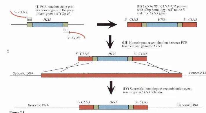
2.11.5 Gene replacement 43
2.11.6 Growth synchronisation methods 46
2.11.7 Determination o f budding index and cell numbers 47
2.11.8 DAPI staining for fluorescent microscopy 47
2.11.9 Relative cell volume comparisons by FACS analysis 48
2.11.10 Cell volume determination by microscopy 48
2.11.11 Luciferase activity assays 48
2.11,13 P-galactosidase assays in yeast colonies 50
RESULTS
Chapter Three
Activation properties of Swi6 and Swi4
Page
3.1 Introduction 51
3.2 LexA-Swi6 is a transcriptional activator 51
3.3 ADH-LexA-Swi6 is functional 53
3.4 LexA-Swi6 can activate transcription in the absence o f M bpl or Swi4 55
3.5 LexA-Swi6 can activate independently o f association with M bpl and Swi4 57
3.6 LexA-Swi6 activity is not dependent upon two potential Cdc28 sites 60
3.7 LexA-Swi4 is a weak transcriptional activator 60
3.8 LexA-Swi4 is functional 62
3.9 Discussion 63
Chapter Four
Developing and using a reporter system for periodic
studies
Page
4.1 Introduction 55
4.3 The development o f a luciferase reporter plasmid 67
4.4 LexA-Swi6 activates luciferase expression 69
4.5 The relationship between luciferase activity and the amount o f total 69
cell protein is linear
4.6 Luciferase activity measurements does not decay rapidly after the 71
addition o f substrate
4.7 Luciferase activity is unstable in yeast 72
4.8 a-factor synchrony shows luciferase activity to fluctuate 74
4.9 dbf2-2 induced synchrony demonstrates that luciferase activity is periodic 75
4.10 Northern analysis o f a dbf2-2 induced synchronous culture showed 77
luciferase mRNA to fluctuate periodically
4.11 Discussion 79
Chapter Five
Transcriptional activation by Swi6 requires the G1 cyclin
Cln3
Page
5.1 Introduction 82
5.2 MBF activity is dependent on Cln3 82
5.3 SBF activity is dependent on Cln3 83
5.4 The need for a plasmid expressing low levels o f LexA-Swi6 83
5.5 GLU-LexA-Swi6 is functional 85
5.6 GLU-LexA-Swi6 activity is dependent on Cln3 86
5.7 The dependency o f Cln3 is specific to Swi6 88
Chapter Six
Investigating the activation properties of Cln3
Page
6.1 Introduction 92
6.2 LexA-ClnS is a very weak transcriptional activator 92
6.3 Stable LexA-Cln3 is a potent transcriptional activator 94
6.4 LexA-Cln3 APEST activity is dependent on the cyclin box 94
6.5 LexA-Cln3 is dependent upon Swi6 for transcriptional activation 96
6.6 Functionally tests on LexA-Cln3 constructs 98
6.7 Discussion 98
Chapter Seven
Cln3 interaction with SBF is nrimarilv through Swi6
Page
7.1 Introduction 101
7.2 ADH-LexA-Swi6A345 interacts with Gal4ACT-Cln3 103
7.3 Gal4ACT-Clns are functionally expressed 105
7.4 Full length LexA-Swi6 interacts with Gal4ACT-Cln3 106
7.5 Deletion analysis o f LexA-Swi6 interactions with Gal4ACT-Cln3 106
7.6 Two-hybrid assay between Swi4 and G1 cyclins 109
Chapter Eight
Further analysis of Swi6 and Cln3 interdenendencv
Page
8.1 Introduction 113
8.2 Swi6 activation domain 1 is Cln3 dependent for transcriptional activation 113
8.3 Functional dependence o f Cln3 and Swi6 115
8.4 Discussion 118
Chapter Nine
Bck2 and its effect on transcription
Page
9.1 Introduction 121
9.2 SBF and MBF activation is Bck2 dependent 123
9.3 LexA-Swi6 activity is dependent on Bck2 123
9.4 The TA R l region o f Swi6 is dependent on Bck2 for activity 125
9.5 LexA-Cln3APEST activity is not dependent on Bck2 126
9.6 Reduction in transcriptional activation in a bck2 mutant is not 126
specific to Swi6
9.7 ADH-LexA-Bck2 can activate transcription 128
9.8 Transcriptional activation by ADH-LexA-Bck2 is dependent on 130
Cln3 but not Swi6
Chapter Ten
General discussion
Page
10.1 General discussion 136
Appendixl
Plasmid maps 143
References
153List of figures
Page
Figure 1.1 The cell cycle of Saccharomyces cerevisiae 2
Figure 1.2 A model illustrating the mode of action of SBF and MBF 11
Figure 1.3 Conserved and related features of the Swi6 family of 14
transcription factors
Figure 1.4 A simplified model for eukaryotic gene transcription 21
Figure 2.1 PGR based gene knock out in yeast 45
Figure 3.1 The “one-hybrid” assay for transcriptional activation 52
Figure 3.2a ADH-LexA-Swi6 can activate transcription of a lexAop-lacZ 54
reporter plasmid in yeast strain W 303-la
ADH-LexA-Swi6AP and ADH-LexA-Swi6A691
Figure 3.3 Functionality test on ADH-LexA-Swi6 plasmids 56
Figure 3.4a Transcription by ADH-LexA-Swi6 is only reduced slightly in 58
the absence of Swi4 or M bpl
Figure 3.4b Reporter gene activation by LexA-p53 and LexA-B2F is only 58
slightly reduced in the absence of Swi4 or M bpl
Figure 3.5 ADH-LexA-Swi4 is a weak transcriptional activator 58
Figure 3.6 ADH-LexA-Swi4 fusion is functional 61
Figure 4.1a lacZ mRNA is transcribed to the same extent in cells expressing 66
LexA-Swi6 or the LexA- only control
Figure 4.1b In the absence of a LexA DNA binding protein, the levels of 66
lacZ mRNA are elevated further
Figure 4.2 ADH-LexA-Swi6 can activate a /^jcAop-luciferase reporter gene 68
Figure 4.3 The relationship between luciferase activity and cell extract 70
is linear
Figure 4.4 Activity of luciferase is constant for up to 5min after addition 70
of substrate
Figure 4.5a Luciferase activity is reduced after the addition of a-factor 73
Figure 4.5b A successful G1 arrest is characterised by a population of 73
unbudded cells
Figure 4.6 G1 synchronised cells show ADH-LexA-Swi6 activity to fluctuate 76
Figure 4.7 Mitotically synchronised cells show ADH-LexA-Swi6 activity 76
to be cell cycle regulated
Figure 4.8 Expression of luciferase mRNA is cell cycle regulated 78
Figure 5.1 MBF activity is Cln3 dependent 84
Figure 5.2 SBF activity is Cln3 dependent 84
Figure 5.3 GLU-LexA-Swi6 fusion is functional 87
Figure 5.4 GLU-LexA-Swi6 activity is Cln3 dependent 87
Figure 5.5 Levels of GLU-LexA-Swi6 protein are unchanged in G1 cyclin 89
Figure 5.6a LexA-p53 activity is not Cln3 dependent 89
Figure 5.6b LexA-pE2F activity is not Cln3 dependent 89
Figure 6.1 ADH-LexA-Cln3 is a weak transcriptional activator 93
Figure 6.2 Stabilised ADH-LexA-Cln3 by deletion of the PEST sequence
can activate transcription, and its activity is cyclin box dependent 93
Figure 6.3 Drawings of LexA-Cln3 constructs used 95
Figure 6.4a The activity of ADH-LexA-Cln3APEST is Swi6 dependent 97
Figure 6.4b The activity of LexA-E2F and LexA-p53 is high in a Swi6 mutant 97
Figure 6.5 Functionality tests on LexA-Cln3 construct 97
Figure 7.1 A drawing of the Two-hybrid' system 102
Figure 7.2 C-terminal deletion of Swi6 interacts with Cln3 104
Figure 7.3 Cyclin construct proteins are expressed 104
Figure 7.4 Full length Swi6 interacts with Cln3 104
Figure 7.5 Drawing of LexA-Swi6 deletions used in Cln3 interactions 107
Figure 7.6 Deletions of LexA-Swi6 reveal a Cln3 interaction domain 108
Figure 7.7 LexA-Swi4 does not interacts with the G1 cyclins 108
Figure 8.1 Transcriptional activation region 1 of Swi6 is Cln3 dependent
and requires Cln2 for optimal activity
114
Figure 8.2 Transcriptional activation region 1 and Cln3 interaction domain
of Swi6 correspond to the same region
117
Figure 9.1a MBF requires Bck2 for optimal activity 122
Figure 9.1b SBF requires Bck2 for optimal activity 122
Figure 9.2 GLU-LexA-Swi6 activity is Bck2 dependent 124
Figure 9.3 Plasmid expression Bck2 under its promoter is functional 124
Figure 9.4 Transcriptional activation region 1 of Swi6 is Bck2 dependent 127
Figure 9.5 LexA-Cln3 activity is not Bck2 dependent 127
Bck2 for activity
Figure 9.7 ADH-LexA-Bck2 can activate transcription 129
Figure 9.8 ADH-LexA-Bck2 is functional 131
Figure 9.9 ADH-LexA-Bck2 activity is dependent on Cln3 but not Swi6 131
Figure 9.10 Transcriptional interdependency between Swi6, Cln3 and Bck2 135
List of tables
Page
Table 2.1 Yeast Strains used in this study
Table 2.2 Plasmids used in this study
Table 2.3 DNA fragments used for northern hybridisation
Table 7.1 Expression of Gal4act-CIn fusions are functional
Table 8.1 Cln3 can not accelerate Start in the absence
25
30
37
105
118
Abbreviations
ADH Alcohol dehydrogenase
p-gal p-galactosidase
bp Base Pair
B SA Bovine serum albumin
Cdk Cyclin dependent kinase
Cdki Cyclin dependent kinase inhibitor
CPM Counts per minute
C-terminal Carboxyl-terminal
DAPI Diamidino-2-phenylindole
DNA Deoxyribonucleicacid
FACS Fluorescence activated cell sorter
g Gram
Gal4ACT Transcriptional activation domain o f t
h Hour
Kb Kilobase
lacZ E. coli p-galactosidase gene
M Molar
MBF MCB binding factor
MCB Mlu\ cell cycle box
mg Milligram
mid-log Mid-logarithmic
min Minute
ml Millilitre
mM Millimolar
mRNA Messenger RNA
OD Optical density
ONPG o-nitrophenol-p-D-galactopyranoside
ORF Open reading frame
PBS Phosphate buffered saline
PCR Polymerase chain reaction
RNA Ribonucleicacid
RT Room temperature
SCB Swi4/6 cell cycle box
S. cerevisiae Saccharomyces cerevisiae
SBF SCB binding factor
SDS Sodium dodecylsulphate
TCP Total cell protein
Tris Tris (hydroxymethyl) aminomethane
ts Temperature sensitive
Tween 20 Polyoxrthylenesorbitan monolaurate
WT Wild type
X-gal 5-bromo-4-chloro-3-indolyl-P-D-galactoside
Pg micro gram
CHAPTER ONE
INTRODUCTION
1.1 THE EUKARYOTIC CELL CYCLE
1 .1 .1 Ke y f e a t u r e s o f t h e e u k a r y o t ic c e l l c y c l e
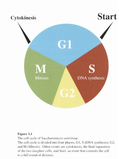
Eukaryotic cells propagate by alternately repeating DNA synthesis (S phase ) and cell
division (M phase). These cellular events are separated by two gaps phases, named G1
and G2 (figure 1.1). Apart from the four phases o f the cell cycle, the majority o f cells in
multicellular organisms are in a state o f quiescence or rest known as GO but can be
induced to re-enter the cell cycle at G1 by growth stimuli. Subsequent passage from G1
into S phase is governed by a complex control mechanism (Murray, 1987), dictating
whether cells should “Start” a new cell cycle or arrest at the G l/S boundary. Once cells
have entered the cell cycle, they make an irreversible commitment to a whole round o f
DNA replication and segregation and cells traverse the G l/S boundary entering into S
phase where they replicate their genome. S phase is followed by G2, in which cells
prepare for the onset o f mitosis. Nuclear and cell division are initiated in M and the two
daughter cells return to G l.
Rigorous control mechanisms known as checkpoints ensure the phases o f the cell
cycle are ordered: for example, it would be detrimental to cell viability if the chromosomes
Cytokinesis
Start
Figure 1.1
T h e cell cycle o f S acch aro m y ces cerevisiae.
entry into S and M phases, hence G l and 0 2 are extremely important in terms o f cell
cycle regulation.
There has been rapid progress in understanding the molecular events that control
the eukaryotic cell cycle in many organisms from yeast to humans. Research has
identified highly conserved control mechanisms common to all eukaryotes.
1.1.2 C o n t r o l o f t h e c e l l c y c l e b y t h e C d c 2 p r o t e i n k i n a s e f a m i l y
The Cdc2-related protein kinase family has come to occupy a central position in our
current understanding o f the eukaryotic cell cycle. The view that Cdc2 is a master
regulator o f cell cycle progression has evolved over the last 10 years as a result o f the
convergence o f work from distantly related organisms. Studies o f the fission yeast
Schizosaccharomyces pombe and frog Xenopus oocytes revealed that the S. pom be cdc2"
gene product was homologous to the p34 subunit o f Xenopus Maturation Promoting
Factor (MPF) (Dunphy et a l, 1988; Gautier et a l, 1988). Mammalian Cdc2 was first
isolated from human cells by phenotypic complementation o f a temperature-sensitive
cdc2' mutant o f the fission yeast (Lee and Nurse, 1987) and is highly homologous in
structure and function to S. pombe Cdc2 and S. cerevisiae Cdc28.
Since the discovery o f the first human Cdc2 homologue, it has become clear that
higher eukaryotes utilise a whole family o f Cdc2-like proteins to regulate the cell cycle.
Different Cdc2-like kinases regulate specific stages o f the cell cycle, whilst family
members are expressed differentially dependent on the cell type (Bates et al., 1994;
1 .1 .3 C d c 2 a n d c y c l in s
Because o f the irreversible nature o f both DNA replication and mitosis, activation o f the
Cdc2 kinases is carefully controlled, being subject to multiple regulations (Nigg, 1 9 9 5 ) .
One important regulatory mechanism is the association with an unstable regulatory
subunit called a cyclin, hence, the Cdc2 family o f protein kinases are known as cyclin
dependent protein kinases (Cdks). Cyclins were initially discovered as proteins whose
abundance oscillates dramatically during the cell cycle o f sea urchin embryos (Evans et al.,
1 9 8 3 ) . Cyclins are now divided into a number o f classes based on sequence similarity and
physiological function. Each Cdk absolutely requires the association with a cyclin
subunit, and binding o f the cyclin to the Cdk causes a conformational change that affects
the active site o f the Cdk, such that the kinase activity is increased (Jeffrey et al., 1 9 9 5 ) .
Association with a cyclin is also thought to contribute towards the Cdk’s substrate
specificity. The majority o f cyclins are only present at particular times o f the cell cycle,
and this is achieved by temporal gene transcription. In addition to cyclin messenger RNA
being transcriptionally regulated by sequence specific transcription factors, cyclins are
subjected to targeted destruction.
1 .1 .4 Ph o s p h o r y l a t i o n o f Cd c2
In addition to cyclin association, the Cdks are regulated by phosphorylation.
Phosphorylation o f a given amino acid in a protein can have a variety o f effects, activating
or inactivating a protein’s enzymatic activity, or increasing or decreasing its affinity for
binding to other proteins. Phosphorylation on one site in Cdc2 inhibits its kinase activity
example the key phosphorylated residues o f fission yeast Cdc2 are tyrosine 15 and
threonine 167. Phosphorylation on tyrosine 15 by the W eel and M ikl kinases
negatively regulates Cdc2 kinase activity (Lundgren et al., 1991; Russell and Nurse,
1987), whereas phosphorylation o f threonine 167 has a positive effect (Gould et al.,
1991). The inhibitory phosphorylation is dominant and hence Cdc2 phosphorylated on
both tyrosine 15 and threonine 167 lacks kinase activity. The positive phosphorylation
on threonine 167 is itself performed by a cyclin/Cdk complex called CAK (Cdk
Activating Kinase), which is composed o f a cyclin named Mcs2 and a Cdk called C rkl
(Buck et al., 1995).
1 .1 .5 In h i b i t o r s o f Cd k a c t iv it y
Besides the availability o f cyclin partners and their phosphorylation status, Cdks are also
regulated by a group o f protein kinases inhibitors, such as Ruml in fission yeast, Sicl o f
budding yeast and the p21 family in mammalian cells (Schwob et al., 1994; Harper et al.,
1993; Lee et al., 1995; Matsuoka et al., 1995; Moreno et al., 1994; Polyak et al., 1994;
Toyoshima and Hunter, 1994; Xiong et al., 1993), these are known as Cdkis (Cyclin
dependent kinases inhibitors). Cdkis can differ in their mode o f action, either by forming
tertiary complexes with cyclin/Cdk or inhibiting cyclin/Cdk complex formation (Guan et
a/., 1994).
1.2 THE BUDDING YEAST CELL CYCLE
Much o f our understanding o f the eukaryotic cell cycle has come from the study o f two
cerevisiae or budding yeast. Study o f the fission yeast has resulted in great progress in
clarifying the control mechanisms o f mitosis, whereas much o f our insight into the G l/S
phase has come from the study o f budding yeast.
1.2.1 Ke y e v e n t s in t h e b u d d i n g y e a s t c e l l c y c l e
The primary control point in budding yeast is exerted at a point in late G l known as Start
(Pringle and Hartwell, 1981), where environmental factors such as nutrient availability
and the presence o f mating pheromones are assessed. In addition. Start cannot be passed
until a critical cell size has been attained. Cells in late G l have three potential fates.
They can continue through the mitotic cell cycle, they can exit the cell cycle into a
quiescent stage (GO), or they can conjugate and mate with cells o f the opposite mating
type and enter the sexual differentiation cycle. However, once a cell has passed Start, it is
committed to the mitotic pathway, it becomes resistant to mating pheromones and can
not become quiescent until cells have passed through a complete cell cycle and re-enters
G l. After the initiation and completion o f DNA synthesis in S phase, the cell enters G2,
the end o f which is marked by the migration o f the replicated nucleus to the neck o f the
budded cell. Mitosis is characterised by the segregation o f the replicated DNA into the
two cells which is followed by cytokinesis, where the new daughter cell buds from the
mother. In budding yeast, the first visible stage o f cell division is taken to be bud
formation which occurs at the G l/S boundary. One consequence o f bud formation is an
asymmetry o f the progeny, one o f the products o f division (the mother cell) being
spends only a short time in 0 1 , requiring less time to overcome the size requirement for
traversing Start (Hartwell and Unger, 1977).
1 .2 .2 Ce l l d i v i s i o n c y c l e m u t a n t s
In the S. cerevisiae cell cycle, both haploid and diploid cells undergo mitosis, permitting
the isolation o f recessive gene mutations in haploids and the analysis o f genes by
complementation in diploids. Using this method, over 70 genes have been identified that
are required for progression through the cell cycle (Pringle and Hartwell, 1981). Cell
Division Cycle (cdc) mutants were originally isolated as conditional mutants which are
temperature-sensitive (ts) for growth. Temperature sensitive mutant strains have a wild
type phenotype at the permissive temperature but arrest at the restrictive temperature.
As originally defined, cdc mutants result in a defect in a particular stage-specific function
o f the cell cycle, this can be seen in an asynchrounsly growing culture. For example, in a
Cdc28ts mutant, cells will arrest at the same point in the cell cycle, at the G l/S boundary,
regardless o f their stage at the time they were shifted from the permissive to the
restrictive temperature.
One o f the main conclusions that has been drawn from the study o f cdc mutants is
that there are only a few major rate-limiting steps in the cell cycle, and only when these
are complete can other dependent events take place (Nurse and Bisset, 1981). Whether a
gene product is involved in a rate-limiting step can be answered empirically by
determining whether speeding up the rate at which the product acts significantly advances
progress through the cycle. For example, isolation o f alleles o f cdc2^ in fission yeast that
step in M phase (Draetta, 1990). Similarly, dominant alleles o f the G1 cyclin CLN3 gene
in budding yeast cause cells to divide at a smaller size (Sudbery et al., 1980), which can
also be achieved by over-expression o f the G1 cyclins (Lew et al., 1992; Richardson et al.,
1989). This mutant phenotype can be viewed as an advance o f Start in cycling
populations, thus implying that the G1 cyclins are rate-limiting for execution o f this
control point.
1.2.3 C d c 2 8 a n d it s a s s o c i a t e d c y c l in s
In budding yeast, a single Cdk, Cdc28, which is homologous to Cdc2 o f fission yeast,
controls the ordered progression through the cell cycle. Cdc28 is thought to
phosphorylate different sets o f target proteins at specific times in the cell cycle. This
temporal target specificity is thought to be controlled by its association with particular
cyclin partners o f which there are at least nine (reviewed in Nasmyth, 1993). Passage
through Start is dependent upon association o f Cdc28 with the G1 cyclins, C ln l, Cln2
and Cln3 (Cross, 1990; Richardson et al., 1989). During S phase, the Cdc28 Cdk is
associated with the B type cyclins, Clb5 and Clb6 (Schwob and Nasmyth, 1993).
Finally, mitosis is regulated by complexes o f Cdc28 and the mitotic cyclins, Clbs 1-4
(Richardson gr a/., 1992; Surana g/ a/., 1991).
1.2.4 C o n t r o l o f S t a r t b y C d c 2 8 / C l n
The G1 cyclins which control the G l/S transition were initially thought to perform
overlapping functions because cells could still grow when any two o f the three CLN genes
were inactivated (Cross, 1990; Richardson et al., 1992), but arrested in G1 if all three
in Start in the absence o f any one o f the Clns (Hadwiger et al., 1989; Nash et a l, 1988).
In turn, over-expression o f any Cln results in a shortened G1 and accelerates passage
through Start (Lew et al., 1992; Richardson et al., 1989). Despite this apparent functional
redundancy, there are distinct differences between the G1 cyclins. Although all three
share some sequence homology, C LN l and CLN2 are far more closely related to each
other than to CLN3 (Hadwiger et al., 1989; Nash et al., 1988). Levels o f C LN l and CLN2
mRNA, protein and associated protein kinase activity all vary through the cell cycle,
rising to a maximum as cells pass Start then decreasing to a comparatively low level in the
remainder o f the cell cycle (Tyers et al., 1993; Wittenberg et al., 1990). In contrast,
amounts o f CLN3 mRNA, protein and associated kinase activity are low and show little
periodicity throughout the cell cycle (Tyers et al., 1993; Tyers et al., 1992; Wittenberg et
a l, 1990).
Because o f the functional redundancy o f the Clns, it was initially proposed that
any one o f the G1 cyclins was sufficient for the Cdk activity required for passage through
Start. The model proposed that once the Cdc28 protein was activated by the G1 cyclins,
this would lead to the transcription o f more Clns via a positive feedback loop and as a
consequence induce the rapid and irreversible execution o f Start (Cross and Tinkelenberg,
1991 ; N asmyth and Dirick, 1991). Recently, the self induction o f Clnl and Cln2 has been
disproved by the observation that the induction o f Clnl and Cln2 is largely Cln3
dependent (Stuart and Wittenberg, 1995). The demise o f the positive feedback model has
led to the proposal that levels o f Cln3 accumulating as cells increase in mass during late
through Start (Dirick et al., 1995; Stuart and Wittenberg, 1995). Thus, the current view is
that Cln3, and not C lnl and Cln2, initiates the execution o f Start. Since Cln3-associated
kinase activity leads to Clnl and Cln2 expression, attention now focuses on whether Cln3
has some regulatory effect on the transcription factors controlling expression o f C lnl and
Cln2.
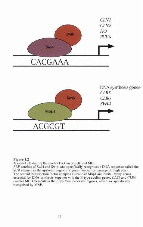
1.2.5 St a r t s p e c if ic g e n e t r a n s c r ip t io n
Two related hetrodimeric transcription factors control the transcription o f many o f the
genes, including C LN l and CLN2, which are periodically expressed as cells pass Start.
The first o f these, SBF (SCB Binding Factor), is composed o f Swi6 and Swi4 (Andrews
and Herskowitz, 1989; Taba et al., 1991) and the second, MBF (M CB Binding Factor), is
composed o f Swi6 and M bpl (Dirick et al., 1992; Koch et al., 1993). Peak expression o f
the C LN l and CLN2 genes is dependent on SBF (Nasmyth and Dirick, 1991; Ogas et al.,
1991). The Swi4 protein o f SBF contains a DNA binding domain which specifically
recognises a DNA m otif called the SCB (Swi4/6 Cell cycle Box) with a consensus
nucleotide sequence o f CACGAAA. This cis-acting regulatory m otif has been identified
in the upstream promoter regions o f CLNl, CLN2, the HO endonuclease gene (which is
involved in mating type switching), some o f the cyclin-like PCL genes (Frohlich et al.,
1991 ; Nasmyth, 1985; Ogas et al., 1991) and in genes involved in cell wall integrity (Igual
et al., 1996). In the second transcription factor, MBF, the DNA binding moiety is M bpl
(Dirick et al., 1992; Koch et al., 1993; Lowndes et al., 1991). M bpl recognises the DNA
m otif ACGCGT otherwise known as MCB (Mwl Cell cycle Box). MCB elements are
CLNl
CLN2
HO
PCL's
Swl6
Swi4
CACGAAA
DNA synthesis genes
CLB5
CLB6
SWI4
ACGCGT
Figure 1.2
A model illustrating the mode of action of SBF and MBF.
SBF consists of Swi4 and Swi6, and specifically recognises a DNA sequence called the SCB element in the upstream regions of genes needed for passage through Start.
The second transcription factor complex is made of M bpl and Swi6. Many genes essential for DNA synthesis, together with the B-type cyclins genes, CLB5 and CLB6
as in the promoter regions o f SWI4 and the B-type cyclin genes, CLB5 and CLB6
(Andrews and Herskowitz, 1990; Breeden, 1988; Epstein and Cross, 1992; Johnston and
Lowndes, 1992; Kuhne and Linder, 1993; Schwob and Nasmyth, 1993). Both SCB and
MCB elements can confer late G 1-specific gene expression to otherwise inactive
promoters (Lowndes et al., 1991; McIntosh, 1993; McIntosh a/., 1991). Figure 1.2
shows the mode o f action o f SBF and MBF.
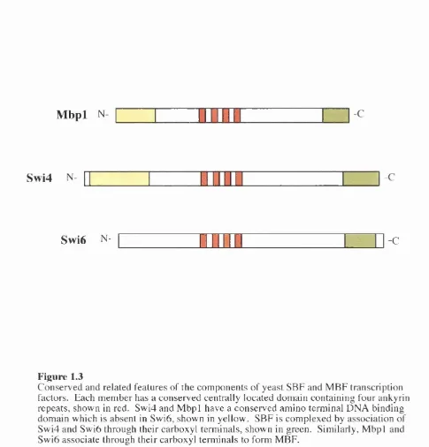
1.2.6 St r u c t u r a l s im il a r it ie s o ft h e Sw i6 f a m il y o f t r a n s c r ip t io n f a c t o r s
There are distinct similarities between Swi6, Swi4 and M bpl, figure 1.3 shows key
features. Swi4 and M bpl both share a conserved amino terminal DNA binding domain,
which is not present in Swi6. Swi6 itself has no specific DNA binding ability, but band
shift assays have demonstrated that Swi6 is needed for the efficient binding o f full length
Swi4 to DNA (Sidorova and Breeden, 1993). Furthermore, it would appear that the
DNA binding domain o f Swi4 can bind DNA independently o f Swi6 (Primig et al., 1992).
Hence, Swi6 may induce some conformation change in Swi4 to allow it to bind its specific
DNA sequences. The region o f Swi6 necessary for Swi4 and M bpl interaction has been
attributed to the carboxyl terminal (Andrews and Moore, 1992; Primig et al., 1992;
Siegmund and Nasmyth, 1996). Similarly, the C-terminals o f Swi4 and M bpl are
sufficient for binding to Swi6 (Andrews and Moore, 1992; Koch et al., 1993; Primig et al.,
1992). In the central region o f each protein are 4, possibly 5, copies o f the ankyrin repeat
motif. This 33 amino acid m otif is evolutionary conserved, it is present in many proteins
with diverse functions (Bork, 1993), and is thought to mediate protein-protein
sensitive phenotype (Ewaskow et al., 1998). The ankyrin repeat domain o f Swi4 is
known to interact with the Clb2-Cdc28 kinase (Siegmund and Nasmyth, 1996), and this
has been postulated to inhibit Swi4-Swi6 transcriptional activity at SCB elements during
the G2/M phase o f the cell cycle.
1.2.7 Al t e r n a t i v e m e c h a n is m s f o r SCB a n d MCB d r i v e n g e n e a c t i v a t i o n
Although SCB containing genes are thought to be regulated by the binding o f SBF and
genes with MCB elements are under the control o f the MBF transcription complex, there
appears to be some redundancy in this simple model. For example, in the absence o f
normal SBF activity the SKN7 gene product can promote expression from a multicopy
SCB-/(3cZ reporter plasmid (Morgan et al., 1995). Furthermore, several other proteins are
able to promote expression from SCB and MCB containing promoters including the
meiotic regulator Rm el (Toone et al., 1995) and Bck2, a protein o f unknown function (Di
Como et al., 1995).
As well as factors other than SBF and MBF which can activate MCB and SCB
mediated gene expression, there appears to be some degree o f cross talk between the
specificity o f SBF and MBF. Several observations support this cross regulation theory.
The DNA binding domain o f Swi4 can bind MCB sites, and the DNA binding domain of
M bpl can bind SCB and MCB sites with equal efficiency (Koch et ah, 1993).
Furthermore, the MCB-dependent Clb5 gene is still cell cycle regulated in a m bpl A (Koch
et al., 1993), similarly, Clnl expression driven by Swi4 occurs from its MCB elements
Mbpl N
-CSwi4
N- -CSwi6
N-
-CFigure 1.3
can compete for MBF complexes, and vice versa. These observations help to explain the
viability o f swi4, m bpl or swi6 strains.
1.2.8 Ac t i v a t i o no fSBF a n d MBF
In S. cerevisiae, several mechanisms for the activation o f SBF and MBF have been
proposed. The periodic expression o f SWI4 (Breeden and Mikesell, 1991) may have
some regulatory role in the activation o f SBF, but C LN l and CLN2 periodic expression
can occur under conditions when Swi4 protein synthesis is blocked (Marini and Reed, _
1992). The periodic binding o f Swi4 to SCB elements has been demonstrated, but not at
the time o f C LN l and CLN2 expression and this binding is not Cdc28 dependent (Koch et
al., 1996). Furthermore, SBF and MBF have been detected at all stages o f the cell cycle
(Dirick et al., 1992; Taba et al., 1991). The activity o f these transcription factors could
therefore be post-translationally regulated, so that they are active in late G1 and inactive
in G2.
Ultimately, Swi6 has emerged as the most attractive candidate for a regulatory role
in the complexes. One reason for this view is the presence o f Swi6 in both transcription
factors. Secondly, in the absence o f Swi6, some genes having SCB and MCB promoter
elements are no longer periodically expressed although they maintain a constitutive and
intermediate level o f expression (Dirick et al., 1992; Foster et al., 1993; Lowndes and
Johnston, 1992; Lowndes et al., 1992). This intermediate level suggests that Swi6 may
have both positive and negative regulatory effects on the expression o f these genes.
Transcriptional activation via SCB elements in the HO promoter requires both Swi6 and
as MCB-regulated genes are also maximally expressed after Cdc28 activation in G1 (Cross
and Tinkelenberg, 1991; Dirick and Nasmyth, 1991; Johnston and Thomas, 1982). These
observations suggest that the periodic activity o f SCB and MCB elements is Cdc28
dependent. Indeed, the Swi6 protein is highly phosphorylated in vivo and has several
potential Cdc28 phosphorylation sites, one o f which. Serine 160, appears to be
periodically phosphorylated (Sidorova et al., 1995). Serine 160 resides within a putative
Cdc28-consensus phosphorylation site and its phosphorylation is required for Swi6 exit
from the nucleus after S phase. In reality, however, phosphorylation at this site is not
Cdc28-dependent nor does it influence G l/S specific transcription. Thus,
phosphorylation o f Swi6 may not be responsible for its transcriptional activity and it
remains unclear what activates the SBF and MBF transcription factors.
1.2.9 Ge n e s t h a t c a n b y p a s s t h e r e q u ir e m e n t f o r G 1 c y c l in s
The requirement for at least one o f the three CLN genes for cell viability appears to
identify a requirement for G1 cyclin in Cdc28 activation. A screen was initiated in an
attempt to identify either genes acting downstream o f the CLN requirement or genes that
activate a parallel pathway making CLN genes unnecessary (Epstein and Cross, 1994). A
mutation in one gene named B Y C l (for bypass for cln requirement) was found to be a
dominant mutation in the previously identified gene S IC l. Sicl is a Cdk inhibitor,
specific to Cdc28/B-type cyclin kinase complexes (Donovan et al., 1994; Mendenhall,
1993; Schwob et al., 1994). After cells pass Start, B-type cyclin/Cdc28 kinases such as
Clb5/Cdc28 and Clb6/Cdc28 must be activated to allow replication o f DNA (Schwob et
initially inactive because o f inhibition by the Sicl protein (Mendenhall, 1993; Schwob et
al., 1994). Activation o f Clb5- and Clb6/Cdc28 occurs after Sicl is targeted for
proteolysis by the ubiquitin-conjugating enzyme Cdc34 (Schwob et al., 1994). As cells
pass Start the kinase activity associated with Cln/Cdc28 phosphorylates S icl, thus
making it a target for Cdc34. Biochemical reconstitution experiments have revealed that
Cdc4, Cdc53 and Skpl constitute a ubiquitin ligase complex that collaborates with the
ubiquitin-conj ugating enzyme, Cdc34, and the G1-specific Cdk, Cln2/Cdc28, to promote
the ubiquitination o f Sicl (Verma et al., 1997). If a major function o f Clns is to promote
proteolysis o f S icl, then Clns should be less important in a sicl mutant. Indeed, a sicl
mutation can suppress the lethality o f a cln l c ln l cln3 triple null mutation (Epstein and
Cross, 1994; Schneider et al., 1996; Tyers, 1996). Thus, the only non-redundant essential
function o f the Clns is to inactivate Sicl.
In addition to the mutation in Sicl which can bypass the requirement for G1
cyclins at Start, the expression o f another gene, BCK2, at low copy can suppress the
growth defect o f a triple cln mutant. The BCK2 gene was originally isolated as a high
copy suppressor o f the temperature sensitive cell lysis defect o f mutations in the protein
kinase C pathway (Lee et al., 1993). Furthermore, in cells lacking BCK2 the additional
loss o f CLN3 or SWI6 resulted in a reduction or loss o f viability respectively (Di Como et
al., 1995). This would suggest that Bck2, functions in a parallel pathway to Cln3 or Swi6
in activating Start. Indeed, the over-expression of BCK2 results in an increase in CLNl
and CLN2 RNA accumulation, but this increase appears to be Swi6 and Swi4 dependent
RNA. Hence, it would appear that bck2 mutants have a C LNl and CLN2 expression
defect. Furthermore, Bck2 appears to be acting through SCB and MCB elements found in
the promoters o f these genes because the synthetic lethality o f bck2 cln3 mutants can be
suppressed by overexpression o f plasmid borne CLN2 expressed under the A D H l
promoter but not its own natural SCB containing promoter. The role that Bck2 plays at
Start is unclear, but it appears to influence CLNl and CLN2 transcription in mainly a
SCB/MCB dependent manner, although there is also evidence for an alternative
independent mechanism.
1.4 GENE TRANSCRIPTION
The Swi6 component o f the two transcription factors SBF and MBF together with the
G1 cyclin Cln3 have been implicated in the regulation o f G l/S specific gene expression
(Dirick et a l , 1995; Stuart and Wittenberg, 1995). However, the way in which regulated
gene expression is affected by these components is unknown. To better understand the
potential mechanisms o f gene regulation, it is necessary to appreciate the basic mechanics
o f transcriptional activation.
1.3.1 Eu k a r y o t i c g e n e a c t i v a t i o n
Eukaryotes contain three distinct RNA polymerase enzymes, each responsible for the
transcription o f nuclear genes. All three polymerases maintain considerable sequence
similarity in their largest subunits (Allison et al., 1985) and actually have five subunits in
common (McKune and Woychik, 1994; Woychik et al., 1990; Young, 1991). Despite
accurate transcription o f a subclass o f nuclear genes. RNA polymerase I only transcribes
genes encoding large ribosomal RNAs. RNA polymerase II transcribes all o f the cell’s
protein-coding messenger RNAs (mRNA) and other small RNAs (snRNAs). RNA
polymerase III synthesises tRNA and the 5S RNA component o f ribosomes.
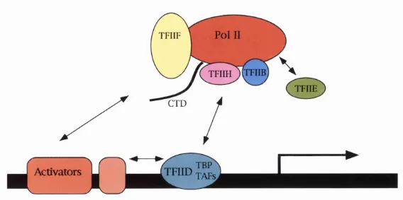
1.3.2 RNA POLYMERASE II
In the RNA polymerase I (Pol I) and Pol III systems, the association o f the polymerase
with the promoter signals the completion o f the assembly process (Buratowski and Zhou,
1992; Lofquist et al., 1993). The Pol II system differs in that even after the polymerase
has been loaded onto the promoter, the complex is not competent to initiate transcription.
This requires the association o f two more general transcription factors (GTFs), TFIIE and
TFIIH. TFIIE is a hetrodimer and TFIIH is a multi-subunit factor. Pol II contains a
unique m otif known as the CTD. CTD refers to a series o f tandem repeats (52 in
humans, 43 in Drosophila, and 26 in yeast), in the C terminal o f the largest subunit o f Pol
II. Phosphorylation o f the serine, threonine, and /or tyrosine residues within the CTD
appears to promote a transition from transcription initiation to elongation. TFIIH
contains a kinase activity capable o f phosphorylating CTD, and purification o f TFIIH
from different organism revealed that a Cdk and a cyclin partner were components o f the
complex.
1.3.3 Po l II a n d g e n e p r o m o t e r r e c o g n it io n
The transcriptional control regions o f eukaryotic protein coding genes can be separated
into at least two categories; a core promoter and upstream (or downstream) regulatory
are recognised by sequence specific DNA-binding factors critical for activating or
repressing transcription initiation (Tjian and Maniatis, 1994). The core promoter
nucleates the assembly o f an initiation complex containing RNA polymerase II and a
number o f accessory factors TFIIA, TFIIB, TFIID, TFIIE, TFIIF, and TFIIH that can
direct a low level o f basal transcription in vitro (Buratowski, 1994; Conaway and
Conaway, 1993; Zawel and Reinberg, 1992) (figure 1.4 shows Pol II holoenzyme). Most
o f the genes transcribed by Pol II have a DNA sequence called the TATA box as the core
promoter element, whose sequence is recognised by the TATA box-binding protein
(TBP) subunit o f TFIID. Detailed biochemical studies o f TFIID has revealed that this
transcription factor consists o f TBP and a number o f T B P-^sociated factors (TAFs)
(Tjian and Maniatis, 1994).
1.3.4 Ye a s t RNA Po l y m e r a s e II
The mechanism o f eukaryotic transcription activation and repression is complicated by
the large number o f proteins involved in the process. Prior to promoter assembly. Yeast
RNA polymerase II is associated with a number of proteins, including the general
transcription factors TFIIB, TFIIH and TFIIF. Polymerase II is not, however, associated
with the TATA-box binding protein (TBP) and its associated factors (TAFs) or TFIIE.
Transcription by polymerase II involves an activator protein binding to specific sites,
often present in multiple copies, upstream o f the TATA-box. The binding o f these
activators may promote transcription by recruiting the polymerase complex to a
K)
Pol II
TFIIFTFIIH ) TFIIB
TFIIE
TFIID TAFS
Activators
Figure 1.4
1 .3 .5 T B P -a s s o c i a t e d f a c t o r s
Although the essential role o f TBP o f different eukaryotes has been extensively analysed
in vivo and in vitro (Hernandez, 1993; Struhl, 1995), the function o f TAFs is less clear. In
vitro, TAFs are dispensable for basal transcription but are required for the response to
activators (Tjian and Maniatis, 1994). This tentative link to activator dependent
transcription suggests that TAFs may act as molecular bridges between particular
activators and the general transcription machinery (Chen et a l, 1994; Sauer et al., 1995).
Indeed, the budding yeast Tafnl45 and its higher eukaryotic homologue, TAFn250, are
known to contact TBP directly.
Recently it has been suggested that TAFs may be required to perform specialised
functions within the transcription complexes on specific promoters. Indeed, its has been
demonstrated that yeast TAFs are not needed for general transcription (Moqtaderi et al.,
1996; Walker era/., 1997).
The most extensively studied yeast TAF is Taful45 which encodes an essential
gene. Cells containing a temperature sensitive mutant o f Tafnl45 arrest as large unbudded
cells at the restrictive temperature, typical o f a mutation in a gene required at Start
(Walker et al., 1997). This cJc-like phenotype is likely to result from the reduced C LN l
and CLN2 transcription observed in these cells. This apparent link between Tafnl45 and
the cell cycle is extremely interesting and suggest a possible crude mechanism for
Cdk/cyclin and SBF/MBF gene activation at Start. It is possible that Tafnl45 could
activate transcription by recruiting the specific factors necessary to the promoter region.
histories, for Tafnl45 has histone acetyltransferase activity (Mizzen et al., 1996). The
past year has seen much progress in the understanding o f chromatin and transcription,
showing that different histone acétylation states play a role in gene activation (Gregory
and Horz, 1998).
1.4 SUMMARY
Recent work has suggested pivotal roles for Cln3 and Swi6 in the control o f the G l/S
transition point in budding yeast. This report attempts to clarify our present
understanding o f Start in this yeast, and the mechanisms which control gene expression at
CHAPTER TWO
MATERIALS AND METHODS
2.1 BACTERIAL STRAINS
The bacterial strain used throughout this study was E. coli D H 5a F ' ^80dlacZAM15
(lacZYA-argF) U169 endAl recA l hsdR17 frk'mk^) deoR thi-1 supE44' gyrA96 relA l. In
addition, bacterial strains used for transpositional disruption o f plasmid genes were
according to Morgan et a l, (1996).
2.2 YEAST STRAINS
Details o f yeast strains used in this study are given below, and list o f strains used is in
Table 2.1
Most strains are derivatives o f W 303-la: MATa ade2-l trpl-1 canl-100 leu2-3,112,
his3-11,15 ura3 GAL p s F ssdl-d. Those marked with an asterisk are derived from L181-6B:
MATa dhf2-2 ura3 leu2, trpl. Strains marked with a double asterisk are derived from
CTY10-5d: MATa trp l leu2, his3 gal4 gal80 URA3::LexAop-LacZ. The strain MDS4:
ade2-l trpl-1 leu2, his3 m bpl::URA3 swi4ts, was a kind gift from Mark Toone. Strain
K2003 is MATa ade2 his3 mat trpl ura3 swi4ts swi6::TRPl . Strain BF305-15d is
HIS3 and TRPl respectivly. CLN3 is under the control o f G ALl promoter, intergrated at
the URA3 locus. Strain BF411-2C is adel URA3::GAL-CLN3 ura3 clnl::H IS3
cln2::LEU2 trpl arg5,6. This strain is ura' due to selection on 5F0A .
T able 2.1
Strain Source
W 303-la
YAT2, cln l ::HIS3.
YAT3, cln l ::URA3.
YAT4, cln2 ::LEU2.
YAT5, cln3 ::URA3.
YAT7, cln3 :: LEU2
YAT8, cln3 ::HIS3 .
YAT9, clnl ::HIS3 cln2 ::LEU2.
YAT13, m bpl point mutation.
YAT12, swi4 ::ADE2.
Y A T U , swi6 ::TRPL
YAT18, bck2::LEU2.
YAT68, bck2 ::HIS3.
L181-6B*
CTY10-5d**
YAT21**,c/«7 ::URA3.
YAT22**, cln2 ::LEU2.
YAT24**, cln3 ::LEU2.
YAT25**, swi6 ::HIS3.
Lab stock
Mark Toone
This study
Mark Toone
Mark Toone
This study
This study
Kate Kramer
Nicolas Bouquin
Kate Kramer
Gary Merrill
This study
This study
(Toyn and Johnston, 1993)
(Bartel gr <3/., 1993)
This study
This study
This study
YAT71**, .-.•//ZS'J. This study
M DS4, m bpl ::URA3 swi4^^ Mark Toone
K2003 swi6::TRPl swi4ts (Nasmyth and Dirick, 1991)
BF305-15d no.21 {X iongetal., 1991)
B F 4 11 -2C Bruce Futcher
2.3 MEDIA AND GROWTH CONDITIONS
2.3.1 E. C P U
E. coli strain D H 5a was grown in Luria-Bertani broth (1% bacto-tryptone, 0.5% yeast
extract, 1% NaCl pH7.5) with the addition o f 50p-g/ml ampicillin and SOjig/ml methicillin
from filter sterilised stock solution for the selection o f plasmids. Liquid cultures were
grown at 37°C with continuous agitation. For solid media, 1.5% Difco agar was added
and the agar plates incubated in a constant temperature incubator at 37°C. Strains were
stored short-term at 4°C on LB agar (with ampicillin as required) and for long term
storage, were grown to stationary phase in LB and kept at -70°C in 20% sterile glycerol.
2.3.2 S. CEREVISIAE
Cells were grown in YPD rich medium (1% yeast extract, 2% bacto-peptone, 2%
glucose) or, for plasmid selection, in synthetic medium (0.67% yeast nitrogen base, 2%
glucose) supplemented with the appropriate amino acids at 20p,g/ml. In the case o f gene
induction from the G A L l-10 promoter, synthetic medium was supplemented with 2%
galactose instead o f glucose. Liquid cultures were grown with continuous agitation; for
incubator. The standard growth temperature for wild type yeast strains was 30°C.
Temperature sensitive strains were grown either at the permissive temperature o f 25°C or
the restrictive temperature o f 37°C.
Yeast strains were stored short-term on YPD or minimal agar plates at 4°C. For
long term storage, freshly grown cells were removed from agar plates, inoculated into 1 ml
o f storage medium (1% yeast extract, 1% bacto-peptone, 2% glucose and 25% glycerol)
and kept at -70°C.
2.4 STANDARD BUFFERS
TE lOmM Tris HCl, ImM EDTA, pH8.0
TAE 40mM Tris base, 20mM glacial acetic acid, ImM EDTA pH8.3
DNA loading buffer 0.1% bromophenol blue, 50% glycerol, 50mM EDTA
RNA loading buffer 0.4% bromophenol blue, 50% glycerol, 0.0IM PO4 (pH6.5)
2.5 ISOTOPES
Radiolabelled isotope [a-^^P] dCTP 11 ITbq/mol (3000Ci/mol) was obtained from
2.6 DNA MANIPULATIONS
2.6.1 Re s t r ic t io n e n d o n u c l e a s e s a n d DNA m o d if y in g e n z y m e s
DNA was incubated with restriction endonucleases (BRL, NEB) in the appropriate
restriction endonuclease buffer at the recommended temperature, with the addition o f
IxBSA according to manufacturer’s recommendation. Klenow Taq polymerase
(Amersham), T4 DNA ligase (BRL) and calf intestinal phosphatase (Boehringer
M annheim) were used according to the manufacturers’ recommendations.
2.6.2 DNA LIGATIONS
Ligation o f DNA fragments was carried out in a 20|il volume in Ix ligation buffer (50mM
Tris-HCl pH7.6, lOmM MgCh, ImM ATP, ImM DTT, 5%(w/v) polyethylene glycol-
8000) and 0.4 units o f T4 DNA ligase (GIBCO BRL). Ligations were incubated at room
temperature for 2h, or alternatively overnight at 16°C before transformation into E. coli.
2 . 6 .3 Re c o v e r y o fDNA f r a g m e n t s f r o m a g a r o s e g e l s
Glass milk extraction was used to remove fragments o f DNA greater than 200bp from
TAE agarose gels following the recommendations o f the manufacturer (BIO 101,
G enecleanll).
2.6.4 Ag a r o s e g e l e l e c t r o p h o r e s is
Agarose gel electrophoresis was carried out in 0.8% w/v agarose gels with TAE
electrophoresis buffer. DNA was loaded onto gels with 1/6 volume DNA loading buffer
and electrophoresed with a constant current o f 50-100mA. DNA was stained by
under short wave (nm) UV using a Uniscience TF-20M transilluminator. The molecular
size o f DNA fragments was determined by comparison to DNA size markers (Life
Technologies).
2.6.5 So u t h e r n h y b r i d i s a t io n
Samples (2-5)Lig) o f genomic DNA were digested with appropriate restriction enzymes
and separated by electrophoresis through a 0.8% agarose gel. DNA transfer and
hybridisation to specific probes was carried out according to Sambrook et a l, (1989).
GeneScreen transfer membranes (Dupont, NEN Research Products) were used as
described in the manufacturer’s instructions.
2 . 6 . 6 Po l y m e r a s e c h a in r e a c t io n
Reactions were carried out in 100|il total volume, 0.5-2|ig o f genomic or 5ng plasmid
DNA, l|xl o f lOmM dNTP stock (Pharmacia Biotech), Ijxl o f 0.1-0.5nMol/)Lil o f each
primer, Ix PCR buffer containing 15mM MgCh (Perkin Elmer) and 2 Units o f Taq
Polymerase (Perkin Elmer). All PCR reactions were performed on a Biometra TRIO-
Therobloc. Reactions were incubated using the same general conditions namely; 94°C
5min, 29(94°C Imin, 50°C 2min, 72°C 3min), 72°C 7min, then held at 20°C for up to
1 2h.
2 . 6 . 7 Tr a n s p o s i t i o n -m e d i a t e d p l a s m i d m a n ip u l a t io n
Plasmid genes were disrupted by transposition according to Morgan et a l, (1996).
Additionally, the same method was used to produce truncated protein products from
2.7 PLASMIDS
The plasmids used in this study are summarised in table 2.2.
Details o f plasmids constructed in this study are given in section 2.7.1 to 2.7.7. Maps o f
some plasmid are in the plasmid appendix on page 145.
Table 2.2
Plasmids Features
pSH18-34
pRS316
pLU C l
pB H l
pTR27
pTR28
pTR432
pTR40
pTR434
pTR437
pTR439
/ûfcZ reporter plasmid: %lexA motifs, G ALl-10 mimmdX
promoter: URAS, 2ji; (Yocum et al., 1984).
URA3, CENcloning vector; (Sikorski and Hieter, 1989).
Contains the firefly luciferase gene a gift from Andrew Furley;
(de Wet et a i, 1987).
As pRS316 with, GALl-10:: SlexA promoter cloned upstream
o f the luciferase coding sequence.
LexA DNA binding domain: TRPl, 2fi.
As pTR27, expressing LexA-Swi6 fusion.
As pTR27, expressing LexA-Bck2 fusion.
As pTR27, expressing LexA-Swi4 fusion.
As pTR27, expressing LexA-Cln3 fusion.
As pTR434 expressing C-terminal PEST deletion mutant Cln3.
pACTI
pACTIII
GAL4 activation domain: LEU2, 2/1; (Durfee et al., 1993).
As pACTI with altered polylinker sequence
(CATATGGCCATGGAGGCCCCGGGGATCT ATGCATTGGGATCCGGATATCAAGATCC pTR290 pTR291 pTR212 pTR83 pTR241 pTR228 pV44ER pTR240 pAP pA691 GAATTCGAAGCTCGAGAGATCT).
As pACTIII, expressing Gal4 -Clnl fusion.
As pACTIII, expressing Gal4-Cln2 fusion.
As pACTI, expressing Gal4-Cln3 fusion.
MCQ-lacZ: URA3 2]i. MCB denotes 3 Mlu\ restriction sites.
As pTR83 with mutated MCB.
SCB-/flrcZ.- HIS3, 2}JL SCB denotes bp -501 to -367 o f the HO
regulatory region encompassing four SBF binding sites.
G ALl-10 promoter upstream o f sequence encoding the LexA
DNA binding domain: TRPl ΠA (Jay aram an et al., 1994).
As pV44ER expressing LexA-Swi6 fusion.
pTR28-based, Cdc28 sites o f Swi6 mutated, S160A S228A.
pTR28-based transposition with TnHIS resulting in a truncation at
residue 691 o f Swi6.
pA345 pTR28-based with transpositional insertion o f TNHIS3 resulting in
a truncation at residue 345 o f Swi6.
a truncation at residue 145 o f Swi6.
pTR29 pTR28-based with internal Nsil region o f SWI6 deleted resulting
in the expression o f a protein with the central region o f Swi6
deleted between residues 103-729.
pTR250 pTR249 pTR345 pTR433 YCp/flcl81 pTR476
As pTR27 expressing LexA-E2F fusion.
As pTR27 expressing LexA-SLN 1 fusion.
As pTR27 expressing LexA-p53 fusion.
TRP1 ::TnHIS3 derivative o f Y E placl 12-CLN3.
(Y E placl 12-CLN3 was a kind gift from C Wittenberg and has a
single copy o f CLN3 under the control o f its own promoter).
Shuttle vector; (Gietz and Sugino, 1988).
As YCp/acl81 with Sphl-Xbal 4.5kb fragment containing
BCK2 gene and flanking sequences cloned from YE^352-BC K 2;
(Lee et aL, 1993).
pGEM/flcZ pSE738 pYA301 TRTl pSD06a pTR303
Contains BamEE-BamEil lacZ fragment, a kind gift from Miguel
Manzanares.
Plasmid containing 7 gene; (Elledge and Davis, 1990)
A C T l containing plasmid; (Gallwitz and Sures, 1980).
Plasmid containing H2B gene; (Hereford et al., 1979)
CYC-GAL-VP16 (Dalton and Treisman, 1992).
B am R l-X bal sites o f pSD06a.
pTR304 BglU-Bagll CLN2 PCR fragment cloned into
BamHl-BamHl sites o f pSD06a.
pTR304 BamHl-Hindlll CLN3 PCR fragment cloned into
BamHl-Hindlll sites o f pSD06a.
pTR477 pTR28 based HIS3::swi6 disruption plasmid.
pTR251 CLN2 under control o f S. pombe A D H promoter (LEU2);
(Fernandez Sarabiae^fl/., 1992).
2.7.1 Co n s t r u c t i o n o f a l u c if e r a s e r e p o r t e r p l a s m i d
pB H l was derived from the CEN URA3 plasmid pRS316 (Sikorski and Hieter, 1989) and
carries the firefly luciferase gene positioned downstream from the G ALl-10 minimal
promoter and LexA binding site sequences. The firefly luciferase gene was excised from
pLU C l (a gift from Andrew Furley, from de Wet et a l, 1987) on a H indlll-Kpnl
restriction fragment whilst a BamUl-Hindlll fragment containing the G A Ll-10 minimal
promoter with the lexAop was purified from pSH l 8-34 (Yocum et al., 1984). A three-
way ligation o f these fragments with pRS316 cut with BamUl-Kpnl produced the
lexAop-GALl-lO-ineiferase reporter plasmid, pB H l.
2.7.2 Co n s t r u c t i o n o f t h e g a l a c t o s e i n d u c i b l e Le xA - Sw i6 p l a s m i d
pTR240 was made by cloning the SWI6 gene downstream o f the DNA sequence coding
for the bacterial LexA DNA binding protein. The CEN vector pV44ER (Jayaraman et al.,
2409bp BamV{{ restriction fragment containing the SWI6 coding sequence was purified
and cloned into the BgR\ site in pV44ER, in frame with the LexA coding sequence.
2.7.3 C o n s t r u c t i o n o ft h e L e x A - B c k 2 p l a s m i d
pTR432 was made by cloning the BCK2 gene downstream o f the DNA sequence coding
for the bacterial LexA DNA binding protein. A 2.56kb PCR product containing the
BCK2 gene flanked by 5'BcR and V X h ol sites was digested with these enzymes, purified
through agarose and cloned into the BarnWl and Sail sites o f the vector pTR27 to produce
an in-frame LexA-Bck2 fusion.
2.7.4 C o n s t r u c t i o n o f a G a l a c t o s e i n d u c i b l e C LN l p l a s m i d
pTR303 was made by cloning a PCR fragment encoding the C LN l gene into the pSD06
(Dalton and Treisman, 1992). pSD06a was cut with BamYil and Xba\ resulting in the
excision o f the VP 16 coding region. A PCR fragment coding for C LN l with 5’ BamYil
and 3 ’ Xba\ ends was digested and cloned into prepared pSD06a vector, resulting in
C LN l under the control o f the GAL-CYC promoter.
2.7.5 Co n s t r u c t i o n o fa Ga l a c t o s e i n d u c ib l e CLN2 p l a s m i d
pTR304 was made by cloning a restriction fragment encoding the CLN2 gene into the
pSD06a. pTR291 was digested with BgR\ yielding a 1.6kb restriction fragment encoding
the CLN2 gene. This fragment was cloned into pSD06a cut with BamYil. A stop codon
before the sequence encoding VP 16 resulting in CLN2 under the control o f the GAL-CYC
2.7.6 Co n s t r u c t i o n o fa Ga l a c t o s e i n d u c i b l e CLN3 p l a s m id
pTR303 was made by cloning a PCR fragment encoding the CLNS gene into the pSD06a.
pSD06a was cut with Bam Rl and HindUl resulting in the excision o f the VP 16 coding
region. A PCR fragment coding for CLN3 with 5’ Bam Rl and 3’ H indlll ends was
digested and cloned into prepared pSD06a vector, resulting in CLN3 under the control o f
the GAL-CYC promoter.
2.7.7 Co n s t r u c t i o n o f a BCK2 g e n o m ic c l o n e e x p r e s s i n g p l a s m i d
The plasmid pTR476, expressing BCK2 under its own promoter was constructed. A
4.5kb BCK2 containing Sphl-Xbal restriction fragment was cloned from YEp352-BCK2
(Lee et al., 1993) into the yeast shuttle vector, YEp/acl81 (Gietz and Sugino, 1988),
digested with Sphl-Xbal. The resulting plasmids marker gene was compatible with the
CTY10-5d reporter strain.
2.8 RNA MANIPULATION
2.8.1 RNA EXTRACTION
1 0* synchronously or asynchronously mid-logarithmically growing cells were harvested,
washed in 0.9% cold saline solution and rapidly frozen in dry ice. Total RNA was
prepared by the hot phenol extraction method (Aves et a l, 1985). The quality and
quantity o f total RNA was assessed by UV spectrometry at Azeo using a Shimadzu UV-