R E S E A R C H A R T I C L E
Open Access
Expression and function of
phosphodiesterases (PDEs) in the rat
urinary bladder
Xiaofei Zhu
1†, Kui Zhai
2*†, Yue Mi
3†and Guangju Ji
2*Abstract
Background:It has been shown that hosphodiesterases (PDEs) play an important role in mediating the smooth muscle tone of rat urinary bladder. However, the gene expression profiles of them were still unknown.
Methods:Urinary bladder Strips were obtained from both neonatal and adult Sprague-Dawley rats. RT-PCR/western blot and organ bath were used to measure the expression and function of PDEs.
Results:Adult rat urinary bladder expressed various PDE mRNA with the following rank order:
PDE5A≈PDE9A≈PDE10A > PDE2A≈PDE4A≈PDE4D > PDE4B≈PDE3B≈PDE8B≈PDE7A≈PDE7B > PDE1A. PDE1B, PDE1C, PDE3A, PDE4C, PDE8A, and PDE11A were not detected. Of interest, the mRNA and protein of PDE3A were significantly decreased in adult rat urinary bladder compared to neonatal rat urinary bladder. Cilostamide, a specific inhibitor for PDE3, significantly inhibited the amplitude and frequency of carbachol-enhanced phasic contractions of neonatal rat bladder strips by 38.8% and 12.1%, respectively. Compared to the neonatal rat bladder, the effect of cilostamide was significantly blunted in adult rat urinary bladder: the amplitude and frequency of carbachol-enhanced phasic contractions were decreased by 13.4% (P< 0.01 vs neonatal rat bladder) and 4.4%, respectively. However, the mRNA and the protein levels of PDE3B were similar between neonatal and adult rat bladder.
Conclusion:We found that several PDE isoforms were expressed in the rat urinary bladder with distinct levels. Moreover, we showed that the function of PDE3 was blunted in adult rat bladder likely due to the decreased expression of PDE3A.
Keywords:Gene expression, Phasic contraction, Phosphodiesterases, Rat urinary bladder
Background
Through mediating cAMP and/or cGMP levels, phos-phodiesterases (PDEs) are involved in several physio-logical processes of urinary bladder. Artim et al. showed that PDE5 mediated the phasic contractions of neonatal rat bladder through nitric oxide-cGMP- protein kinase G pathway [1]. Our previous study found that PDE3 and PDE4 regulated the phasic contractions of neonatal rat bladder through PKG and PKA pathways, respectively [2]. To date, 11 PDE families have been identified [3]: 3 families (PDEs 4, 7, and 8) selectively hydrolyze cAMP, 3 families selectively hydrolyze cGMP (PDEs 5, 6, and 9),
and 5 families (PDEs 1, 2, 3, 10, and 11) hydrolyze both cyclic nucleotides with varying efficacies. As some of these PDE families consist of more than one gene and some genes are alternatively spliced, 21 different isoforms and more than 50 variants have been identified [4].
The urinary bladder is the organ that collects urine ex-creted from kidneys before disposal by urination. One of the most important features of the urinary detrusor smooth muscle is its ability to generate considerable phasic contractions [1, 2, 5]. These spontaneous phasic contractions change markedly with age [5]: during the first 2–3 weeks of life, they are characterized by
high-amplitude low-frequency spontaneous contractions;
then, they turn into low-amplitude high-frequency pat-tern characteristic of normal adult rat. The spontaneous * Correspondence:[email protected];[email protected]
†Equal contributors
2National Laboratory of Biomacromolecules, Institute of Biophysics, Chinese
Academy of Sciences, 15 Datun Road, Beijing 100101, China Full list of author information is available at the end of the article
activities in neonatal rat bladder are likely to be myo-genic [6] and can also be modulated by the interstitial cell network [7–9].
In this study, we tested the expression profiles of all PDEs families except PDE6 (its expression is restricted to the retina) in adult rat urinary bladder with RT-PCR techniques. Moreover, we evaluated the functional role of PDE3 in mediating the phasic contraction of
neonatal and adult rat bladder strips through
pharmacological method.
Methods
Animals and regents
Neonatal (10-day old) and adult (90-day old) Sprague-Dawley rats were purchased from Vital River Laborator-ies (Beijing, China). All animal procedures described in this study were performed according to the Guide for the Care and Use of Laboratory Animals published by the US National Institutes of Health (NIH Publication No. 85–23, revised 1996), and with approval from the Institute of Biophysics Committee on Animal Care. The rat was anesthetized by 5% chloral hydrate and the whole bladder was removed, placed into cold Tyrode so-lution composed of (mM): 137 NaCl, 5.4 KCl, 1.8 CaCl2, 1.0 MgCl2, 10 glucose, 10 HEPES, (pH 7.4). After the cleaning of the fat tissues, the bladder was cut into two longitudinal pieces by using a fine dissecting scissors along the axis from the neck to the fundus.
Cilostamide (Cil) was from Tocris Bioscience (Bristol, UK). All other reagents were from Sigma Aldrich unless mentioned.
Polymerase chain reaction (PCR)
Reverse transcriptional-PCR were performed as previ-ously reported [2, 10]. Standard PCR was performed using GoldStar Taq DNA Polymerase (CWBIO, Beijing, China) according to the manufacturer’s instructions. The primers sequences were shown in Table 1. PCR conditions were set up as follows: 94 °C for 2 min, then the following three steps for 34 cycles (94 °C for 30 s, 55 °C for 30 s, 72 °C for 1 min), and a final step of cool-ing to 4 °C. PCR products were run on a 1.7% agarose gel and visualized under UV light using ethidium brom-ide staining. qRT-PCR was performed on a Corbett
Rotor-Gene 6600 QPCR system machine using
TransScript™Green qRT-PCR SuperMix (TRANSGENE
BIOTECH, Beijing, China) according to manufacturer’s instructions. The sequences of gene-specific primers
were 5′TCACAGGGCCTTAACTTACAC3′ (forward)
and 5′GGAGCAAGAATTGGTTTGTCC3′ (reverse)
for PDE3A, and 5′GCAAGAGAGAGGCCCTCAG3′
(forward) and 5′TGTGAGGGAGATGCTCAGTG3′
(reverse) for GAPDH. Amplifications were performed with a 10 min template denaturation step at 95 °C,
followed by three steps for 35 cycles (denaturation at 95 °C for 30 s, annealing at 58 °C for 30 s, and exten-sion at 72 °C for 30 s). All samples were amplified in triplicate and the mean was obtained for further calcu-lations. The comparative 2-ΔΔCt method was used to analyze mRNA fold changes between neonatal and Table 1The primer sequences of PDE isoforms
Name Sequences
PDE1A (Forward, F) 5′CCACTTTGTGATCGGAAGTC3′
PDE1A (Reverse, R) 5′TTCTGCTGAATGATGTCCACC3′
PDE1B (F) 5′CAGGGTGACAAGGAGGCAGAG3′
PDE1B (R) 5′GACATCTGGTTGGTGATGCC3′
PDE1C (F) 5′TCTCAAAGGATGACTGGAGG3′
PDE1C (R) 5′GCTTCTCTGTCACCCTGTC3′
PDE2A (F) 5′CCTCCTGTGACCTCTCTGACC3′
PDE2A (R) 5′TGAACTTGTGGGACACCTTGG3′
PDE3A (F) 5′TCACAGGGCCTTAACTTACAC3′
PDE3A (R) 5′GGAGCAAGAATTGGTTTGTCC3′
PDE3B (F) 5′CCTCAGGCAGTTTTATACAATG3′
PDE3B (R) 5′TGCTTCTTCATCTCCCTGCTC3′
PDE4A (F) 5′GTGGAGAAGTCTCAGGTGGG3′
PDE4A (R) 5′TGGAACTTGTCAGGCAGGG3′
PDE4B (F) 5′TAGAAGATAACAGGAACTGG3′
PDE4B (R) 5′GCAATGTCTATGTCAGTCTC3′
PDE4C (F) 5′ACGTGGCGTACCACAACAGC3′
PDE4C (R) 5′TACCGCGAGGTGATGGTTCTC3′
PDE4D (F) 5′GGATAATGGAGGAGTTCTTCC3′
PDE4D (R) 5′CGATTGTCCTCCAAAGTGTCC3′
PDE5A (F) 5′CCCTGGCCTATTCAACAACGG3′
PDE5A (R) 5′GTGGGTCAGGGCCTCATACAG3′
PDE7A (F) 5′TGGACAAGCCAAGTGTATGCTG3′
PDE7A (R) 5′TTTAAGTAACAGTGCATGGCC3′
PDE7B (F) 5′AAAGCCCAGTGGAAGAGC3′
PDE7B (R) 5′CGAAGGGAGGTGGTAAATG3′
PDE8A (F) 5′CAACAAGCCTCTGAAAGC3′
PDE8A (R) 5′TCGGTCTGGGAGAAATAC3′
PDE8B (F) 5′ACCACAACTCCACCCATG3′
PDE8B (R) 5′AGAGGCTTGTTGATGCTG3′
PDE9A (F) 5′TGGGTGGACTGTTTACTGG3′
PDE9A (R) 5′CGGTCTTCATTGTCTTTCG3′
PDE10A (F) 5′TGCTTGGTGGCGTTTGTTAG3′
PDE10A (R) 5′TTCTCTGATGCCTGGGATGTAC3′
PDE11A (F) 5′TTTAGCGGTGATTGTGGG3′
PDE11A (R) 5′TCTCGAAGTACAGCGTGAGG3′
GAPDH (F) 5′CAAGTTCAACGGCACAGTCAAG3′
adult rat. Then the calculated 2-ΔΔCt was transformed into a percentage using the control as 100% to show the mRNA expression difference.
Western blots
Western blots were performed as previously described [2]. In brief, 10μg protein of each sample was separated by SDS-polyacrylamide gel electrophoresis using 12% gels. The resolved proteins were transferred to PVDF membranes (Millipore) at 300 mA for 45 min or 1 h. The membranes were blocked for 1 h with Tris-buffered saline-Tween 20 (TBST) containing 5% BSA at room temperature with slow shaking. The blocked membranes were probed with polyclonal anti-PDE3A antibody (Santa Cruz Biotechnology, California, USA, 1:1000), polyclonal anti-PDE3B antibody (Santa Cruz Biotechnol-ogy, California, USA, 1:3000), and monoclonal anti-GAPDH antibody (Sigma-Aldrich, St. Louis, MO, 1:10,000) at 4 °C overnight. After washing, the mem-brane was incubated with peroxidase-conjugated IgG (ZSJQ-BIO, Beijing, China, 1:10,000) for 1 h at room temperature. Final detection was performed using en-hanced chemiluminescence detection solution 1 and 2 (1:1) (ECL, Millipore, Billerica, MA). Densitometry was used to measure the expression of PDE3A and PDE3B relative to GAPDH orα-actin with ImageJ (NIH).
Contractile measurement of rat bladder strips
Contractile measurement of rat bladder strips was per-formed as previously reported [2]. The bladder strips (with intact mucosa) were tied up and mounted in a classical isolated organ bath (coupled the model BL-420F acquisition system, Chengdu TME Technology Co, Ltd., Sichuan, China) to measure isometric tension. The bladder strips were allowed to equilibrate in physio-logical salt solution [in mM: NaCl 119, KCl 4.7, CaCl2 2.5, MgSO41.17, KH2PO41.18, glucose 11, NaHCO325] for 1.5 h before drug testing. After equilibration, 100 nM carbachol was applied to enhance the amplitude and fre-quency of phasic contractions, without significantly
changing the baseline tension, as previously described [1]. The effects of the PDE3 inhibitors, Cil (1μM), were studied on the carbachol-enhanced phasic contraction. In experiments performed in the presence of pharmaco-logical reagents diluted in dimethylsulfoxide (DMSO), control bladder strips were treated in the presence of the equivalent concentration of DMSO (<0.1%). The mean amplitude and frequency of carbachol-enhanced phasic contractions were during 5 min before or 5 min after ap-plication of Cil or DMSO (<0.1%). The effect of Cil on both parameters was expressed in percentage of the amplitude and frequency of carbachol-enhanced phasic contractions measured before drug application. Baseline tone was not appreciably changed by various treatments and therefore was not subjected to detailed analysis.
Data analysis
Data were represented as mean ± SEM of n strips in
organ bath experiments or N rats in RT-PCR/western
blotting experiments. Significant differences were
determined by Student’s t-test. Only results with values of P< 0.05 were considered significant.
Results
Using RT-PCR technique, we determined the mRNA levels of 18 different PDE isoforms in adult rat urinary bladder. As shown in Fig. 1 and Table 2, PDE5A, PDE9A and PDE10A were most highly expressed; PDE2A, PDE4A, PDE4B, PDE4D, and PDE8B were moderately detected; PDE3B, PDE7A, and PDE7B were slightly found; while PDE1B, PDE1C, PDE3A, PDE4C, PDE8A, and PDE11A were absent in all samples tested. Taken together, the mRNA of several PDE isoforms were expressed in rat urinary bladder with distinct levels.
In contrast to our previous finding that PDE3A was highly expressed in neonatal rat bladder [2], PDE3A mRNA was not detected in adult rat urinary bladder. To further verify this, we took qRT-PCR on mRNA from both neonatal and adult rat urinary bladder. As shown in Fig. 2a, the mRNA levels of PDE3A was about 60%
decreased in adult rat urinary bladder compared to neo-natal rat urinary bladder (p< 0.01). Next, we measured its protein levels by using a specific PDE3A antibody. We found that the PDE3A protein was also significantly lowered in adult rat urinary bladder than in neonatal rat urinary bladder (Fig. 2b-c). Our results indicated that PDE3A expression was significantly lower in adult rat urinary bladder than in neonatal rat urinary bladder.
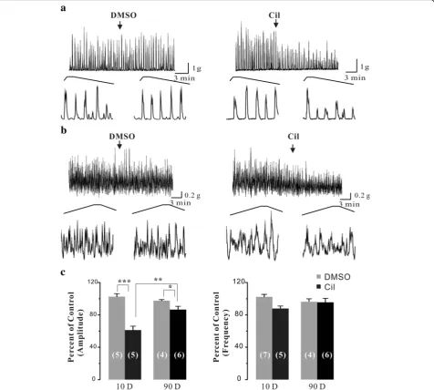
Next, we test the functional roles of PDE3 in the phasic contractions of adult rat urinary bladder smooth muscle. We found that Cil (1 μM), a specific PDE3
in-hibitor, significantly decreased the amplitude of
carbachol-enhanced contractions of adult rat bladder strips by 13.4 ± 4.0% (P< 0.05), and slightly, although not significantly, reduced their frequency by 4.4 ± 4.9%. When comparing to the neonatal bladder smooth muscle strips, Cil-mediated effects on the phasic con-tractions of smooth muscle strips were much smaller in adult rat urinary bladder. In detail, the effects of Cil were decreased by 2.5 fold (from 38.8% to 13.4%, p< 0.01) in amplitude without a significant decrease (12.1% to 4.4%, p> 0.05) in frequency. These results suggested that PDE3-mediated functional role was blunted in adult rat urinary bladder.
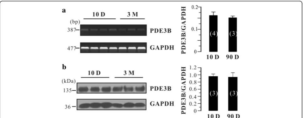
There are two subfamilies of PDE3, PDE3A and PDE3B [11]. They are encoded by different genes and participate in different cellular processes. Is there any re-lationship between the PDE3B expression and the blunted effects of Cil in adult rat bladder? To answer this question, we tested PDE3B mRNA and protein levels in neonatal and adult rat urinary bladder. As
shown in Fig. 3a, PDE3B mRNA was detected in both neonatal (N= 4) and adult (N= 3) rat urinary bladders. However, the expression level was not significantly dif-ferent between those two groups (Fig. 3a). Moreover, PDE3B protein expression in neonatal and adult urinary bladders was not different (N= 3 each; Fig. 3b). Taken together, PDE3B was not altered between neonatal and adult rat urinary bladder, suggesting that it is not in-volved in the blunted effects of Cil in adult rat bladder.
Discussion
Cyclic nucleotide PDEs carry out essential roles in signal transduction by modulating cAMP and cGMP levels and have been recognized as potential targets of several blad-der diseases such as overactive bladblad-der. The presence and activities of PDE1–5 have been tested and con-firmed in the rodent, rabbit, guinea pig, and human urinary bladder by pharmacological, biochemical, immu-nohistochemical and molecular methods [12–16]. By using qPCR technique, Lakics et al. have shown that sev-eral PDE isoform of PDE7–10 were expressed in the hu-man urinary bladder [16]. Moreover, the expression of PDE5 in different cell types of urinary bladder such as the urothelium, suburothelial interstitial cells and blad-der blood smooth muscle cells has been determined in the guinea pig [17]. However, the molecular levels of the whole PDEs family in the rodent urinary bladder are still unknown. Here, we established the mRNA expression levels of 18 different PDE isoforms in rat urinary bladder by RT-PCR. Our results indicated that PDE5 is one of the major isoform expressed in rat urinary bladder, Table 2The expression levels of PDE isoforms in adult rat urinary bladder
PDE
1A 1B 1C 2A 3A 3B 4A 4B 4C 4D 5A 7A 7B 8A 8B 9A 10A 11A
1 + ˗ ˗ ++ ˗ + ++ ++ ˗ ++ +++ + + ˗ + +++ +++ ˗
2 + ˗ ˗ ++ - + ++ ++ ˗ ++ +++ + + ˗ + +++ +++ ˗
3 + ˗ ˗ ++ - + ++ ++ ˗ ++ +++ + + ˗ + +++ +++ ˗
+++ indicates high expression; ++ indicates medium expression; + indicates low expression;−indicates no detection
corresponding with its important role in the urinary bladder [15, 17]. In consistent with the finding in human urinary bladder [14], PDE9A gene was shown to be highly expressed in rat urinary bladder. Meanwhile, Nagasaki et al. showed that PDE9 protein was widely distributed in the urothelial epithelium of the human lower urinary tract including the bladder [18]. Thus, it would be of interest to test its functional roles in the
bladder. Although PDE10A gene was abundantly
expressed, little is known about its role in the rat and human urinary bladder. It has been well known that
PDE1 was a major isoform expressed the in human urin-ary bladder [14]. However, the function of PDE1 in the rat urinary bladder was still controversial. Qiu et al. showed that PDE1 inhibitor had a strong relaxant effect on pre-contracted bladder strips [12]. Our previous study found that PDE1 inhibitor had no effect on spon-taneous phasic contraction of bladder strips [2]. One possibility is that the PDE1 inhibitor vinpocetine used in Qiu’s study is not specific to PDE1. It can inhibit voltage-sensitive sodium channels and cause a
dose-dependent decrease in extracellular calcium [19].
Fig. 3Effects of Cil on the carbachol-enhanced phasic contractions of neonatal and adult rat urinary bladder strips.aOriginal traces of carbachol-enhanced phasic contractions in neonatal rat bladder strips before and after the application of DMSO (<0.1%) and the PDE3 inhibitor Cil (1μM).
Moreover, our results indicated that only PDE1A was slightly expressed while both PDE1B and PDE1C were not detected, suggesting that PDE1 expression is varied among species. This difference might partially explain that PDE1 inhibitors exhibit effects in human [20–22] but not in rat bladder [2].
The PDE3 family contains two subfamilies, PDE3A and PDE3B, which are encoded by distinct but related genes [11]. PDE3A and PDE3B isoforms are widely expressed but also exhibit tissue-specific patterns. For example, PDE3A is highly expressed in cardiovascular system, platelets, and oocytes, while PDE3B is mainly expressed in adipocytes, hepatocytes, and pancreatic β-cells [11]. In the present study, we found that both mRNA and protein of PDE3A were dramatically de-creased in adult rat urinary bladder (Fig. 2), whereas PDE3B was not altered (Fig. 3). Although the physio-logical significance underlying in this process are un-known, we speculated that PDE3A might play an important role in the development of rat urinary blad-der. It has been established that PDE3A is involved in smooth muscle phenotype switch [23–25] and regulates VSMC cycle progression through controlling cAMP pools [26]. In addition, neonatal urinary bladder is much more proliferative than adult urinary bladder [27]. Thus, it is not surprised that PDE3A would be an important regulator in the development of urinary bladder. However, this possibility should be further tested.
There is evidence that the characteristics of bladder spontaneous activity are age-dependent [28], and are re-lated to changes in expression of several proteins such as large conductance Ca2+
-activated K+channels [5] and connexin [29]. Our results showed that Cil-mediated ef-fects on the phasic contractions were blunted in adult
rat bladder strips (Fig. 4) likely due to decreased PDE3A expression. Although PDE3B was consistently expressed throughout the development (Fig. 3), it could not com-pensate for the PDE3A-mediated effects. These results suggest that PDE3A and PDE3B may take different roles in mediating urinary bladder smooth muscle tone. Nevertheless, these results supported that the notion that PDE3A and PDE3B exhibit distinct functional roles.
Conclusion
In conclusion, our results for the first time provided a comprehensive view of the gene expression profiles of PDE isoforms in rat urinary bladder. We also found that the function of PDE3 was blunted in adult rat bladder likely due to the decreased expression of PDE3A but not PDE3B, suggesting that PDE3A and PDE3B might play different roles in the urinary bladder.
Abbreviations
Cil:Cilostamide; DMSO: Dimethylsulfoxide; PCR: Polymerase chain reaction; PDE: Phosphodiesterase
Acknowledgements
Not applicable.
Funding
This work was supported by a grant from the National Science Foundation of China (Grant No. 31300956) and the Beijing Nature Science Foundation (Grant No. 7172133).
Availability of data and materials
The data supporting the conclusions of this article are included within the article.
Authors’contributions
Conceived and designed the experiments: KZ GJ. Performed the
Authors’information
Not applicable.
Ethics approval
The experimental protocol was approved by the Institute of Biophysics Committee.
Consent for publication
Not applicable.
Competing interests
The authors declare that they have no competing interests.
Publisher’s Note
Springer Nature remains neutral with regard to jurisdictional claims in published maps and institutional affiliations.
Author details
1Department of Urology, Beijing Jishuitan Hospital, Beijing, China.2National
Laboratory of Biomacromolecules, Institute of Biophysics, Chinese Academy of Sciences, 15 Datun Road, Beijing 100101, China.3Department of Urology, Peking University First Hospital, Beijing, China.
Received: 20 July 2016 Accepted: 29 June 2017
References
1. Artim DE, Kullmann FA, Daugherty SL, Wu HY, de Groat WC. Activation of the nitric oxide-cGMP pathway reduces phasic contractions in neonatal rat bladder strips via protein kinase G. Am J Physiol Renal Physiol. 2009;297(2): F333–40.
2. Zhai K, Chang Y, Wei B, Liu Q, Leblais V, Fischmeister R, Ji G. Phosphodiesterase types 3 and 4 regulate the phasic contraction of neonatal rat bladder smooth myocytes via distinct mechanisms. Cell Signal. 2014;26(5):1001–10.
3. Lugnier C. Cyclic nucleotide phosphodiesterase (PDE) superfamily: a new target for the development of specific therapeutic agents. Pharmacol Ther. 2006;109:366–98.
4. Bender AT, Beavo JA. Cyclic nucleotide phosphodiesterases: molecular regulation to clinical use. Pharmacol Rev. 2006;58(3):488–520. 5. Ng YK, de Groat WC, Wu HY. Smooth muscle and neural mechanisms
contributing to the downregulation of neonatal rat spontaneous bladder contractions during postnatal development. Am J Physiol Regul Integr Comp Physiol. 2007;292(5):R2100–12.
6. Szell EA, Somogyi GT, de Groat WC, Szigeti GP. Developmental changes in spontaneous smooth muscle activity in the neonatal rat urinary bladder. Am J Physiol Regul Integr Comp Physiol. 2003;285(4):R809–16.
7. Gillespie JI, Markerink-van Ittersum M, de Vente J. cGMP-generating cells in the bladder wall: identification of distinct networks of interstitial cells. BJU Int. 2004;94(7):1114–24.
8. Rahnama’i MS, van Koeveringe GA, van Kerrebroeck PE, de Wachter SG. The effect of indomethacin on the muscarinic induced contractions in the isolated normal guinea pig urinary bladder. BMC Urol. 2013;13:8. 9. Finney SM, Stewart LH, Gillespie JI. Cholinergic activation of phasic activity
in the isolated bladder: possible evidence for M3- and M2-dependent components of a motor/sensory system. BJU Int. 2007;100(3):668–78. 10. Zhai K, Hubert F, Nicolas V, Ji G, Fischmeister R, Leblais V. Beta-adrenergic
cAMP signals are predominantly regulated by phosphodiesterase type 4 in cultured adult rat aortic smooth muscle cells. PLoS One. 2012;7(10):e47826. 11. Ahmad F, Degerman E, Manganiello VC. Cyclic nucleotide
phosphodiesterase 3 signaling complexes. Horm Metab Res. 2012;44(10):776–85.
12. Qiu Y, Kraft P, Craig EC, Liu X, Haynes-Johnson D. Identification and functional study of phosphodiesterases in rat urinary bladder. Urol Res. 2001;29(6):388–92.
13. Qiu Y, Kraft P, Craig EC, Liu X, Haynes-Johnson D. Cyclic nucleotide phosphodiesterases in rabbit detrusor smooth muscle. Urology. 2002;59(1):145–9.
14. Lakics V, Karran EH, Boess FG. Quantitative comparison of
phosphodiesterase mRNA distribution in human brain and peripheral tissues. Neuropharmacology. 2010;59(6):367–74.
15. Rahnama’i MS, Uckert S, Hohnen R, van Koeveringe GA. The role of phosphodiesterases in bladder pathophysiology. Nat Rev Urol. 2013;10(7):414–24.
16. Rahnama’i MS, Hohnen R, Van Kerrebroeck PE, van Koeveringe GA. Phosphodiesterase type 2 distribution in the guinea pig urinary bladder. World J Urol. 2015;33(10):1623–33.
17. Rahnama’i MS, van Koeveringe GA, Hohnen R, Ona S, van Kerrebroeck PE, de Wachter SG. Distribution of phosphodiesterase type 5 (PDE5) in the lateral wall of the guinea pig urinary bladder. BJU Int. 2013;112(2):246–57. 18. Nagasaki S, Nakano Y, Masuda M, Ono K, Miki Y, Shibahara Y, Sasano H.
Phosphodiesterase type 9 (PDE9) in the human lower urinary tract: an immunohistochemical study. BJU Int. 2012;109(6):934–40.
19. Sitges M, Galvan E, Nekrassov V. Vinpocetine blockade of sodium channels inhibits the rise in sodium and calcium induced by 4-aminopyridine in synaptosomes. Neurochem Int. 2005;46(7):533–40.
20. Uckert S, Stief CG, Odenthal KP, Becker AJ, Truss MC, Jonas U. Comparison of the effects of various spasmolytic drugs on isolated human and porcine detrusor smooth muscle. Arzneimittelforschung. 1998;48(8):836–9. 21. Truss MC, Uckert S, Stief CG, Kuczyk M, Jonas U. Cyclic nucleotide
phosphodiesterase (PDE) isoenzymes in the human detrusor smooth muscle. I. Identification and characterization. Urol Res. 1996;24(3):123–8. 22. Truss MC, Uckert S, Stief CG, Forssmann WG, Jonas U. Cyclic nucleotide phosphodiesterase (PDE) isoenzymes in the human detrusor smooth muscle. II. Effect of various PDE inhibitors on smooth muscle tone and cyclic nucleotide levels in vitro. Urol Res. 1996;24(3):129–34. 23. Tilley DG, Maurice DH. Vascular smooth muscle cell phosphodiesterase
(PDE) 3 and PDE4 activities and levels are regulated by cyclic AMP in vivo. Mol Pharmacol. 2002;62(3):497–506.
24. Dunkerley HA, Tilley DG, Palmer D, Liu H, Jimmo SL, Maurice DH. Reduced phosphodiesterase 3 activity and phosphodiesterase 3A level in synthetic vascular smooth muscle cells: implications for use of phosphodiesterase 3 inhibitors in cardiovascular tissues. Mol Pharmacol. 2002;61(5):1033–40. 25. Palmer D, Maurice DH. Dual expression and differential regulation of
phosphodiesterase 3A and phosphodiesterase 3B in human vascular smooth muscle: implications for phosphodiesterase 3 inhibition in human cardiovascular tissues. Mol Pharmacol. 2000;58:247–52.
26. Begum N, Hockman S, Manganiello VC. Phosphodiesterase 3A (PDE3A) deletion suppresses proliferation of cultured murine vascular smooth muscle cells (VSMCS) via inhibition of mitogen activated protein kinase (MAPK) signaling and alterations in critical cell cycle regulatory proteins. J Biol Chem. 2011;286(29):26238–49.
27. Jost SP. Cell cycle of normal bladder urothelium in developing and adult mice. Virchows Arch B Cell Pathol Incl Mol Pathol. 1989;57(1):27–36. 28. Szigeti GP, Somogyi GT, Csernoch L, Szell EA. Age-dependence of the
spontaneous activity of the rat urinary bladder. J Muscle Res Cell Motil. 2005;26(1):23–9.
29. Ikeda Y, Fry C, Hayashi F, Stolz D, Griffiths D, Kanai A. Role of gap junctions in spontaneous activity of the rat bladder. Am J Physiol Renal Physiol. 2007; 293(4):F1018–25.
• We accept pre-submission inquiries
• Our selector tool helps you to find the most relevant journal
• We provide round the clock customer support
• Convenient online submission
• Thorough peer review
• Inclusion in PubMed and all major indexing services
• Maximum visibility for your research
Submit your manuscript at www.biomedcentral.com/submit