Open Access
Research article
The study of expanded tri-lobed flap in a rabbit model: possible flap
model in ear reconstruction?
Eray Copcu*
1, Mesut Yazici
2, Barlas Etensel
2, Yakup Yüreklý
3and
Muharrem Balkaya
4Address: 1Department of Plastic and Reconstructive Surgery, Medical Faculty, Adnan Menderes University, Aydin, TURKEY, 2Department of Pediatric Surgery, Medical Faculty, Adnan Menderes University, Aydin, TURKEY, 3Department of Nuclear Medicine, Medical Faculty, Adnan Menderes University, Aydin, TURKEY and 4Department of Physiology, Veterinary Faculty, Adnan Menderes University, Aydin, TURKEY Email: Eray Copcu* - [email protected]; Mesut Yazici - [email protected]; Barlas Etensel - [email protected];
Yakup Yüreklý - [email protected]; Muharrem Balkaya - [email protected] * Corresponding author
Abstract
Background: Local flaps are widely used in reconstructive surgery. Tri-lobed skin flap is a relatively new flap and there has been no experimental model of this flap. This flap can be used for repair of full thickness defects in the face, ears and alar region. Based on the size of ears in a rabbit, we designed a model of ear reconstruction using expanded tri-lobed flap. Local flaps are more advantageous in that they provide excellent color and texture matching up with those of the face, adequately restore ear contour, place scars in a favorable location and ideally accomplish these goals in a single stage with minimal donor site morbidity.
Methods: Eight adult New Zealand rabbits were divided into two groups. 50 ml round tissue expander were implanted to four rabbits. After completion of the expansion, a superiorly based tri-lobed flap was elevated and a new ear was created from the superior dorsal skin of each rabbit. Scintigraphy with Technetium-99m pertecnetate was performed to evaluate flap viability.
Results: Subtotal flap necrosis was seen in all animals in non-expanded group. New ear in dimensions of the original ear was created in expanded group without complication. Perfusion and viability of the flaps were proved by Technetium-99m pertecnetate scintigraphy.
Conclusion: According to our knowledge this study is the first to demonstrate animal model in tri-lobed flap. Also, our technique is the first application of the trilobed flap to the possible ear reconstruction. We speculated that this flap may be used mastoid based without hair, in human. Also, tri-lobed flap may be an alternative in reconstruction of cylindrical organs such as penis or finger.
Background
Although local flaps are widely used, tri-lobed skin flap is not popular. Tri-lobed flaps as a modification of bi-lobed flaps were first reported by Harashina in 1977, and Weerda presented his experiences with this flap in facial
defects in 1978 [1,2]. As far as we know, there have been less than 10 reports on the flap. Ohtsuka used this flap in facial reconstruction in a patient with hemangioma [3]. Since tri-lobed flap has a right angle, Tanabe and Pompei used this flap for reconstruction of inverted nipple [4,5].
Published: 16 December 2003
BMC Surgery 2003, 3:13
Received: 23 July 2003 Accepted: 16 December 2003
This article is available from: http://www.biomedcentral.com/1471-2482/3/13
BMC Surgery 2003, 3 http://www.biomedcentral.com/1471-2482/3/13
Masakazu used perforator-based tri-lobed flap for cover-age of lumbosacral defects [6]. Copcu proposed this flap for reconstruction of the posterior sulcus of the ear because the angled, erect shape of the posterior ear can be obtained with this flap [7].
Ear reconstruction (total or subtotal) is one of most diffi-cult tasks in Plastic Surgery. The requisites for a successful total ear reconstruction are twofold. One is the construc-tion of a genuine cartilage framework and the other is the provision of durable yet thin coverage [8]. An ideal flap for ear reconstruction should provide color and texture matching with the recipient area, be thin for excellent cov-erage, safe and resistant to traumas. Moreover, it should create a sufficient angle as in the original shape of the ear. Although more than twenty flaps have been described for ear reconstruction, there is no animal model for flaps in ear reconstruction. Tissue expanders are the indispensable materials for plastic surgeons to get a new and durable tis-sue. Tissue expansion has given surgeons the ability to harvest large flaps of color-, thickness-, and texture-matched skin while simultaneously minimizing donor-site defects [9]. The superior match of color and texture achieved with tissue expanders as compared with the results achieved with conventional methods of microtia repair is one of the most important merits of the expan-sion procedure [10]. The aim of this study was to reveal that expanded tri-lobed flap elevated from the peri-auric-ular area could be a model for creating of a new ear in rabbits.
Methods
This study was carried out in Laboratory of Experimental Studies, Adnan Menderes University. We used eight adult New Zealand white rabbits weighing between 2–3 kg. Each rabbit was put in a separate cage. After they adapted to the environment, the experiments were done. The whole back was shaved in all rabbits. The animals were anesthetized with a combination of intramuscularly injected ketamin hydrochloride (35 mg/kg) and xylazine (5 mg/kg). Rabbits were randomly divided into two groups. The operations were performed under aseptic conditions.
Flap design
Tri-lobed flap was designed for ear reconstruction. Sizes of flaps were the same as in the original rabbit ear. Lengths of the arms of the flap were also the same as in the original ear of each rabbit. First, sizes of the ears of each rabbit were calculated. Bases of the flaps were located between the two ears of the rabbits. All lobes of the flaps were ellip-tical, and there was a 45° angle between them (Figure 1). The three arms of the skin flaps were elevated from the deep fascia. The last two arms (arms B and C) were ele-vated from the tip to the end of the base and these two
arms were sutured with 4/0 prolene to create a new ear (Figure 2 and 3). The defect caused by the elevations of the arms B and C were closed by transposition of the arm A (Figure 4).
Preparation of the tri lobed flap: Landmarks of the flap
Figure 1
Preparation of the tri lobed flap: Landmarks of the flap.
Elevation of the flap
Figure 2
Application of tissue expanders
Tissue expanders, which were 50 ml and round (Nagor Ltd, England), were implanted to the upper dorsal area of the four rabbits in Group 1 before the elevation of the flap. (Figures 5, 6, 7) For implantation of tissue expand-ers, 2 cm long incisions were made perpendicular to the areas where the expanders were to be placed and pockets of the sufficient size to allow insertion of a 50 ml round
expanders were created between the skin (panniculus car-nosus was included) and the deep fascia by blunt dissec-tion. 10 ml isotonic saline was injected into the expanders intra-operatively. The second saline injection was per-formed in the post-operative fifth day and 10 ml isotonic was given. Later, each expander was inflated at three days intervals with 10 ml isotonic saline under aseptic condi-tions. The last expansion was done in the 16th
postopera-tive day, and all expanders were inflated with total 50 ml saline.
Creating of new ear
Figure 3
Creating of new ear.
Closing of the donor area
Figure 4
Closing of the donor area.
Design of the flap in superior dorsal skin of the rabbit
Figure 5
Design of the flap in superior dorsal skin of the rabbit.
Inflated tissue expander
Figure 6
BMC Surgery 2003, 3 http://www.biomedcentral.com/1471-2482/3/13
Application of alloplastic material
Porous polyethylene (Medpor®) implant in 80 mm length
and 3 mm width was applied in one rabbit in the expanded group. Base of this implant was sutured to the deep fascia and located between the flaps. This rabbit was followed for one more month.
Macroscopic evaluation
Viable parts of the flaps were determined and percentages of viable flaps were estimated by a computer-assisted analysis of the images. Since the demarcation between viable and necrotic tissues was easily identified, it was not necessary to use vital dyes to assess flap necrosis. Views of the flaps were transferred to a computer with a digital camera and percentages of necrotic areas were calculated using a computer-assisted analysis (with Adobe Pho-toshop 6.0 software). The formula below was used: Viable flap area (mm2) / Total flap area (mm2) × 100
Evaluation of perfusion
Perfusion of the flaps was evaluated by nuclear medicine techniques. Static images were acquired by using a gamma camera (Siemens e.Cam Single, Illinois, USA) to observe the perfusion visually after injection of 0.5 mCi Techne-tium-99m pertecnetate. For quantitative evaluation, 0.1 mCi of Technetium-99m pertecnetate was injected subcutaneously to the flap and dynamic imaging was per-formed by acquisition of 1 second frames for 10 minutes. Data were processed to obtain a decay corrected time-activity curve of radiotracer washout. Benzylpenicillin 1 g was given intramuscularly for five days postoperatively.
Since the number of animals was small, statistical analysis could not be performed.
Results
No rabbit died of the procedures, and they were still alive 21 days after the procedures.
Non-expanded group
Flaps were created at the same time as in the expanded group. Subtotal flap necrosis was seen after the elevation of the flap. Two flaps had necrosis of more than 50 % per-cent in post-operative 7th day and the last two flaps also
had necrosis of 50 % of the flap in post-operative 21st day.
These flaps were not evaluated with Technetium-99m scintigraphy.
Expanded group
Minimal tip necrosis was seen in one rabbit around the suture line. All flaps were intact in post-operative 21st day.
Results confirmed the perfusion and viability of the flaps (Figures 8 and 9). Flaps were well perfused.
We used porous polyethylene (Medpor®) implant in 80
mm length and 3 mm width in one rabbit in expanded group. Base of this implant was sutured to the deep fascia and located between the flaps. The rabbit was followed for one month and there was no complication. An erect flap was created with this alloplastic material (Figure 10).
Results of macroscopic evaluation were listed in Table 1.
Discussion
Tri-lobed skin flap is a relatively new flap. Although many local flaps were reported with success in reconstructive surgery there are few reports related with tri-lobed flap and no animal model was presented in the literature.
Iida presented two kinds of tri-lobed flap: type I flap, which could be moved mainly by rotation, and type II, which compared with type I could serve as an advanced flap or transposed flap. Both flaps consisted of three trian-gular flaps. Flaps described in our study were totally differ-ent from the flaps of Iida. Our flaps were elliptical and more simple and easy to perform. Furthermore, they can be considered as modification of bi-lobed flaps. Bi-lobed flaps are often used when the primary closure of a skin defect is difficult, but there have been few reports on its method of design [12]. Esser first described the bi-lobed flap in 1918 [13]. Esser's initial bilobed flap design depicted two transposition flaps in equal size transferred to cover distal nasal tip wounds. McGregor and Soutar theoretically demonstrated a method for designing the bi-lobed flap [14]. They roughly classified bi-bi-lobed flaps into type I and type II flaps. One of the most detailed reports was presented by Cook [15]. Cook stated that, depending
New ear which was reconstructed with tri-lobed flap
Figure 7
on the flap's anatomic location, the surgeon had a great ability to vary the size of each lobe of this flap.
In this study we tried to perform an animal model with expanded tri-lobed skin flap. We were inspired by sizes of rabbit ears and aimed to obtain long but narrow flaps safely using an expander. As far as we know, there has not been an animal model for reconstruction of ears.
Reconstruction of the ear is still a challenge in plastic sur-gery since ear has a special and complex anatomy. The normal external ear forms an angel about 23 degrees with temporal area [4]. Diverse materials and methods have been used in auricular region. The earliest account of auricular reconstruction appears to have been written in about 600 B.C. by Sushruta and a slightly more profes-sional procedure than that of Sushruta was recommended in about 300 A.D. by Celsus, who described the repair of an auricle by creating quadrangular advancement flaps and the treatment of concomitant atresia by drilling the bone and plunging the hole. As early as 1597, the Italian surgeon Tagliacozzi described and illustrated repair of both upper and lower ear deformities with skin flaps from behind the auricular region [5]. Until today, more flaps, techniques and implantation materials have been described for reconstruction of the ear [6-9]. Generally multiple surgical procedures were required in reconstruc-tion of congenital microtia. The surgical reconstrucreconstruc-tion procedure for microtia was established with the advent of
Tanzer's operation [7]. Excellent tissue is required for cov-ering the framework in all techniques. Since this tissue should have similar original texture, color and hair bear-ing characteristics of the original ear, local flaps gained more popularity [10,11].
Neumann first applied the concept of tissue expansion to ear reconstruction and introduced the idea of expansion to the field of surgery [12]. One of the largest series was presented by Brent in 1992 [13]. Tanino and Miyasaka reported 14% exposure of the expander in microtia recon-struction as a complication [14]. Hata et al. also presented their experiences in ear reconstruction with tissue expander [3]. The Chiba University schedule for total ear reconstruction includes three stages [15]. Tissue expander is used in the first stage. The superior match of color and texture achieved with the tissue expander as compared with the results achieved with conventional methods of microtia repair is one of the most important merits of the expansion procedure [3]. Another important merit of the expansion procedure is the recovery and maintenance of skin sensation; however, because the expanded fascia and grafted skin cover the posterior surface of the auricle in graft, sensation in this region will completely disappear postoperatively.
The ear is a very complex organ and there is no animal which has ears similar to those of human beings. There are two major considerations for successful total-ear
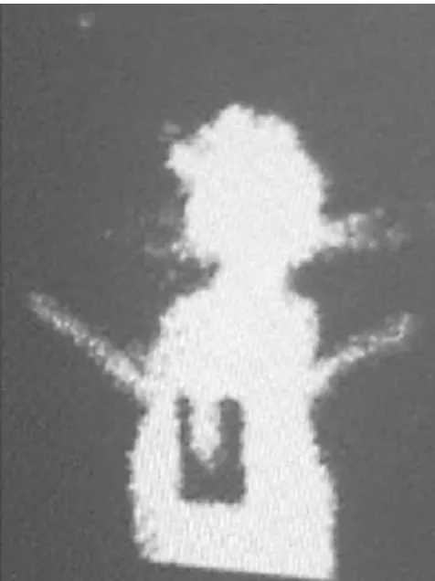
Evaluation of the flap with Technetium-99m scintigraphy: Static view of the flap
Figure 8
BMC Surgery 2003, 3 http://www.biomedcentral.com/1471-2482/3/13
Evaluation of the flap with Technetium-99m scintigraphy: Dynamic view of the flap
Figure 9
Evaluation of the flap with Technetium-99m scintigraphy: Dynamic view of the flap. Time-activity curve of radiotracer washout.
Erectly view of the tri-lobed flap with porous polyethylene implant
Figure 10
Erectly view of the tri-lobed flap with porous polyethylene implant.
Table 1: Macroscopic evaluations of the flaps:
Group Rabbit No: Post-operative day 7 th Post-operative day 14 th Post-operative day 21st
Non-expanded group 1 72 % 60 % 41 % (*)
2 45 % (*) 3 40 % (*)
4 68 % 63 % 40 % (*)
Expanded group 5 100 % 95 % 95 %
6 100 % 100 % 100 %
7 100 % 100 % 100 %
8 100% 98 % 98 %
(5) erect cylindrical tissue can be obtained,
(6) since the new ear is reconstructed with two lobes of the flap, edges of the ear could be created easily,
(7) design of the flap is not complex and does not require geometrical knowledge.
The aim of this study was to obtain a reliable flap and thus create an alternative for ear reconstruction. The most important part of ear reconstruction is creation of a frame-work to give a shape to the new ear. We also investigated whether the flap was resistant to alloplastic materials in one rabbit and obtained promising results.
Conclusion
It can be concluded that tri-lobed flaps can be obtained successfully although they cause some risks due to their sizes. Since tri-lobed flap can achieve an epithelized tissue in two sides of an organ as in the ears, they can be alterna-tive in partial or total ear reconstruction.
Any process without a framework can not be a model for ear reconstruction. Although we had only one rabbit with aloplastic material in expanded group, we speculated that this flap may be used with autogenous and alloplastic materials.
A new ear which is erect, sufficiently angled and thin and thus easily adapted to the shape of the framework, and resistant to external traumas could be reconstructed with this flap without any donor site problems. Finally, this flap is not only suitable for ear reconstruction but also it may be used in reconstruction of cylindrical organs such as penis or finger.
Competing interests
None declared.
Authors' contributions
E. Copcu designed the study, performed the operations and prepared the manuscript. M. Yazici and B. Etensel par-ticipated in the performing of the operations. Y. Yurekli performed the scintigraphy. M. Balkaya participated in the design of study and performed the statistical analysis. All authors read and approved the final manuscript.
References
1. Harashina T, Maruyama Y, Kitamura K: The trilobed flap. Case report.Plast Reconstr Surg 1977, 60:623-624.
2. Weerda H: [A trilobed rotation cheek flap for closure of facial defects (author's transl)].HNO 1979, 27:358-359.
3. Ohtsuka H, Miki Y, Shioya N: Trilobed flap in facial
reconstruction.Br J Plast Surg 1982, 35:493-497.
4. Tanabe HY, Tai Y, Kiyokawa K, Yamauchi T: Nipple-areola recon-struction with a dermal-fat flap and rolled auricular cartilage.Plast Reconstr Surg 1997, 100:431-438.
5. Pompei S, Tedesco M: A new surgical technique for the correc-tion of the inverted nipple.Plast Reconstr Surg 1999, 23:371-374. 6. Ao M, Mae O, Namba Y, Asagoe K: Perforator-based flap for
cov-erage of lumbosacral defects. Plast Reconstr Surg 1998,
101:987-991.
7. Copcu E: Postauricular trilobed skin flap for reconstruction of postauricular sulcus.Eur J Plast Surg 2002, 25:341-343.
8. Park C, Suk Roh T: Total ear reconstruction in the devascular-ized temporoparietal region: I. Use of the contralateral tem-poroparietal fascial free flap. Plast Reconstr Surg 2001,
108:1145-1153.
9. Radovan C: Tissue expansion in soft-tissue reconstruction.
Plast Reconstr Surg 1984, 74:482-492.
10. Hata Y, Hosokawa K, Yano K, Matsuka K, Ito O: Correction of con-genital microtia using the tissue expander.Plast Reconst Surg
1989, 84:741-751.
11. Iida N, Ohsumi N, Sakai M: A trilobed flap for reconstruction of nasal skin defects.Plast Reconstr Surg 1997, 100:991-995. 12. Iida N, Ohsumi N, Tonegawa M, Tsutsumi K: Simple method of
designing a bilobed flap.Plast Reconstr Surg 1999, 104:495-499. 13. Esser JFS: Gestielite lokale Nasenplastik mit zweizipfligem
Lappen, Deckung des sekundaren Defktes vom ersten Zipfel durch den zweiten.Dtsch Z Chir 1918, 143:385-90.
14. McGregor JC, Soutar DS: A critical assessment of the bilobed flap.Br J Plast Surg 1981, 34:197-205.
15. Cook JL: A review of the bilobed flap's design with particular emphasis on the minimization of alar displacement.Dermatol Surg 2000, 26:354-62.
16. Farkas LG: Anthropometry of the Head and Face.New York: Raven 1984.
17. Aguilar EF 3rd: Auricular reconstruction of congenital micro-tia (grade III).Laryngoscope 1996, 106:1-26.
18. Nagata S: Reconstruction of the auricle.Plast Reconstr Surg 1994,
93:225-231.
19. Tanzer RC: Total reconstruction of the auricle. The evolution of a plan of treatment.Plast Reconstr Surg 1971, 47:523-533. 20. Brent B: Earlobe construction with an auriculomastoid flap.
Plast Reconstr Surg 1976, 57:389-391.
21. Brent B: Technical advances in ear reconstruction with autog-enous rib cartilage grafts – personal experience with 1,200 cases.Plast Reconstr Surg 1999, 104:319-334.
22. Masson JK: A simple island flap for reconstruction of concha-helix defects.Br J Plast Surg 1972, 25:399-403.
23. Renard A: Postauricular flap based on a dermal pedicle fýr ear reconstruction.Plast Reconstr Surg 1981, 68:159-165.
24. Neumann CG: The expansion of an area of skin by progressive distension of a subcutaneous balloon.Plast Reconstr Surg 1957,
19:124-127.
25. Brent B: Auricular repair with autogenous rib cartilage grafts: Two decades of experience with 600 cases.Plast Reconstr Surg
1992, 90:355-374.
26. Tanino R, Miyasaka M: Reconstruction of microtia using tissue expander.Clin Plast Surg 1990, 17:339-353.
27. Danino AM, Yoshimoto S, Ichinose M, Kuroki T: The Chiba Uni-versity chronology for total ear reconstruction.Plast Reconstr Surg 2000, 106:218-219.
Pre-publication history
The pre-publication history for this paper can be accessed here: