REVIEW
Methods for the extraction, storage, amplification and sequencing of DNA from
environmental samples
Gavin Lear
1*, Ian Dickie
2, Jonathan Banks
3, Stephane Boyer
4, Hannah L. Buckley
5,
Thomas R. Buckley
1,6, Rob Cruickshank
7, Andrew Dopheide
6, Kim M. Handley
1, Syrie Hermans
1,
Janine Kamke
1, Charles K. Lee
8, Robin MacDiarmid
9, Sergio E. Morales
10, David A. Orlovich
11,
Rob Smissen
12, Jamie Wood
12and Robert Holdaway
121School of Biological Sciences, University of Auckland, 3a Symonds Street, Auckland 1010, New Zealand 2School of Biological Sciences, University of Canterbury, Christchurch 8140, New Zealand
3The Cawthron Institute, Nelson 7010, New Zealand
4Insect Biology Research Institute (IRBI) – UMR 7261 CNRS / Université François-Rabelais de Tours, Parc Grandmont,
37200 Tours, France
5School of Science, Auckland University of Technology, Auckland 1010, New Zealand 6Landcare Research, Auckland 1142, New Zealand
7Department of Ecology, Lincoln University, Lincoln 7647, New Zealand 8School of Sciences, The University of Waikato, Hamilton 3240, New Zealand 9Plant and Food Research, Auckland 1025, New Zealand
10Microbiology and Immunology, University of Otago, Dunedin 9054, New Zealand 11Department of Botany, University of Otago, Dunedin 9054, New Zealand
12Landcare Research, Lincoln 7640, New Zealand
*Author for correspondence (Email: [email protected])
Published online: 11 December 2017
Abstract: Advances in the sequencing of DNA extracted from media such as soil and water offer huge opportunities for biodiversity monitoring and assessment, particularly where the collection or identification of whole organisms is impractical. However, there are myriad methods for the extraction, storage, amplification and sequencing of DNA from environmental samples. To help overcome potential biases that may impede the effective comparison of biodiversity data collected by different researchers, we propose a standardised set of procedures for use on different taxa and sample media, largely based on recent trends in their use. Our recommendations describe important steps for sample pre-processing and include the use of (a) Qiagen DNeasy PowerSoil® and PowerMax® kits for extraction of DNA from soil, sediment, faeces and leaf litter; (b) DNeasy
PowerSoil® for extraction of DNA from plant tissue; (c) DNeasy Blood and Tissue kits for extraction of DNA
from animal tissue; (d) DNeasy Blood and Tissue kits for extraction of DNA from macroorganisms in water and ice; and (e) DNeasy PowerWater® kits for extraction of DNA from microorganisms in water and ice. Based on
key parameters, including the specificity and inclusivity of the primers for the target sequence, we recommend the use of the following primer pairs to amplify DNA for analysis by Illumina MiSeq DNA sequencing: (a) 515f and 806RB to target bacterial 16S rRNA genes (including regions V3 and V4); (b) #3 and #5RC to target eukaryote 18S rRNA genes (including regions V7 and V8); (c) #3 and #5RC are also recommended for the routine analysis of protist community DNA; (d) ITS6F and ITS7R to target the chromistan ITS1 internal transcribed spacer region; (e) S2F and S3R to target the ITS2 internal transcribed spacer in terrestrial plants; (f) fITS7 or gITS7, and ITS4 to target the fungal ITS2 region; (g) NS31 and AML2 to target glomeromycota 18S rRNA genes; and (h) mICOIintF and jgHCO2198 to target cytochrome c oxidase subunit I (COI) genes in animals. More research is currently required to confirm primers suitable for the selective amplification of DNA from specific vertebrate taxa such as fish. Combined, these recommendations represent a framework for efficient, comprehensive and robust DNA-based investigations of biodiversity, applicable to most taxa and ecosystems. The adoption of standardised protocols for biodiversity assessment and monitoring using DNA extracted from environmental samples will enable more informative comparisons among datasets, generating significant benefits for ecological science and biosecurity applications.
Key words: biological heritage; biodiversity monitoring; community profiling; DNA primers; DNA sequencing; eDNA; environmental DNA; Illumina; metabarcoding; metagenomics; molecular ecology
Introduction
The first widely-used DNA sequencing approach (Sanger et al. 1977) was sufficient to sequence the human genome (Venter et al. 2001), but the limited throughput of this technique remains a major constraint on its use for the analysis of complex DNA pools. Even following the release of high-throughput pyrosequencing platforms such as the 454 Genome Sequencer Instrument, capable of generating 25 million bases in a single 4-hour run (Margulies et al. 2005), DNA-based analyses of biological communities in multiple samples remained problematic due to difficulties associated with combining and later identifying DNA originating from many different samples. More recently, multiplex primer labelling approaches have been developed that, after DNA sequencing, allow the user to determine which DNA sequences originated from each of multiple DNA samples combined in a single solution prior to their analysis. Today, DNA from many hundreds of samples can be combined and analysed in parallel (Barberan et al. 2014, 2015; Shokralla et al. 2015). This ability to generate large amounts of sequence data from numerous samples in parallel offers huge potential for using DNA to monitor the biological diversity in any sample from which DNA can be extracted. Significantly, these methods are applicable to all organisms (e.g. Archaea, Bacteria, Protista, Fungi, Animalia and Plantae), all genetic markers (e.g. 16S, 18S, ITS, COI and rbcL) and all sample media (e.g. soil, water, air and tissue) such that DNA analysis protocols could provide a universal tool for future biodiversity and biosecurity assessments. The combination of the mass amplification of these genetic markers or ‘DNA barcodes’ by PCR, with high-throughput DNA sequencing to identify a mixture of organisms is most commonly referred to as ‘metabarcoding’. We seek to overcome the currently fragmented understanding of the identity and distribution of both native and introduced species, using a unified DNA metabarcoding approach for high throughput assessments of communities across all domains of life.
While DNA-based biodiversity assessment methods are not yet in widespread use beyond the microbial world, there are many potential benefits, as well as uncertainties, resulting from their application to a far wider range of organisms, as reviewed in Holdaway et al. (2017). For example, traditional observational techniques for biodiversity monitoring can be highly dependent on (and biased by) taxonomic and diagnostic expertise that is in scarce supply worldwide (Paknia et al. 2015). Cryptic species can be misidentified and whole taxa may be underrepresented or overlooked due to factors including their small size, nocturnal habits, occurrence in less-accessible habitats (e.g. below ground), or the non-random movement of study organisms in response to disturbance while being surveyed (Watson et al. 1995). In contrast, samples for DNA metabarcoding may be collected by non-specialists and may not require invasive sampling protocols since sampling or capture of whole, individual organisms is typically not required. Many environmental substrates are easy to sample (e.g. soils) and contain significant populations of micro-organisms, as well as small invertebrates, which will be represented in the DNA extracted from these substrates. In addition to the cellular DNA that may be directly extracted from communities of organisms, large quantities of ‘environmental DNA’ are continually excreted and shed in the environment by living organisms. For example, animals can be detected based on DNA excreted into environments from their urine (Valiere & Taberlet 2000), faeces (Kurose et al. 2005), hair and skin
(Henry & Russello 2011). Similarly, plant DNA originating from roots, root exudates and litter can provide information about plant community composition (Yoccoz et al. 2012). Consequently, DNA extracted from samples of soil, water or other material may simultaneously provide information about the occurrence, distribution and diversity of organisms and communities, across multiple branches of life. The detection of organisms from environmental DNA has an additional benefit in allowing the detection of transient organisms, which may be missed by traditional observational sampling. Once collected, environmental DNA can be stored for long periods, providing a library of sample material that can be accessed at any time for re-analysis. This library creates opportunities for investigations of taxa, genes, and hypotheses that were not considered in the original study. The DNA sequence analysis of large sample numbers using metabarcoding approaches is becoming a more cost-competitive method for biodiversity and biosecurity monitoring as sequencing technologies advance. With a plethora of DNA extraction, storage, amplification and sequencing methods available it is not possible to provide the exact costs associated with these procedures. As an indicative cost, and not including the costs of human resources, the processing and analysis of over 300 samples is achievable (as of September 2017) for under NZ$12 000, excluding general sales tax (~NZ$3500 for DNA extraction; ~NZ$600 for DNA amplification; ~NZ$600 for PCR purification, ~NZ$7000 for analysis of 384 samples on an Illumina MiSeq DNA sequencing machine).
While DNA sampling and analysis holds much promise for use in biological heritage monitoring and assessment, a plethora of techniques are currently being used, not only for sample collection, but also the extraction, amplification, sequencing and storage of DNA from environmental samples. These methods, which vary among research groups focusing on different taxa and sample media, and even between individual researchers within these groups, mean that comparisons of data across studies are subject to multiple, poorly quantified biases. Additionally, it can take some time for new researchers to select from the often daunting list of sample processing options before commencing their own analysis. To address these shortcomings, we propose that standardised protocols should be promoted for the extraction, storage, amplification and sequencing of environmental DNA. These will help to reduce biases associated with the comparison of sample data collected by different researchers, and could provide additional opportunities for collaboration and sharing of data. In addition to providing a framework that existing researchers can choose to follow, we recommend here a standard set of methodologies that may also help to make DNA metabarcoding protocols more accessible to less experienced users.
amplicon data. The recommendation of a standardised set of methodologies provides additional impetus for researchers to test the impact and implications of their use of alternative methods for assessments of community composition including error rates for the false positive and negative detection of organisms from sample media. Thus, the importance of biases
associated with various aspects of DNA metabarcoding may be better quantified and understood. A number of reviews have recently been published to synthesise achievements in environmental DNA research (Goldberg et al. 2016) and to highlight the advantages and limitations of these methods for biodiversity assessment, but largely focused on assessments of
Glossary of terms
Amplicon: A piece of DNA that is the source or product of natural or artificial replication events, such as DNA fragments generated during PCR. Typically PCR amplicon lengths are no greater than 5000 nucleotide bases in length, although longer PCR products may be generated using specialised DNA polymerases.
Blocking primer: These are short DNA sequences, or ‘primers’ used to block the amplification of specific DNA fragments. They are most commonly used to maximise the detection of low-abundance DNA sequences. For example, to increase the detection rates of animal DNA in the gut of a carnivore, blocking primers may be used to inhibit amplification of the host carnivores’ DNA. A blocking primer is typically designed to bind to the unwanted DNA sequence, but is modified in such a way that it does not prime amplification during PCR.
Chimeras: In the context of molecular DNA studies, a chimera refers to any sequence that is formed when two or more sequences are joined together during PCR. Chimeric DNA sequences can artificially inflate diversity estimates and must be removed during bioinformatics analysis.
Deep-sequencing: The ‘depth’ of sequencing refers to the number of reads obtained by DNA sequencing. Deep-sequencing will provide many sequences per sample, whereas shallow-sequencing provides fewer sequences per sample, usually due to more samples being combined in a single multiplexed sequencing run.
Degenerate primers: A mixture of similar, but not identical primers where one or more of the nucleotide bases (A, C, G, T) in the primer sequence varies. Degenerate primers are used to increase primer universality.
DNA barcode: a short DNA sequence found in an organism that can be used to identify it.
DNA extraction: A process whereby the DNA is separated from the sample media. This is commonly achieved using both physical and chemical methods to lyse cells and to separate the DNA from any contaminants or inhibitory substances associated with the sample material.
DNA methylation: A method used by cells to control gene expression. A methyl (CH3) group is added to a DNA strand, effectively fixing the gene in an ‘off’ position with regards to gene expression, but without changing the DNA sequence. Environmental DNA (eDNA): DNA that is collected from an environment, rather than from an individual. Most commonly, this term is used to describe DNA that is no longer located in living cells (e.g. excreted DNA and DNA within cellular debris), although frequently it is also used to describe DNA extracted from environmental media such as soil including DNA extracted from micro- and macro-organisms within the sample.
False priming: When one or both of the primers used bind to a region of DNA outside of the target area, leading to amplification of unintended gene or non-gene regions. This result is often caused by non-specific binding by one or more of the bases in the primer sequences.
Flow cell: In the context of this review, flow cells are a surface on which sequencing chemistry occurs and over which sequencing polymerases, nucleotides and buffers can be pumped. DNA may be hybridised to flow cells in low molar quantities before sequencing en masse.
GC-content: A term which refers to the portion of guanine or cytosine bases that are present in a genome, gene or gene region. High GC-content DNA is more stable and tolerant to high temperatures than low GC-content DNA, due to the triple hydrogen bonds associated with the GC base pairing.
Hairpin: A U-shaped loop that is created when base pairs are formed between two different sections of the same DNA or RNA strand.
Metabarcoding: A molecular biodiversity detection method that uses short genetic markers, or DNA barcodes, to identify the presence of, and distinguish between, organisms in a sample.
Metagenomics: The study of the all the genetic material associated with an environment or sample. Microbiome: The genetic material of all the microorganisms present in a particular environment or sample.
Mock communities: Cells or DNA derived from a pre-defined range of organisms, often mixed in known concentrations. Mock communities and mock community DNA are usually provided in the form of a solution that can be incorporated into experiments to determine the accuracy of DNA sequencing approaches.
Next-generation sequencing (NGS): High-throughput DNA sequencing technologies that allow highly multiplexed sequencing, without the need for a cloning step.
Nuclear ribosomal DNA (nrDNA): DNA regions that originate in the nucleus of organisms, as opposed to plastid or mitochondrial DNA.
Paired end read: Paired ends refer to the two ends of the same DNA molecule. In paired end sequencing, one end of the DNA sequence is read before DNA sequence analysis is initiated from the other end of the DNA molecule.
Paraphyletic: A taxonomic term that refers to organisms descended from a common evolutionary ancestor, but does not include all the descendants.
PCR purification/PCR clean-up: The process of purifying amplicons for downstream analyses. During this step, components left over from the PCR that were not incorporated into the amplicons (e.g. residual primers, primer dimers, dNTPs and polymerases) are removed.
Plastid sequences: Originate from the plastids of organisms, rather than the nucleus. Plastids are organelles that have their own DNA and ribosomes, and are found in the cells of plants, algae and some protists.
Polymerase chain reaction (PCR): A molecular method whereby a specific DNA sequence is amplified across several orders of magnitude. The product of this reaction is called an amplicon. A common variant on this approach, quantitative PCR (qPCR), allows quantification of the number of target DNA sequences present in the original sample.
Polymerase: An enzyme that synthesises DNA. Modified versions of this enzyme are available and are used for PCR. Polyphyletic: A group of organisms composed of unrelated organisms descended from more than one ancestor.
Primer universality: A characteristic of a primer pair that determines how suitable it is to amplify the same gene region in a wide variety of different species. Increased primer universality means the DNA of a greater variety of organisms may be amplified by PCR.
Primer: A short strand of DNA that is required to initiate DNA synthesis. In PCR, a forward and reverse primer are used in combination to target a specific gene region for amplification.
Sequence identity: The extent to which one DNA sequence matches another, usually presented as the percentage of nucleotides (A, C, G, T), that correspond between the two sequences.
Sequencing: A process whereby the order of nucleotides within a DNA sequence is determined.
Sequencing read length: The number of nucleotide bases reported for a fragment of DNA following DNA sequence analysis. Single Nucleotide Polymorphism (SNP): When a single nucleotide (A, T, C or G) in one genome varies from that in another genome.
Template: The DNA, obtained through DNA extraction from a sample, that is added to a PCR. It will normally contain the region of interest to be amplified.
Virion: The infectious form of a virus, which transports the viral genome between cells. Virome: The genetic material from all the viruses associated with a host or environment.
specific taxa (Elbrecht & Leese 2015; Aylagas et al. 2016) or sample media (Drummond et al. 2015; Klymus et al. 2107). The aims of this review include, but are not restricted to, the following: (i) to review recent practices for the extraction, storage, amplification and sequencing of DNA from a broad range of environmental samples for the detection of a broad range of taxa, (ii) to recommend standard procedures for DNA extraction, amplification and sequencing of key taxa from the broadest range of environmental samples, and (iii) to identify emerging methods, such as new DNA sequencing approaches and shotgun metagenomics of relevance for future biodiversity assessments using DNA metabarcoding.
Review of current practices for extraction,
storage, amplification and sequencing of DNA
from environmental samples
Overview
We performed a literature review to summarise recent
Methods
We performed a search of the ISI Web of Science Core Collection in August 2015 (www.webofknowledge.com). We searched the literature for articles containing the terms ‘environmental DNA’ or ‘eDNA’ in the title, keywords or abstract. We refined our search terms to include only science and technology research published in the English language since 2010 and excluded books and conference proceedings by selecting only ‘articles’. In total, we reviewed the full text of 584 articles. Articles that were determined to be reviews and perspectives were removed from the larger database. Articles
0 5 10 15 20 25 30 35 40 45 50
2010 2011 2012 2013 2014 2015
Num
be
r o
f s
tudi
es
Publication year
Figure 1
Figure 1. Number of eDNA studies published each year that were captured by our search of the ISI Web of Science Core Collection in August 2015 containing the terms ‘environmental DNA’ or ‘eDNA’. We refined our search terms to include only science and technology research published in the English language since 2010 and excluded books and conference proceedings by selecting only ‘articles’. Articles that were considered to be reviews and perspectives were removed from the larger database. Articles focused on ‘extracellular DNA’ were also removed.
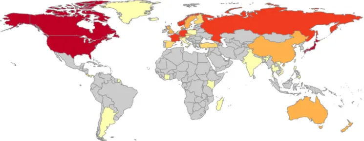
Figure 2. Geographic locations of the 167 studies reviewed. Colours on map indicate the number of studies conducted in each country: () ≥10; () 8–9; (no countries belong to this category) 6–7; () 4–5; () 2–3; () = 1; () no data. Three studies were conducted that focused on samples taken from the Antarctic land mass (data not shown). The exact number of studies conducted in each country is shown in Table S1 in Supplementary Material.
focused on ‘extracellular DNA’ were similarly removed leaving a total of 167 articles for in-depth review.
The terms ‘eDNA’ and ‘environmental DNA’ are largely redundant in studies of microbial communities and are used only infrequently by microbial researchers. As a consequence, we expect that studies on microbial DNA will be underrepresented due to our choice of search terms, even though almost all molecular studies on complex microbial communities can be considered as research on eDNA. Thus, it is important to note that our search terms were used to generate a varied dataset of eDNA-based studies focusing on a broad range of taxa and were never intended to capture all eDNA research. For example, there are 129 papers in the ISI Web of Science Core Collection up to 2015 that use the phrase ‘metabarcoding’ but not our search terms. Additionally, studies using alternative or more descriptive terms such as paleoenvironmental DNA (Rawlence et al. 2014) were not captured by our search terms meaning that research focusing on specific aspects of environmental DNA, such as ancient DNA may be underrepresented in our analysis.
Summary of international eDNA research undertaken prior to August 2015
Of the 167 articles reviewed, ~75% were published in the previous 3 years; only six were published in 2010 (Fig. 1). Locations of prior investigations
Organisms studied
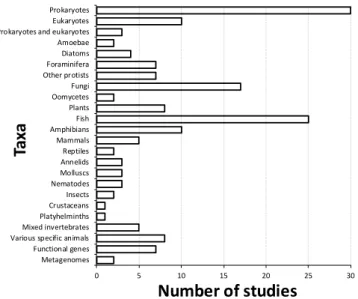
Studies of prokaryote communities (largely of Bacteria and Cyanobacteria) were most common (e.g. Costa et al. 2015; Dong et al. 2015; Pal et al. 2015), followed by investigations of fish (e.g. Jerde et al. 2013; Takahara & Minimoto 2013; Janosik & Johnston 2015; Fig. 3). Ten studies focused on a broad range of eukaryotic biodiversity (e.g. Pawlowski et al. 2011; Baldwin et al. 2013), whereas only three of the studies targeted both prokaryotic and eukaryotic organisms (Xiao et al. 2014; Young et al. 2014; Kowallik et al. 2015). Just over 10% of the studies investigated fungi (e.g. Rao et al. 2012; Lazarus & James 2015). Research on micro-eukaryotes was relatively common, with multiple studies being conducted on the foraminifera, as well as diatoms and a number of other protist taxa (e.g. Bradford et al. 2013; Lejzerowicz et al. 2014; Zimmermann et al. 2015). Various invertebrate taxa were investigated in 18 studies (e.g. Bienert et al. 2012; Yu et al. 2012; Deiner et al. 2015). Plants were investigated in just eight studies (e.g. Parducci et al. 2013; Pansu et al. 2015b), mammals in five (e.g. Nichols & Spong 2014; van Bleijswijk et al. 2014) and birds (in combination with other organisms) in just one study (Thomsen et al. 2012b).
Studies conducted on invasive organisms
A total of 26 studies were identified as using environmental DNA to investigate the presence of invasive organisms (See Table S2 in Supplementary Material). With the exception of one study investigating a fungal pathogen (Guignardia sp.) of citrus (Hu et al. 2014), all investigations were undertaken in aquatic environments. A majority of these studies investigated the presence of exotic fish such as common carp (Cyprinus carpio; Eichmiller et al. 2014; Takahara et al. 2015), silver and bighead carp (Hypophthalmichthys molitrix and nobilis; Jerde et al. 2013; Farrington et al. 2015), African jewelfish (Hemichromis lifalili; Moyer et al. 2014), bluegill sunfish (Lepomis macrochirus; Takahara & Minimoto 2013) and zebrafish (Danio rerio; Collins et al. 2013).
Multiple studies highlight the advantages of sampling environmental DNA for improving occurrence and detection
Figure 3. Taxa targeted in 167 studies reviewed. ‘Functional genes’ refers to the amplification of single genes encoding for functional processes (e.g. nitrogen fixation), ‘Metagenomes’ refers to gene data obtained by shotgun metagenomics.
0 5 10 15 20 25 30
Prokaryotes Eukaryotes Prokaryotes and eukaryotes Amoebae Diatoms Foraminifera Other protists Fungi Oomycetes Plants Fish Amphibians Mammals Reptiles Annelids Molluscs Nematodes Insects Crustaceans Platyhelminths Mixed invertebrates Various specific animals Functional genes Metagenomes
Number of studies
Ta
xa
Figure 3
estimates for invasive organisms. For example, Hunter et al. (2015) estimated detection probabilities in excess of 91% for Burmese python (Python bivittatus) with positive results reported outside of the leading northern edge of the organism’s known range. The analysis of environmental DNA is thought to offer substantial cost benefits over traditional methods for invasive organism detection. For example, Jerde et al. (2011) achieved a positive detection result for silver carp in just a few hours from the analysis of environmental DNA, compared to 93 days of effort the authors predicted would be required to obtain the same result by electrofishing. The authors were also able to confirm that the invasive carp were closer to the invasion of upstream lake systems than had previously been detected by traditional methods. Others have similarly reported that the analysis of environmental DNA is a superior approach compared to traditional survey methods for assessing the presence of invasive aquatic animals such as the bluegill sunfish (Takahara & Minimoto 2013) and the Chinese giant salamander (Andrias davidianus; Fukumoto et al. 2015).
Studies conducted on rare organisms
A total of 8% of studies were identified as using environmental DNA to investigate rare or threatened organisms (Table S2). Most of these studies investigated the presence of fish, including Chinook salmon (Oncorhynchus tshawytscha; Laramie et al. 2015), brook and bull trout (Salvelinus fontinalis and S. confluentus; Wilcox et al. 2013), and slackwater darter (Etheostoma boschungi; Janosik & Johnston 2015), as well as amphibians such as the great crested newt (Triturus cristatus; Rees et al. 2014; Biggs et al. 2015), Idaho salamander (Dicamptodon aterrimus), Rocky Mountain tailed frog (Ascaphus montanus; Goldberg et al. 2011; Pilliod et al. 2013) and eastern hellbender (Cryptobranchus alleganiensis alleganiensis; Olson et al. 2012; Spear et al. 2015). With the exception of the bull trout and slackwater darter, which are respectively classified as vulnerable and endangered (IUCN Red List version 2.3), these organisms may be locally rare, but are otherwise classified as low risk in terms of their conservation status. All of the studies that used environmental DNA to detect rare organisms took place in aquatic habitats. Although the analysis of DNA from environmental samples is not in widespread use for the detection of rare organisms, authors point to multiple potential advantages for its use, such as non-invasive and greatly reduced sampling efforts, and in some cases confirming meaningful relationships between organism density and DNA amplification (Pilliod et al. 2013). In one example, eastern hellbenders (Cryptobranchus a. alleganiensis) were successfully detected using environmental DNA at densities approaching the lowest reported natural population densities (Olson et al. 2012). In many cases, the likelihood of false positive detection is reported to be low; potential biases for the incomplete detection of DNA can be quantified by formal estimation of DNA detection probabilities under occupancy modelling frameworks, as used by Moyer et al. (2014) and more recently by Furlan et al. (2016).
Study environments
Material). These water column studies targeted a broad range of taxa including prokaryotes (e.g. Barberan & Casamayor 2014; Mao et al. 2014), fish (e.g. Jerde et al. 2013; Takahara & Minimoto 2013), amphibians (e.g. Goldberg et al. 2011; Olson et al. 2012; Rees et al. 2014), and invertebrates (e.g. Goldberg et al. 2013; Machler et al. 2014). In contrast, slightly more studies conducted in marine habitats focused on sediments (e.g. Nagahama et al. 2011; Pawlowski et al. 2011; Dong et al. 2015) compared to water samples (e.g. Stoeck et al. 2010; Thomsen et al. 2012a; Pochon et al. 2013). Micro-eukaryotes and fungi were the most common targets of marine sediment studies (e.g. Singh et al. 2012; Lejzerowicz et al. 2014), while the marine water column studies variously targeted prokaryotes (e.g. Cottrell & Kirchman 2012) or eukaryote organisms including fish (e.g. Thomsen et al. 2012a) and invertebrates (e.g. Thomsen et al. 2012a; Pochon et al. 2013). Terrestrial samples – mainly soil – were studied almost as often as marine samples, with the most common focus of these studies being the analysis of fungi (e.g. Teasdale et al. 2013; Lazarus & James 2015; Song et al. 2015) followed by prokaryotes (e.g. Lin et al. 2010; Kanokratana et al. 2011). Several of the soil-based DNA metabarcoding studies targeted earthworms (Bienert et al. 2012; Ficotela et al. 2015; Pansu et al. 2015a) or large vertebrates (Andersen et al. 2012). Gut and faecal material, collected in 5% of the studies, has been used to analyse the diets of herbivores (Hibert et al. 2013), carnivores (Boyer et al. 2015) and carrion feeders (Calvignac-Spencer et al. 2013). Several of the 167 DNA metabarcoding studies analysed pools of invertebrate specimens collected in malaise traps (Yu et al. 2012; Ji et al. 2013; Liu et al. 2013; Yang et al. 2014) or from soil samples (Yang et al. 2014), while others targeted invertebrate DNA extracted directly from soil (McGee & Eaton 2015) or aquatic habitats (Pochon et al. 2013; Cowart et al. 2015).
Summary of DNA extraction methods
For the analysis of community composition from environmental DNA, the DNA must first be separated from the cellular material and the sample media (e.g. from soil particles) which can contain a wide variety of contaminants that may inhibit PCR. The choice of DNA extraction approach varied depending on the media from which the DNA was extracted, and the target organism under study (Fig. 4). PowerSoil® and
PowerMax® Soil DNA isolation kits (now rebranded as DNeasy
0 20 40 60 80
Water Soils and sediments Animal tissue Plant material Gut contents, faeces Biofilm Other substrates
Number of studies
Sam
ple
M
ed
ia
DNeasy Blood & Tissue (Qiagen) DNeasy Plant (Qiagen)
QIAmp kits (Qiagen) DNeasy PowerMax (Qiagen) DNeasy PowerSoil (Qiagen) DNeasy PowerWater (Qiagen) Ultraclean Soil (MO BIO) FastDNA Spin (MP Biomedicals) Nucleospin (Macherey-Nagel) Other kits
Manual methods
Figure 4
Figure 4. DNA extraction kits and methods used for different sample media. Details of ‘other kits’ used for DNA isolation are provided in Table S4 in Supplementary Material.
PowerSoil and DNeasy PowerMax by Qiagen, Carlsbad, USA), as recommended by the Earth Microbiome Project (EMP; www.earthmicrobiome.org), were used in almost half of studies examining DNA from soil or sediment material (e.g. Pawlowski et al. 2011; Andersen et al. 2012; Teasdale et al. 2013; Lejzerowicz et al. 2014; Dong et al. 2015). In contrast, DNeasy Blood and Tissue kits (Qiagen) were commonly used to isolate and extract DNA from both marine and freshwater (e.g. Thomsen et al. 2012a; Goldberg et al. 2013; Spear et al. 2015; Takahara et al. 2015). DNeasy kits are available in a 96 well extraction format, making them more attractive for the analysis of a large number of samples. DNeasy PowerWater®
DNA isolation kits (Qiagen) were used in ~20% of freshwater studies (e.g. Olson et al. 2012; Jerde et al. 2013; Deiner et al. 2015; Janosik & Johnston 2015). Although the number of plant-based studies captured by our analysis of the literature was low, DNeasy Plant Mini kits and QIAmp DNA Investigator kits (Qiagen) were used in multiple cases for the extraction of non-plant DNA (e.g. fungal DNA) from plant material (e.g. Bazzicalupo et al. 2013; Nichols & Spong 2014). A wide variety of manual (i.e. non-commercialised) methods were used across all types of sample media.
Some of the biases in the choice of methods to extract DNA from different sample media likely reflect differences in the organisms targeted from these different media (Fig. 5). Studies targeting macro-organisms, including fish and amphibians, most commonly extracted DNA using DNeasy Blood and Tissue kits (e.g. Takahara & Minimoto 2013; Laramie et al. 2015; Spear et al. 2015). In contrast, DNeasy PowerSoil® and PowerMax® kits were most commonly used in
studies targeting the DNA of both prokaryotic and eukaryotic micro-organisms (e.g. Pawlowski et al. 2011; Lejzerowicz et al. 2014; Costa et al. 2015; Dong et al. 2015). Studies using Dneasy PowerWater® kits targeted DNA from fish (e.g. Jerde
et al. 2013; Keskin 2014), but also amphibians (e.g. Olson et al. 2012) and an aquatic reptile (Burmese python; Hunter et al. 2015). Although DNeasy PowerSoil® kits were commonly
0 5 10 15 20 25 30 35 Prokaryotes
Eukaryotes Prokaryotes and eukaryotes Amoebae Diatoms Foraminifera Other protists Fungi Oomycetes Plants Fish Amphibians Mammals Reptiles Annelids Molluscs Nematodes Insects Crustaceans Platyhelminths Mixed invertebrates Various specific animals Functional genes Metagenomes
Number of studies
Ta
xa
DNeasy Blood & Tissue (Qiagen) DNeasy Plant (Qiagen)
QIAmp kits (Qiagen) DNeasy PowerMax (Qiagen) DNeasy PowerSoil (Qiagen) DNeasy PowerWater (Qiagen) Ultraclean Soil (MO BIO) FastDNA Spin (MP Biomedicals) Nucleospin (Macherey-Nagel) Other kits
Manual methods
Figure 5
Figure 5. DNA extraction kits and methods used in studies focused on the analysis of environmental DNA originating from different taxa. DNA in a number of studies, but only twice since 2014 (Merlin
et al. 2014; Wasaki et al. 2015).
Summary of gene regions used in prior analyses of environmental DNA
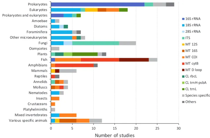
Polymerase chain reaction (PCR) is a method used to amplify a single or a few copies of a target piece of DNA, potentially to generate thousands to millions of copies. To initiate a PCR, short strands of DNA, or primers, are required. These short DNA sequences are chosen to bind either side of the gene region to be amplified, and therefore the choice of DNA primers dictates which DNA region is multiplied during PCR. As expected, the gene regions selected for amplification varied according to the target taxa (Fig. 6). Regions of the prokaryote 16S rRNA gene were used in almost all studies targeting the DNA of Bacteria and Archaea (e.g. Barberan & Casamayor 2014; Mao et al. 2014; Dong et al. 2015), unless primers were designed to detect individual species and genera (e.g. Aeromonas hydrophilia strain Vah; Griffin et al. 2013). Five studies also chose to use shotgun metagenomic methods, which do not require the use of DNA primers, to identify prokaryote DNA from metagenomes (not shown in Fig. 6; Delmont et al. 2011; Nakai et al. 2011; Inskeep et al. 2013; Costa et al. 2015; Owen et al. 2015).
Investigations of amoebae, diatoms, and other micro-eukaryotes often used regions of the 18S rRNA gene as the DNA barcode (e.g. Pawlowski et al. 2011; Bradford et al. 2013; Zimmermann et al. 2015), which was also targeted in nematode-focused (e.g. Bhadury & Austen 2010; Kanzaki et al. 2012) and some arthropod-focused studies (e.g. Pochon et al. 2013; Yang et al. 2014). The 18S rRNA gene region was similarly amplified in several fungal analyses (e.g. Nagahama et al. 2011; Lazarus & James 2015), but the internal transcribed
spacer (ITS) region 1 (ITS1, located between 18S and 5.8S rRNA genes) was the more commonly targeted (e.g. Bellemain et al. 2010; Singh et al. 2012; Bazzicalupo et al. 2013; Song et al. 2015), with more recent studies switching to the ITS2 region (between 5.8S and 28S rRNA genes; Bazzicalupo et al. 2013). A majority of studies of plant DNA were based on the trnL UAA intron within the chloroplast transfer RNA, or tRNA gene (e.g. Hibert et al. 2013; Parducci et al. 2013; Pedersen et al. 2013). The DNA of arthropods was most commonly targeted by amplification of mitochondrial cytochrome c oxidase subunit I (COI) DNA (e.g. Yu et al. 2012; Ji et al. 2013; Machler et al. 2014), whereas mitochondrial cytochrome b (cyt-b) gene regions were the most frequent target for the detection and identification of amphibians (e.g. Goldberg et al. 2011; Olson et al. 2012; Pilliod et al. 2013; Spear et al. 2015).
0 5 10 15 20 25 30 Prokaryotes
Eukaryotes Prokaryotes and eukaryotes Amoebae Diatoms Foraminifera Other microeukaryotes Fungi Oomycetes Plants Fish Amphibians Mammals Reptiles Annelids Molluscs Nematodes Insects Crustaceans Platyhelminths Mixed invertebrates Various specific animals
Number of studies
Ta
xa
16S rRNA 18S rRNA 28S rRNA ITS MT 12S MT 16S MT COI MT cytB MT D loop CL rbcL CL trnH-psbA CL trnL Species specific Others
Figure 6
Figure 6. Gene regions targeted in studies focused on different taxa (excluding microarray or metagenomic studies). rRNA indicates ribosomal RNA genes, MT indicates mitochondrial genes, and CL indicates chloroplast genes.
Summary of PCR product purification approaches Following PCR, the reaction mixture must be ‘purified’ to remove remaining primers as well as PCR enzymes and salts. A variety of PCR purification approaches were used. The most commonly-adopted PCR purification approaches were the Agencourt AMPure XP system (e.g. Bazzicalupo et al. 2013; Pochon et al. 2015b; Song et al. 2015), the Qiagen MinElute PCR purification kit (e.g. Stoeck et al. 2010; Thomsen et al. 2012a; Calvignac-Spencer et al. 2013) and the Promega Wizard SV Gel and PCR Clean-Up system (e.g. Bhadury & Austen 2010; Callejas et al. 2011; Keskin 2014). The AMPure XP method was used only in high-throughput sequencing studies and the Promega Wizard system only in Sanger sequencing studies, whereas the Qiagen MinElute method was used for a range of applications.
Summary of DNA sequence analysis methods used
DNA sequencing is used to determine the order of nucleotides (A, C, G and T) within a DNA molecule and may be used to identify the genes of interest in a sample and potentially the organism from which the gene originated. Sanger sequencing remained the most common sequencing approach from 2010 through to 2015 (e.g. Bhadury & Austen 2010; Jerde et al. 2011; Minamoto et al. 2012; Collins et al. 2013; Keskin 2014; Janosik & Johnston 2015), despite the increasing availability and performance of high-throughput sequencing technologies (Fig. 7). While not providing any detailed community-based information, qPCR was used with increasing frequency for DNA detection and quantification (e.g. Pilliod et al. 2013; Takahara & Minimoto 2013; Merlin et al. 2014; Moyer et al. 2014; Farrington et al. 2015; Laramie et al. 2015; Spear et
al. 2015). The 454 pyrosequencing platform was used with increasing frequency prior to 2013 but declined in popularity after this date (e.g. Stoeck et al. 2010; Pawlowski et al. 2011; Yu et al. 2012; Parducci et al. 2013; Pochon et al. 2013; Yang et al. 2014; Zimmermann et al. 2015), coinciding with a rise in the use of Illumina sequencing platforms for DNA analysis; however, the maximum number of studies using the latter system in any year was just seven (Costa et al. 2015; Deiner et al. 2015; Dong et al. 2015; Ficotela et al. 2015; Pansu et al. 2015a; Pochon et al. 2015b; Song et al. 2015). The Ion Torrent platform was used just three times (all since 2013) among the 167 studies reviewed (Deagle et al. 2013; Young et al. 2014; Zaiko et al. 2015).
0 2 4 6 8 10 12 14 16 18
2010 2011 2012 2013 2014 2015
Num
be
r
of
s
tudi
es
Publication year
Sanger qPCR 454 Illumina Ion Torrent End-point PCR Others
Figure 7
Conclusions
Based on our analyses of 167 research papers:
(i) Studies of freshwater habitats (water, sediment or biofilm) were more frequent than studies of marine or terrestrial habitats (45% compared to 23% and 22% of studies, respectively). DNA was extracted from water samples in 46% of the studies, mainly for the purpose of detecting specific animals using qPCR or Sanger sequencing methods. Analyses of DNA extracted from soils and sediments (40% of studies) targeted a wider range of organisms including plants, fungi, micro-eukaryotes, and prokaryotes, typically using either Sanger sequencing or high-throughput sequencing systems.
(ii) DNA was most commonly extracted from soil and sediment samples using DNeasy PowerSoil® and PowerMax®
kits (Qiagen). Extraction of DNA from water samples was most commonly achieved using DNeasy Blood & Tissue kits, followed by DNeasy PowerWater® kits. An assortment of
other kit-based and manual, or non-commercialised methods were also used for DNA extractions from these sample media. Several different methods were used for DNA extractions from other sample media, although DNeasy Blood & Tissue kits and DNeasy Plant kits were respectively used slightly more frequently than other approaches for extractions targeting animal or plant DNA.
(iii) Consistent gene regions were targeted in most studies of prokaryotes (16S rRNA gene), amphibians (mitochondrial cytochrome b, or cyt-b gene) and plants (chloroplast trnL intron); arthropods were usually analysed using primers targeting the mitochondrial COI gene. More studies of fungal communities targeted the internal transcribed spacer ITS1 region than the 18S rRNA gene. However, the 18S rRNA region was used in a variety of studies of micro-eukaryote and animal taxa. There is little consistency in primer targets for the analysis of fish DNA, with equal numbers of studies targeting mitochondrial COI, cyt-b and D-loop regions.
(iv) Our selective review identified only a limited number of studies using next-generation sequencing methods (seven Illumina-based studies, eight 454-based studies, and just one study using Ion Torrent out of 167 studies reviewed in total), with Sanger-based DNA sequencing remaining the most commonly used approach prior to August 2015.
Identification of standard procedures for
DNA extraction, storage, amplification and
sequencing
Sample storage prior to DNA extraction
It is generally best to keep samples cool and to carry out DNA extractions as soon as possible after samples are collected, to limit potential DNA degradation due to the material being removed from its original context. However, it is often impractical to carry out DNA extractions immediately after sample collection, and it may be desirable to retain samples for future analyses. A variety of methods are used to store samples, including cooling to 4, -20, or -80°C (depending on available facilities), drying, freeze-drying, or addition of preservative buffers, such as DMSO-EDTA. The suitability and feasibility of these approaches will depend on the taxa to be investigated, sample media, and the duration of storage. The simplest approach is to use cooling. Lauber et al. (2010) concluded that storage of microbiome samples at temperatures ranging from -20°C to -80°C for up to 14 days
had little impact on the resulting inferences; similar findings have been reported in other studies (e.g. Carroll et al. 2012). Bainard et al. (2010) concluded that drying methods had an adverse impact on recovery of arbuscular fungal DNA, suggesting that samples should instead be frozen to prevent DNA degradation. We recommend the immediate storage of samples at ~4°C following collection. For field studies, this is most easily achieved by the transfer of samples to cool boxes containing ice or other frozen material. Cooled samples should be transferred to -20°C freezers within 48 h for short-term storage (e.g. weeks to months) and to -80°C freezers for longer term storage (e.g. months to years). In situations where the cooling of samples in the field is difficult, such as when sampling in remote locations, ambient temperature sample storage may be considered. Proprietary (e.g. RNAlater® and DNAgard™) and non-proprietary (e.g. DMSO-EDTA) solutions can be used to stabilise nucleic acids for this purpose. A summary of these, and other approaches for storing sample DNA for molecular analyses is provided by Nagy (2010), where details on individual methods are described in terms of their adequacy for field work, optimal storage period (i.e. short-, medium-, or long-term), ease of use, health hazards and associated costs. RNA degradation by RNase enzymes is a significant concern during sampling handling and storage (Chomczynski 1992). Therefore, additional or different sample storage procedures may be required for studies in which the analysis of sample RNA is desirable. Readers are pointed to Kasahara et al. (2006) for more information on appropriate methods for sample storage for RNA preservation.
Sample pre-processing
Before extracting DNA, pre-processing steps are often required, in order to (i) reduce the number of samples from which DNA is to be extracted, e.g. by combining multiple samples together, and (ii) reduce sample volumes required for processing by concentrating the target biomass required for DNA extraction. Procedures to concentrate the target biomass normally require its separation from the sample media and may simultaneously reduce concentrations of PCR-inhibiting substances in samples. Here, we review a number of common approaches used to process samples before and during DNA extraction.
Reducing sample numbers for DNA extraction
Prokaryotes
Eukaryotes
Herbivores
Predators
Increasing organism size
Incr
easing spa
tial homog
eneity of eDNA and
chance of de
tection in a s
tandar
d sample siz
e
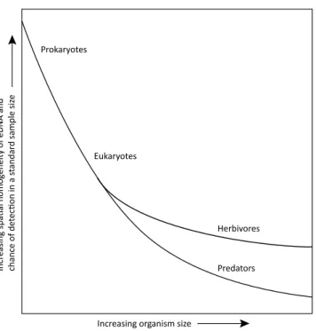
Figure 8. Schematic of the relationship between the size of an organism and the spatial homogeneity of its DNA in the environment, which is positively related to the likelihood of detecting that organism using DNA metabarcoding. Broadly, the spatial distribution of larger organisms such as multicellular eukaryotes, particularly predators, is less homogenous than that of smaller organisms such as prokaryotes, single-celled eukaryotes and herbivores. Consequently, the detection of larger organisms tends to require biomass concentration and/or bigger samples than are necessary for the detection of smaller organisms.
and homogenisation may be applicable where the main aims of a study are to determine the presence of specific taxa in a community, particularly if these organisms are spatially heterogeneous, such as is more likely to be the case for larger organisms and predators (Fig. 8). We recommend the analysis of as many individual samples as is possible, without pooling, particularly when estimates of taxon richness and diversity are key study aims.
Reducing sample volumes for DNA extraction
Biomass and DNA concentration may be appropriate for organisms that have high spatial heterogeneity (i.e. are at low population density relative to the sampled area) and have a correspondingly low chance of detection in a sample (Fig. 8). For example, predatory mites are typically less abundant in soil or leaf litter than herbivorous mites, and therefore may require concentration to achieve a sample representative of the community. Once concentrated samples have been obtained, DNA can be extracted from these using standard protocols.
The method of biomass concentration depends on the organisms or material in question. Where the aim is to concentrate environmental DNA (rather than cellular biomass, live or dead) from arbitrarily large volumes of terrestrial material, such as soil or leaf litter, DNA can be recovered using a saturated phosphate buffer solution (Bienert et al. 2012). After mixing the sample material with the buffer solution, the DNA is washed from the sample material, concentrated and extracted from one or more subsamples of the buffer solution using a conventional extraction kit (the
Macherey-Nagel Nucleospin Soil kit is recommended for this purpose; Taberlet et al. 2012). This simple protocol allows the efficient and economic recovery of homogenised pools of DNA from considerably larger sample volumes than can be processed using conventional extraction approaches. Such procedures inevitably bias against the extraction of DNA from more adherent organisms (e.g. biofilm dwelling bacteria may be underrepresented compared to their free-living counterparts; Garrett et al. 2008). Nevertheless, this approach is particularly relevant for the detection of large and/or sparsely distributed organisms and removes the need to process multiple small samples for DNA extraction and analysis. Where the aim is to concentrate cellular biomass, rather than extracellular DNA, methods of biomass concentration vary depending on the target organisms for analysis. Examples of biomass concentration methods for different taxa include the following.
Trapping and collecting live organisms. The composition of soil invertebrates is commonly assessed by first extracting the organisms from the soil using a combination of approaches including pitfall traps (Drummond et al. 2015) and modified Tullgren and Baermann funnels (Bao et al. 2012), each of which rely on the capture of organisms during active movement. Similarly, aerial insects may be captured using malaise traps (Yu et al. 2012). Aquatic invertebrates are commonly concentrated during capture using kick-net samplers (Machler et al. 2014). DNA is then extracted from the concentrated biomass.
Flotation. Flotation is used for the extraction of micro-arthropods from sandy soils, particularly for less-active taxa such as podurid Collembola (Geurs et al. 1991). This method is largely based on the difference in density of the animals and the flotation fluid (commonly heptane; Geurs et al. 1991).
Centrifugation. Centrifugation aids the collection of buoyant biomass (e.g. nematode cysts; Bellvert et al. 2008) from complex media such as soil. Density gradient centrifugation may also be used to separate small organisms such as bacterial cells from complex media, prior to downstream molecular processing (Dichosa et al. 2014).
Filtration. The biomass of microorganisms in both water (Lear et al. 2014) and air (DeLeon-Rodriguez et al. 2012) are commonly concentrated by filtration prior to extraction.
Manual sorting. Biological material such as roots, leaf fragments, and invertebrates may be manually picked from substrates such as soil, to increase the abundance of target biomass. Unwanted materials may be removed in a similar way, for example by sieving to exclude larger particles or inorganic material (e.g. stones) from the extraction process.
DNA extraction protocols
General recommendations for the extraction of DNA from environmental samples
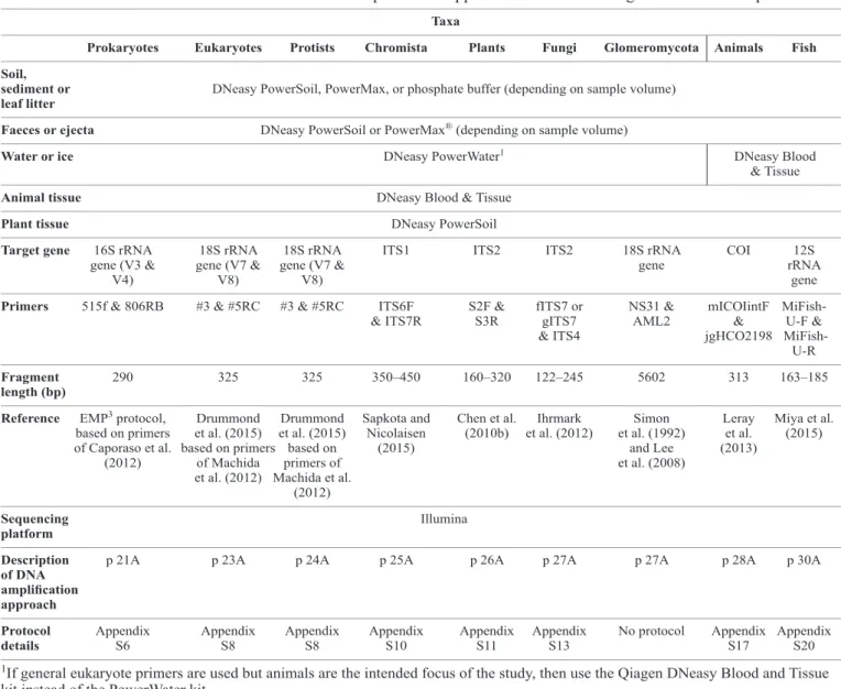
As highlighted by our literature review, different protocols are used for the extraction of DNA depending on the sample medium. A variety of pre-treatment options are also used, depending on the sample media and volume (Table 1). Based on these observations, we make some broad recommendations for standard procedures for the extraction of DNA from different sample media. DNeasy PowerSoil® DNA Isolation
kits, with a capacity of up to 0.25 g, are widely used in studies of soil, sediment, faecal material and leaf litter, and have been recommended for use by a number of international standards consortia following comparisons with a range of other methods (Gilbert et al. 2014). Consequently, we recommend the PowerSoil® DNA Isolation kit for extractions from the
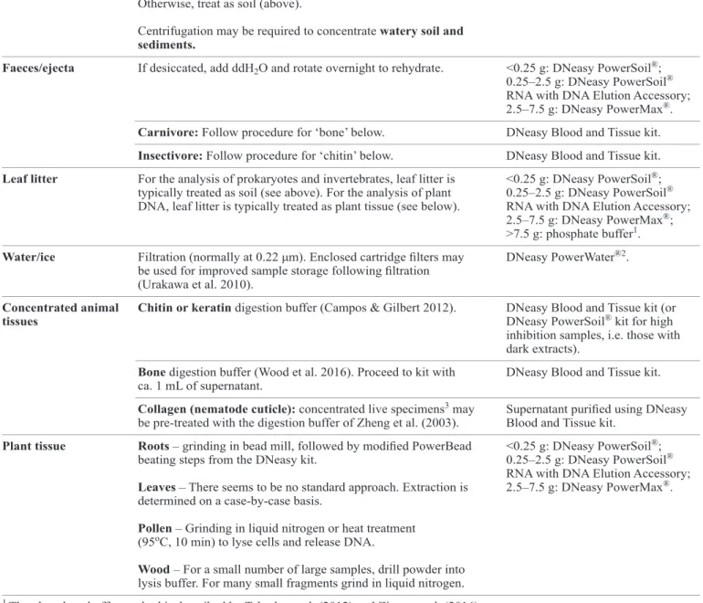
Table 1. Suggested pre-treatment and DNA extraction protocols for a range of sample media.
__________________________________________________________________________________________________________________________________________________________________
Extraction media Pre-treatment options Recommended extraction kit and ‘wet’ sample volumes
__________________________________________________________________________________________________________________________________________________________________
Soils/peats/sediments If sampling from large volumes of soil, sample material may first <0.25 g: DNeasy PowerSoil®;
be mixed. This is most commonly done by ‘hand’ or using kitchen 0.25–2.5 g: DNeasy PowerSoil®
blenders to homogenise soil slurries (Tien et al. 1999). Alternatively, RNA with DNA Elution Accessory; environmental DNA may be recovered from large soil volumes 2.5–7.5 g: DNeasy PowerMax®;
using the saturated phosphate buffer method (Bienert et al. 2012). >7.5 g: phosphate buffer1.
Allophanic soils such as andisols bind environmental DNA very efficiently and may require a two-step pre-treatment
(Huang et al. 2016).
Other difficult samples can benefit from the addition of lytic enzymes such as proteinase K, as in Bulat et al. (1998).
If desiccated, add ddH2O and rotate overnight to rehydrate.
Otherwise, treat as soil (above).
Centrifugation may be required to concentrate watery soil and sediments.
__________________________________________________________________________________________________________________________________________________________________
Faeces/ejecta If desiccated, add ddH2O and rotate overnight to rehydrate. <0.25 g: DNeasy PowerSoil®;
0.25–2.5 g: DNeasy PowerSoil®
RNA with DNA Elution Accessory; 2.5–7.5 g: DNeasy PowerMax®. __________________________________________________________________________________________________________________________________________________________________
Carnivore: Follow procedure for ‘bone’ below. DNeasy Blood and Tissue kit.
__________________________________________________________________________________________________________________________________________________________________
Insectivore: Follow procedure for ‘chitin’ below. DNeasy Blood and Tissue kit.
__________________________________________________________________________________________________________________________________________________________________
Leaf litter For the analysis of prokaryotes and invertebrates, leaf litter is <0.25 g: DNeasy PowerSoil®;
typically treated as soil (see above). For the analysis of plant 0.25–2.5 g: DNeasy PowerSoil®
DNA, leaf litter is typically treated as plant tissue (see below). RNA with DNA Elution Accessory; 2.5–7.5 g: DNeasy PowerMax®;
>7.5 g: phosphate buffer1.
__________________________________________________________________________________________________________________________________________________________________
Water/ice Filtration (normally at 0.22 μm). Enclosed cartridge filters may DNeasy PowerWater®2.
be used for improved sample storage following filtration (Urakawa et al. 2010).
__________________________________________________________________________________________________________________________________________________________________
Concentrated animal Chitin or keratin digestion buffer (Campos & Gilbert 2012). DNeasy Blood and Tissue kit (or
tissues DNeasy PowerSoil® kit for high
inhibition samples, i.e. those with dark extracts).
__________________________________________________________________________________________________________________________________________________________________
Bone digestion buffer (Wood et al. 2016). Proceed to kit with DNeasy Blood and Tissue kit. ca. 1 mL of supernatant.
__________________________________________________________________________________________________________________________________________________________________
Collagen(nematode cuticle): concentrated live specimens3 may Supernatant purified using DNeasy
be pre-treated with the digestion buffer of Zheng et al. (2003). Blood and Tissue kit.
__________________________________________________________________________________________________________________________________________________________________
Plant tissue Roots – grinding in bead mill, followed by modified PowerBead <0.25 g: DNeasy PowerSoil®;
beating steps from the DNeasy kit. 0.25–2.5 g: DNeasy PowerSoil®
RNA with DNA Elution Accessory;
Leaves – There seems to be no standard approach. Extraction is 2.5–7.5 g: DNeasy PowerMax®.
determined on a case-by-case basis.
Pollen – Grinding in liquid nitrogen or heat treatment (95oC, 10 min) to lyse cells and release DNA.
Wood – For a small number of large samples, drill powder into lysis buffer. For many small fragments grind in liquid nitrogen.
__________________________________________________________________________________________________________________________________________________________________ 1 The phosphate buffer method is described by Taberlet et al. (2012) and Zinger et al. (2016).
2 Where animal or plant derived DNA is the principal target, DNeasy Blood and Tissue or DNeasy Plant Tissue kits should be used
instead.
accommodated by the DNeasy PowerSoil® RNA Isolation kit
means that samples of up to 2.5 g can be processed, when used in combination with an RNeasy DNA Elution Accessory kit to co-isolate DNA from the sample material. For even larger samples, the DNeasy PowerMax® Soil DNA Isolation kit,
with a capacity of up to 5–10 g, is recommended. Terrestrial samples that exceed the capacity of the PowerMax® kit (>5–10
g) can be processed using the saturated phosphate buffer method (Bienert et al. 2012). A variety of methods, including PowerSoil® kits, have been used for DNA extraction from gut
contents, faeces and plant tissues and yield high concentrations of quality DNA (Dineen et al. 2010; Wagner Mackenzie et al. 2015). Consequently, we also suggest the use of PowerSoil® or
PowerMax®kits for these sample types, for consistency across
different analyses. We recommend the use of PowerWater® kits
for the recovery of DNA from microbial communities in both marine and freshwater, since this approach is widely used and is most similar to the PowerSoil® approach recommended for
a variety of terrestrial media. However, where animal or plant-derived DNA is a main target of extractions from water, DNeasy Blood & Tissue and DNeasy PowerSoil® extraction approaches
are suggested, respectively, allowing for better comparisons with sequence data collected directly from biological tissue. The preferential use of these and similar kits reduces the lab-to-lab variation that can arise from the use of non-commercial (i.e. non kit-based) methods, allowing access to comparable data from a wider group of users and creating consistency across studies carried out around the world. Alternative methods should nevertheless be used, where necessary, to maximise the quantity and quality of DNA extracted from difficult samples (e.g. soils containing elevated concentrations of heavy metals or humic organic matter).
Extraction of DNA from soil, sediment and leaf litter General considerations
Early soil biodiversity studies relied on cell extractions prior to DNA isolation, followed by the removal of humic material by means of chromatography (Faegri et al. 1977; Torsvik 1980). These so called ‘indirect methods’ laid the groundwork for modern amplicon sequencing and metagenomics (Lane et al. 1985; Pace et al. 1986) and opened a window into the poorly-explored biodiversity of soils (Torsvik et al. 1990; Deagle et al. 2009). Subsequent method development, principally by Ogram et al. (1987), saw a shift towards ‘direct methods’ of extraction, which are the current standard. These methodologies frequently rely on the physical or chemical lysis of cellular material present in the sample media, combined with column-based DNA purification. The direct extraction of DNA from the original sample medium can result in higher (greater than an order of magnitude) DNA yields than indirect methods while retaining the molecular size of the DNA fragments within a range deemed suitable for DNA sequencing analysis (Miller et al. 1999; Miller 2001). However, DNA from other sources (e.g. plant debris) is frequently co-extracted in high concentration. Many comparisons of DNA extraction methods for soil, sediment and leaf litter samples have been published (Frostegard et al. 1999; Miller et al. 1999; Martin-Laurent et al. 2001; Miller 2001; Dineen et al. 2010; Mahmoudi et al. 2011) including for the recovery of ancient DNA (Haile 2012), each highlighting differences among methods in the community composition detected.
A wide variety of sample pre-processing steps are recommended before the extraction of DNA from soil and similar media. The selective removal of non-target material
such as stones, leaf litter or coarse root material, or alternatively the removal of sample material by size (i.e. by sample sieving) is common. As a minimum, the removal of larger inanimate material is advised (e.g. stones) to maximise the yields of DNA extracted. However, the details of sample processing approaches will vary depending on the research question as well as the target organism(s). For example, coarse root material would frequently be removed in studies where plant community DNA, or the DNA of plant endosymbionts and pathogens, is not under investigation.
DNA extraction is particularly difficult from samples containing high concentrations of clay and humic material. DNA binds strongly to clay particles (Frostegard et al. 1999; Cai et al. 2006) preventing the isolation of DNA into the extraction supernatant. Humic material has a similar size and charge to DNA, resulting in co-purification, as may be evidenced by the brown colour of some DNA extracts. The presence of humic material in DNA extracts inhibits the activity of some enzymes including DNA polymerases (Dong et al. 2006). Additionally, the co-extraction of humic material may interfere with DNA quantification by spectrophotometry, since both DNA and humic material exhibit optimal absorbance at both 230 and 260 nm. Fluorometric methods such as Qubit (ThermoFisher Scientific) are less affected by humic material, tending to provide more accurate estimates of DNA concentration in soil extracts. A number of commercial kits, including PowerSoil®
DNA Isolation kits, are designed to remove PCR inhibitors from soil and similar material, including recalcitrant humic substances. In the case of DNEasy PowerMax® kits, addition of
phenol to the extraction column may further improve the DNA recovery from clay-rich samples processed using this method (e.g. in step 1 of the standard protocol, add 10 ml PowerBead Solution with 5 ml phenol (phenol:chloroform:isoamyl alcohol pH 7–8); Charlotte Jordans, Geneworks Pty Ltd., pers. comm.). Alternative strategies for the removal of humic substances from soil DNA include the use of aluminium sulphate (Dong et al. 2006) and Sephadex columns (Tsai & Olson 1992b).
Extraction requirements and recommendations
DNeasy PowerSoil® kits are widely used and have been shown to be well optimised for DNA extraction from a variety of soils, including compost, sediment, clay and acidic soils (Roose-Amsaleg et al. 2001; Tedersoo et al. 2014). The same kits are also used for the extraction of DNA from leaf litter (Voříšková & Baldrian 2013). We recommend use of DNeasy PowerSoil® and PowerMax® kits for the extraction of DNA from soil, sediment and leaf litter (Table 1). Additional steps such as sample dilution or supplementing PCR mixtures with adjuvents such as BSA may be required for sample media containing elevated concentrations of PCR inhibitory substances.
Extraction of DNA from faeces and ejecta General considerations
ecology of the depositor as a way of sampling the biodiversity in its environment (Kuch et al. 2002). This third approach envisions depositors as ‘environmental samplers’ and in effect faeces are ‘biodiversity capsules’ (Boyer et al. 2015) containing concentrated DNA from taxa consumed by the depositing species. While faecal DNA analysis can introduce sampling biases for biodiversity assessments, and requires an understanding of the ecology of the depositing species, it can be an excellent approach for detecting rare species in the environment. Each of these approaches has different assumptions and it is important that researchers are clear from the outset about what questions they aim to address. Care should be taken when sampling faeces, as many diseases and parasites that are transmissible to humans can exist in animal dung. Such precautions can involve simply wearing latex gloves when handling faeces and avoiding inhaling dust from dry faeces (wearing a dust mask). In exceptional circumstances (such as sampling bat guano from caves) it may be advisable to wear a respirator.
As the gastrointestinal tracts of most animals are excellent mixers, there may be no need to homogenise individual faecal samples. However, if small samples from multiple specimens are being combined for DNA extraction (e.g. invertebrate frass), they should be well mixed, using either a bead beater, or pestle and mortar. Larger volume samples may be mixed as a slurry with lysis buffer using, for example, stomacher laboratory paddle blenders (Abu Al-Soud & Rådström 1998).
If detecting the diet of the depositing species is of interest then the outside layer of each dung bolus represents a contamination risk because it has been in contact with the soil or other external substrate, and needs to be carefully removed (see the dung subsampling procedure of Wood and Wilmshurst (2011)). Obviously, this becomes more difficult for smaller specimens, and may be impossible for invertebrate frass, although UV light irradiation of the specimens may assist in reducing surface contamination in such instances (Cone & Fairfax 1993).
Extraction requirements and recommendations
Faecal DNA extraction is relatively straightforward, but differs for humic-rich (herbivore) and humic-poor (large carnivore) faeces (Table 1). In most cases, it is appropriate to extract DNA from faecal material using DNeasy PowerSoil®
or PowerMax® kits (following rehydration if specimens are
dry). However, we recommend the use of DNeasy Blood and Tissue kits for the analysis of carnivore faeces, allowing for better comparisons with sequence data collected directly from biological tissue. These sample materials may require decalcification and digestion steps prior to DNA extraction and isolation. Similarly, insectivore dung requires a chitin digestion step followed by use of a DNeasy Blood and Tissue kit, or DNeasy Powersoil® kit, if the sample media is also suspected
to contain high concentrations of humic material (Table 1).
Extraction of DNA from water and ice General considerations
A key issue with the extraction and amplification of DNA from water is low DNA concentration, which can require the filtration of many litres of water to obtain sufficient DNA from a single sample (Wilcox et al. 2016). Additionally, in lentic systems in particular, environmental DNA appears to be distributed somewhat patchily (Lear et al. 2014) which could yield false negative results for a given species of interest. For example, Furlanand Gleeson(2016) found that they needed to collect
up to 12 two litre water samples from each sampling station to detect redfin perch (Perca fluviatilis) that was known to be present in a lake. The mixing of running water will reduce spatial variability in the composition of environmental DNA; however, the downstream transport of DNA in lotic systems suggests that DNA sequence data may not only represent species present in the vicinity of sampling, but potentially from long distances upstream (Deiner et al. 2015).
Extraction requirements and recommendations
There are two distinct methods to collect and concentrate environmental DNA from water; precipitation followed by centrifugation (Turner et al. 2015), or filtration. Comparisons of the two methods on the same water samples have shown that higher concentrations of DNA are obtained by filtration methods (Deiner et al. 2015). However, there is a trade-off between filter pore size and the volume of water that can be filtered before the filter clogs. For this reason, it may be preferable to filter multiple small volumes of water through separate filters and later combine the DNA collected from each filter via ethanol precipitation (Santas et al. 2013). The optimal filter pore size is generally suggested to lie between 0.6 µm and 1.5 µm (Eichmiller et al. 2016; Minamoto et al. 2016). We suggest using 1.5 µm glass fibre filters (Type 934‐AH) to filter water for assessments of vertebrate DNA but smaller filter sizes (e.g. 0.2 μm) are recommended for the capture of microbial biomass (Lear et al. 2014).
Once the water is collected, it should be filtered and stored as soon as possible since the quality and quantity of DNA present in water decreases rapidly (Thomsen et al. 2012a; Maruyama et al. 2014). For example, Maruyama et al. (2014) observed detectable concentrations of bluegill fish (Lepomis macrochirus) to decline by half in under 7 h at 20oC.
Even when unfiltered water is frozen at -20oC, reductions of
amplifiable DNA as great as ten-fold are reported in the literature (Cornelisen et al. 2012). Therefore, it is preferable to filter samples as soon as possible after sampling, and to preserve the filtered material at low temperature; studies have shown filters (and associated DNA) can be stored at -20oC for later DNA
extraction without significant loss of amplifiable DNA (Gilpin et al. 2013). Other research suggests the fixation of filtered DNA with ethanol may allow sample material to be preserved at room temperature for many days (Minamoto et al. 2012; Thomsen et al. 2012a; Minamoto et al. 2016). However, since this approach is yet to gain widespread acceptance, its use for the preservation of rare target DNA is not recommended. For the collection of DNA or microbial biomass from small (i.e. less than one litre) volumes, on site-filtration is recommended using devices such as Sterivex filters (Wright et al. 2009) or customised portable filtration devices, such as those described by Yamanaka et al. (2016).
Comparative studies of DNA extraction effectiveness from filters suggest that the Qiagen DNeasy PowerWater®
DNA extraction kit (a bead beating method) was less likely to extract PCR inhibitors along with the DNA compared with the Qiagen DNeasy Blood and Tissue kit, although the latter obtained higher concentrations of total DNA (Eichmiller et al. 2016). Thus, we recommend the use of the PowerWater® kit
Extraction of DNA from animal tissue General considerations
The extraordinary morphological and ecological diversity of animals presents some particular issues for DNA extraction. Many invertebrates (i.e. ecdysozoans, such as nematodes and arthropods) have hard, waterproof outer cuticles that may represent a barrier for the spread of DNA into the surrounding environment (Goldberg et al. 2013). This will be a particular problem for highly-sclerotised arthropods, such as weevils, which as a consequence may be under-represented in environmental DNA samples. Therefore, the DNA of animals, and invertebrates in particular, is often targeted directly from homogenised animal tissue rather than from true environmental