Volume 17 Number 5 pp. 383–389 CThe Authors 2014 doi:10.1017/thg.2014.44
Case Report of Multiple Valve Disease Found
in Triplets
Andrea ´A. Moln ´ar,1,∗Attila Kov ´acs,2,∗Astrid Apor,2 ´Ad ´am D. T ´arnoki,3D ´avid L. T ´arnoki,3
Tam ´as Horv ´ath,2P ´al Maurovich-Horvat,2R ´obert G. Kiss,1Gy ¨orgy Jermendy,4and B ´ela Merkely2 1Department of Cardiology, Military Hospital, Budapest, Hungary
2Cardiovascular Imaging Research Group, Heart and Vascular Center, Semmelweis University, Budapest, Hungary 3Department of Radiology and Oncotherapy, Semmelweis University, Budapest, Hungary
43rd Department of Internal Medicine, Bajcsy-Zsilinszky Hospital, Budapest, Hungary
Valvular heart disease is a multifactorial disorder. Twin studies may help to better understand both genetic and environmental determinants contributing to the development of valve lesions. We describe the case of a 45-year-old female asymptomatic triplet with multiple valvular heart lesions, with a somewhat differ-ent pattern between the dizygotic twin pairs compared with the monozygotic twin pair. After thorough assessment of medical history and physical examination, the triplet underwent two- and three-dimensional transthoracic and transesophageal echocardiographic examinations to assess the pathomechanism and severity of their heart valve lesions. The monozygotic twin pair (second-born twin B and third-born twin C) showed the same pattern of valvular lesions: mild mitral, tricuspidal, and aortic regurgitation of the same pathomechanism (posterior mitral valve cleft and aortosclerosis). Interestingly, the examination of first-born twin (twin A), who was dizygotic to twins B and C, revealed mild protosystolic mitral and mild tricuspidal regurgitation, but neither aortic insufficiency nor mitral cleft or indentation could be detected. Beyond the genetic effect, we presume that the intrauterine twinning process might also play a role in the development of congenital valvular heart disease. In order to verify this, further investigation should be performed on larger twin populations. Nevertheless, when one twin is affected, the other asymptomatic twin should also be examined for valvular heart disease.
Keywords:twin, triplet, valvular heart disease, mitral cleft, echocardiography
Heart valves are thin membranes regulating blood flow by constant opening and closing. The four valves in the heart are between the atria and ventricules (called mitral and tri-cuspid valves), and in the arteries leaving the heart (called aortic and pulmonary valves). Regurgitation occurs when a valve malfunctions and allows some blood to flow in the wrong direction leading to valvular heart disease (Lancel-lotti et al.,2010b). Valvular heart disease is a multifacto-rial disorder determined by both genetic and environmen-tal factors. In clinical practice the most frequent cause of mitral and tricuspid regurgitation is ischemic heart dis-ease, while the aortic valve is mostly affected by the de-generative calcification (Lancellotti et al., 2010a, 2010b). However, valve lesions defined dominantly by genetic fac-tors are also known; for example, in Marfan syndrome, Ehlers–Danlos syndrome, and other connective tissue dis-orders (Boudoulas et al.,1994; Grau et al.,2007). Deeper understanding of genetic or environmental influences in the development of functional and organic valve lesions is of high importance. Recent advances in echocardiography
can help to accurately assess the pathomechanism and the severity of valvular disorders, while twin studies are used to evaluate the ratio of underlying genetic and environmental components. Investigation of a triplet set, including both monozygotic and dizygotic pairs, is a unique opportunity to highlight these issues.
Case Presentation
We report the case of a 45-year-old asymptomatic Hungar-ian female triplet. The first-born twin (twin A) was dizygotic with the others, the second- (twin B) and third-born twins (twin C) were monozygotic with each other. Zygosity was
RECEIVED6 May 2014;ACCEPTED17 June 2014. First published online 5 August 2014.
ADDRESS FOR CORRESPONDENCE: A. ´A. Moln´ar, Department of Cardiology, Military Hospital, Budapest, Hungary. E-mail:
FIGURE 1
(Colour online) Two-dimensional transthoracic color Doppler image of parasternal long axis view of Twin B showing mild aortic and mitral regurgitation (A image), and four-chamber view demonstrating central mitral and tricuspid regurgitant jet (B and C images).AR: aortic regurgitation; MR: mitral regurgita-tion; TR: tricuspidal regurgitaregurgita-tion; Ao: aorta; LA: left atrium; LV: left ventricule; RA: right atrium; RV: right ventricule.
determinated by a standard multiple-choice questionnaire (Heath et al.,2003). The triplets were recruited by the Hun-garian Twin Registry and examined as part of our twin study, using multiple cardiovascular imaging modalities (Littvay et al.,2013). Twins provided their informed consent before entering the study. First, medical history and anthropo-metrical data were collected (Table 1). According to their medical records, all of the siblings had a separate amniotic sac. The birth weight of twin A was 2,100 g, twin B was 1,850 g, and twin C was 1,900 g. All the siblings were diag-nosed and treated for hypertension since 2008, and dyslipi-demia since 2003. Twin C was treated for an autoimmune
disease of the thyroid gland named the Graves–Basedow
disease since 2011. Only twin A was an active smoker since 1988; the other twins have never smoked.
Transthoracic, two-dimensional echocardiographic ex-amination (Philips iE33, S5-1 transducer) revealed multi-ple valvular heart lesions in each twin, but with a differ-ent pattern. Mild aortic, mild holosystolic mitral, and mild tricuspid regurgitation with central regurgitant jet were found during the examination of the monozygotic twin pair (twins B and C;Figure 1). Mild protosystolic mitral and mild tricuspidal regurgitation with central regurgitant jet were also noticed in twin A, but no aortic regurgitation could be detected. Transthoracic echocardiography showed normal dimensions of both ventricules and proximal part of the aorta, and normal left ventricular systolic function (Table 1). Left ventricular systolic function was character-ized by ejection fraction, which represented the volumetric fraction of blood pumped out of the left ventricle with each heartbeat (Table 1).
TABLE 1
Clinical Characteristics and Echocardiographic Data
Twin A Twin B Twin C
Anamnestical data
Hypertension + + +
Diabetes mellitus - -
-Dyslipidaemia + + +
Smoking + -
-Graves–Basedow disease - - +
Antropometrical data
Weight at birth (g) 2,100 1,850 1,900
Weight in 2014 (kg) 61.1 59.2 74.3
Height in 2014 (cm) 160 158 159
BMI in 2014 (kg/m2) 23.9 23.7 29.4
Echocardiographic parameters
AOD (mm) 17.7 14 14.1
LA volume index (ml/m2) 28.7 28.6 37.7
RA volume index (ml/m2) 26.4 24.6 52.1
LV EDV (ml) 87.9 54.6 97.3
LV ESV (ml) 37.7 17.6 43.4
EF (%) 60 65 67
RV basal diameter (mm) 30 28.3 30.6
Mitral annulus end systolic dimensions (mm) 19×34×2.7 24×31×3.3 25×3×3.5
MR vena contracta area (cm2) 0.05 0.05 0.04
TR vena contracta area (cm2) 0.2 0.14 0.1
AR vena contracta area (cm2) - 0.03 0.04
Mitral valve cleft - Posterior leaflet Posterior leaflet
Note: AOD: aortic annulus diameter; LA: left atrium; RA: right atrium; LV EDV: left ventricular end diastolic volume; LV ESV: left ventricular end systolic volume; EF: ejection fraction of left ventricule; RV: right ventricule; MR: mitral regurgitation; TR: tricuspidal regurgitation; AR: aortic regurgitation.
FIGURE 2
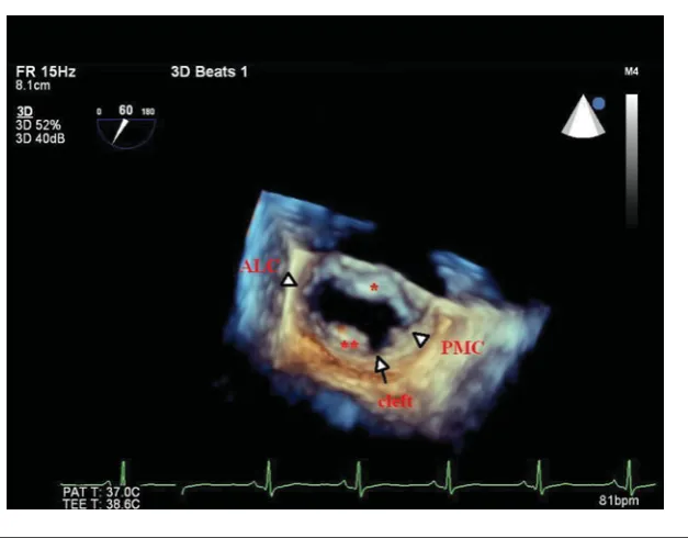
(Colour online) Three-dimensional transesophageal image of the mitral valve as viewed from the left atrium demonstrating the posterior mitral leaflet cleft (arrow) in twin C. ALC: antero-lateral commissure (arrowhead); PMC: postero-medial commissure (arrowhead);∗anterior mitral leaflet;∗∗posterior mitral leaflet.
et al., 2010a). Cleft of mitral valve leaflet is an anatomic lesion defined as a slit-like hole that extends to the mitral annulus and can be accompanied by mitral valve prolapse, atrial septal defect, counterclockwise rotation of the pap-illary muscles, and the presence of an accessory pappap-illary muscle or mitral valve leaflet (Di Segni & Edwards,1983).
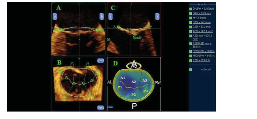
FIGURE 3
(Colour online) Three-dimensional morphologic analysis and model of the mitral valve in twin C. (A) The mitral annulus is manually defined in multiple rotational planes, (B) yielding a resultant three-dimensional contour superimposed on the en face view of the valve. (C) The mitral valve leaflets are then manually traced in multiple parallel planes, resulting in (D) a line of coaptation displayed on a color-coded, three-dimension-rendered valve surface. A: anterior; P: posterior; AL: anterolateral; PM: posteromedial; Ao: aorta; A1, A2, A3: scallops of the anterior mitral valve leaflet; P1, P2, P3: scallops of the posterior mitral valve leaflet.
A: mitral leaflets were unimpaired and the central regur-gitation jet was detected only in the protosystolic phase at the onset of coaptation. None of the twins had mitral valve prolapse.
The aortic valve consists of three cusps. Their associ-ation with the respective coronary artery identifies them as left, right, and non-coronary cusp. The most common congenital abnormality of the heart is the bicuspid aortic valve leading to aortic valve malfunction. In this condi-tion, instead of three cusps, the aortic valve has two cusps (Lancellotti et al.,2010b; Martin et al.,2007). In our case, the three-dimensional transesophageal echocardiography proved that the aortic valve included three cusps in all of the twins. The pathomechanism of central mild aortic re-gurgitation found in the monozygotic twin pair was early stage aortosclerosis. Signs of early stage calcification were detected in the right aortic cusp in twin B and non-coronary aortic cusp in twin C. Calcification restricts the mobility of aortic valve and causes valve closure malfunction, leading to regurgitation (Lancellotti et al.,2010b). The aortic valve in twin A was structurally and functionally normal. All leaflets of the tricuspid valve were structurally unimpaired.
Quantification of the severity of valve regurgitation is es-sential for therapeutic management. The vena contracta is a quantitative method to assess the severity of regurgitation, and is defined as the narrowest portion of the regurgitant jet downstream from the regurgitant orifice (Enriquez-Sarano et al.,2005). In our case, vena contracta areas of valve regur-gitations were measured using multiplanar reconstruction of the three-dimensional full-volume color Doppler data set in all of the twins (Table 1andFigure 4). All valve lesions were classified as mild in all the twins. The number and
location of the papillary muscles were normal and no atrial septal defect could be detected.
Discussion
Our report shows a unique case regarding a triplet with mul-tivalvular disease. The monozygotic pair showed the same pattern and pathomechanism of valvular lesions: mild mi-tral, tricuspidal and aortic regurgitation, as well. Isolated cleft of the posterior mitral valve leaflet and early-stage aortosclerosis were explored in the background of valvular lesions in both monozygotic twins. Interestingly, the ex-amination of twin A, who was dizygotic to twins B and C, revealed no aortic insufficiency, and the characteristics and pathomechanism of mitral valve regurgitation were differ-ent. Early-stage calcification of aortic cusps was found only in the monozygotic twins, despite the same cardiovascular risk factors being present in all the twins. This may lead us to the assumption that genetic factors might play a role in the development of aortic valve calcification. Limited information is available regarding genetic determinants of valvular calcification, which is an important precursor of clinical valvular disease. In a genomewide association study, Thanassoulis and coworkers (2013) have identified a s
ingle-nucleotide polymorphism in the lipoprotein(a) locus that
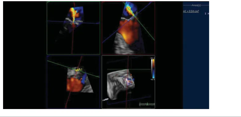
FIGURE 4.
(Colour online) Quantification of mitral valve regurgitation in twin C using multiplanar reconstruction of the three-dimensional full-volume color Doppler dataset to assess the vena contracta area. A1: vena contracta area.
by two-dimensional echocardiography can be difficult, and sometimes a patient with an isolated cleft may remain un-diagnosed (Van Praagh et al., 2003). Wyss and coworkers (2009) reported that the prevalence of isolated cleft of the posterior mitral leaflet was 0.11% (n=22 out of 19,320 two-dimensional transthoracic echocardiograms). In our case, two-dimensional transthoracic echocardiography did not reveal the mechanism of mitral regurgitation. Using the three-dimensional transesophageal echocardiography tech-nique enabled us to identify the cleft of the posterior mitral valve leaflet. Assessment of the mechanism and severity of mitral valve regurgitation is of paramount importance for therapeutic management. Three-dimensional echocardio-graphy has improved both morphological and functional assessment of valvular heart disease. It provides additional morphologic information of the components of mitral valve apparatus, which leads to better understanding of the mech-anism of mitral regurgitation (Cai & Ahmad,2012; Lancel-lotti et al.,2010a).
Surgical correction is recommended in cases of severe mitral regurgitation in symptomatic patients with or with-out left ventricular dysfunction, and for asymptomatic pa-tients with left ventricular dilatation and/or left ventricular ejection fraction<50% (Bonow et al.,2008). Because of the asymptomatic status of the twins in our case and the lack of significant regurgitation, regular clinical follow-up and periodical echocardiographic examination were suggested. Early recognition of this rare clinical entity can identify the patients who would benefit from surgical intervention before compensatory left ventricular remodeling and con-tractile dysfunction develop. Besides this, multiple births have an increased risk of birth defects (Li et al.,2003). This raises the question of whether when one twin is affected with valvular lesion, the other asymptomatic twin should be examined as well.
This is the first case to our knowledge to present posterior mitral valve cleft in monozygotic twins within a triplet pair, who are discordant regarding the mitral valve lesion with the other dizygotic twin. There is no evidence for the precise genetic or environmental (intrauterine/extrauterine) back-ground of congenital posterior mitral valve cleft. However, previous publications suggest that twins appear to be as-sociated with an increased risk of congenital heart diseases compared with singletons (Bahtiyar et al.,2007; Campbell et al., 2009). It appears that the interplay of altered pla-centation in conjunction with a genetic predisposition may play some part in this increased congenital heart disease risk (Bahtiyar et al.,2007; Bjarneg˚ard et al.,2013). There-fore, we assume that beyond genetic effects, the altered local intrauterine environmental factors (altered placental hemo-dynamics and vascular factors) might also play a role in the development of mitral valve cleft. In order to verify this, further investigations should be performed on larger twin populations.
Consent
Written informed consent was obtained by all three patients for the publication of this case report and any accompanying images.
Glossary
Aorta: The largest artery in the human body originating
from the left ventricle via the aortic valve.
Aortic annulus: Fibrous ring surrounding the aortic
ori-fice and serves for the attachment of the cusps of the aortic valve.
Aortosclerosis: Arteriosclerosis of the aorta; a pathological
Atrial septal defect: A form of a congenital heart disease that enables blood flow between two compartments of the heart called the left and right atria.
Atrium: A thin walled chamber that allows blood to
re-turn to the heart.
Coaptation of mitral valve: Fitting together process of the
two surfaces of mitral valve leaflets during valve closure.
Color Doppler: Echocardiographic technique that
esti-mates the average velocity of flow within a vessel by color coding the information.
Commisure: The area where the two valve leaflets come
together.
Cordae tendineae: Cord-like tendons that connect the
papillary muscles to the tricuspid and mitral valve.
Dyslipidemia: Abnormal level of lipids (e.g., cholesterol
and/or fat) in the blood.
Ehlers–Danlos syndrome: Inherited connective tissue
dis-order caused by a defect in the structure, production, or processing of collagen or proteins that interact with col-lagen. The manifestations of the disease involve the car-diovascular system (e.g., heart valves), the musculoskeletal system, and skin.
Holosystole: Cardiac systole is the contraction of the
car-diac muscle in response to an electrochemical stimulus to the heart’s cells. Holosystole refers to the entire phase of cardiac systole.
Infective endocarditis: Inflammation of the inner tissue of
the heart (such as valves) caused by infectious agents.
Marfan syndrome: Genetic disorder caused by misfolding
of the protein fibrillin-1, which forms fibers in connective tissue. More than 30 different signs and symptoms are vari-ably associated with the Marfan syndrome. The most serious signs involve the cardiovascular system (e.g., dilatation of the aorta, heart valve disease).
Mitral valve annulus: Fibrous ring around the mitral
valve.
Papillary muscles: Muscles located in the ventricles that
attach to the leaflets of the mitral and tricuspid valves via the cordae tendineae and contract to prevent inversion or prolapse of these valves.
Protosystole: Cardiac systole is the contraction of the
car-diac muscle in response to an electrochemical stimulus to the heart’s cells. Protosystole refers to the early phase of cardiac systole.
Ventricle: The large chamber that collects and expels
blood received from an atrium toward the peripheral beds within the body and lungs.
References
Bahtiyar, M. O., Dulay, A. T., Weeks, B. P., Friedman, A. H., & Copel, J. A. (2007). Prevalence of congenital heart defects in monochorionic/diamniotic twin gestations: A systematic literature review.Journal of Ultrasound Medicine,26, 1491– 1498.
Bjarneg˚ard, M., Enge, M., Norlin, J., Gustafsdottir, S., Fredriksson, S., Abramsson, A., . . . Betsholtz, C. (2013). Endothelium-specific ablation of PDGFB leads to pericyte loss and glomerular, cardiac and placental abnormalities. Development and Dsease,131, 1847–1857.
Bonow, R. O., Carabello, B. A., Chatterjee, K., de Leon, A. C. Jr, Faxon, D. P., Freed, M. D., . . . Shanewise, J. S. (Writ-ing Committee Members, American College of Cardiol-ogy/American Heart Association Task Force). (2008). Fo-cused update incorporated into the ACC/AHA 2006 guide-lines for the management of patients with valvular heart disease: A report of the American College of Cardiol-ogy/American Heart Association Task Force on Practice Guidelines (Writing Committee to Revise the 1998 Guide-lines for the Management of Patients With Valvular Heart Disease).Circulation,118, e523–e661.
Boudoulas, H., Vavuranakis, M., & Wooley, C. F. (1994). Valvu-lar heart disease: The influence of changing etiology on nosology.The Journal of Heart and Valve Disease,3, 516– 526.
Cai, Q., & Ahmad, M. (2012). Three-dimensional echocar-diography in valvular heart disease.Echocardiography,29, 88–97.
Campbell, K. H., Copel, J. A., & Bahtiyar, M. O. (2009). Congenital heart defects in twin gestations. Minerva Ginecologica,61, 239–244.
Di Segni, E., & Edwards, J. E. (1983). Cleft anterior leaflet of the mitral valve with intact septa. A study of 20 cases.American Journal of Cardiology,51, 919–926.
Enriquez-Sarano, M., Avierinos, J. F., Messika-Zeitoun, D., Detaint, D., Capps, M., Nkomo, V., . . . Tajik, A. J. (2005). Quantitative determinants of the outcome of asymptomatic mitral regurgitation.New England Journal of Medicine,352, 875–883.
Grau, J. B., Pirelli, L., Yu, P. J., Galloway, A. C., & Ostrer, H. (2007). The genetics of mitral valve prolapse.Clinical Genetics,72, 288–295.
Heath, A. C., Nyholt, D. R., Neuman, R., Madden, P. A., Bucholz, K. K., Todd, R. D., . . . Martin, N. G. (2003). Zygos-ity diagnosis in the absence of genotypic data: An approach using latent class analysis.Twin Research,6, 22–26. Lancellotti, P., Moura, L., Pierard, L. A., Agricola, E., Popescu,
B. A., Tribouilloy, C., . . . Zamorano, J. L., on behalf of the European Association of Echocardiography. (2010a). European Association of Echocardiography recommenda-tions for the assessment of valvular regurgitation. Part 2: Mitral and tricuspid regurgitation (native valve disease). European Journal of Echocardiography,11, 307–332. Lancellotti, P., Tribouilloy, C., Hagendorff, A., Moura, L.,
Popescu, B. A., Agricola, E., . . . Zamorano, J. L., on be-half of the European Association of Echocardiography. (2010b). European Association of Echocardiography rec-ommendations for the assessment of valvular regurgitation. Part 1: Aortic and pulmonary regurgitation (native valve disease).European Journal of Echocardiography, 11, 223– 244.
Society of Echocardiography and the European Association of Echocardiography. (2012). EAE/ASE recommendations for image acquisition and display using three-dimensional echocardiography.Journal of American Society of Echocar-diography,25, 3–46.
Li, S. J., Ford, N., Meister, K., & Bodurtha, J. (2003). In-creased risk of birth defects among children from multiple births.Birth Defects Research Part A: Clinical and Molecular Teratology,67, 879–885.
Littvay, L., M´etneki, J., Tarnoki, A. D., & Tarnoki, D. L. (2013). The Hungarian Twin Registry.Twin Research and Human Genetics,16, 185–189.
Martin, L. J., Ramachandran, V., Cripe, L. H., Hinton, R. B., Andelfinger, G., Tabangin, M., . . . Benson, D. W. (2007). Evidence in favor of linkage to human chromosomal regions 18q, 5q and 13q for bicuspid aortic valve and associated
cardiovascular malformations.Human Genetics,121, 275– 284.
Thanassoulis, G., Campbell, C. Y., Owens, D. S., Smith, J. G., Smith, A. V., Peloso, G. M., . . . Post, W. S., for the CHARGE Extracoronary Calcium Working Group. (2013). Genetic associations with valvular calcification and aortic stenosis.New England Journal of Medicine,368, 503–512. Van Praagh, S., Porras, F. D., Oppido, G., Geva, T., & Van
Praagh, R. (2003). Cleft mitral valve without ostium pri-mum defect: Anatomical data and surgical considerations based on 41 cases.Annals of Thoracic Surgery,75, 1752– 1762.