ORIGINAL ARTICLE
Optimization of a simple, accurate and low
cost method for starch quantification in green
microalgae
Tze Ching Yong, Chia‑Sheng Chiu and Ching‑Nen Nathan Chen
*Abstract
Background: Lipids and starch are important feedstocks for bioenergy production. Genetic studies on the biosyn‑ theses of lipids and starch in green microalgae have drawn significant attention recently. In these studies, quantifica‑ tions of lipids and starch are required to clarify the causal effects. While lipids are assayed with similar procedures worldwide, starch in green microalgae has been measured using various methods with deficiencies in accuracy or high cost.
Results: A simple, accurate and low cost procedure for routine quantification of starch in green microalgae was developed. This procedure consists of quick‑freezing of the cells, solvent extraction of the pigments, 134 °C autoclav‑ ing and glucoamylase double digestions of starch, followed by a glucose assay using the dinitrosalicylic acid reagent. This procedure was optimized to quantify starch in small volumes of green microalgal culture. The accuracy of starch quantification using this procedure was 102.3 ± 2.5% (mean ± SD, n = 6), as indicated by using cornstarch as internal controls. The working protocol is available at http://dx.doi.org/10.17504 /proto cols.io.2mhgc 36.
Conclusions: This quantification approach overcomes the current problems in the starch quantification of green microalgae such as inaccuracy and high cost. This approach would provide an opportunity to compare the effects of genetic, physiological or cultivation manipulations on the productivity of starch in green microalgae elucidated in different labs, which is essential in the enhancement of lipid productivity studies in microalgae.
Keywords: Microalgae, Photosynthate partitioning, Starch quantification, Biofuels
© The Author(s) 2019. This article is distributed under the terms of the Creative Commons Attribution 4.0 International License (http://creativecommons.org/licenses/by/4.0/), which permits unrestricted use, distribution, and reproduction in any medium, provided you give appropriate credit to the original author(s) and the source, provide a link to the Creative Commons license, and indicate if changes were made.
Background
Biodiesel is superior to bioethanol in terms of produc-tion consideraproduc-tions, and their raw materials are lipids and starch, respectively (Chisti 2008). Although some oleagi-nous green microalgae accumulate high levels of lipids in their cells, a significant drawback found in them is that their energy reservoir includes starch that is less desirable as a feedstock for biofuel production. In the production of bioethanol, the raw materials containing starch have to be hydrolyzed into glucose first, followed by anaerobic fermentation, centrifugation and distillation to produce and concentrate bioethanol. In this procedure, one-third
of the glucose carbon is lost and it takes a significant amount of energy input. On the other hand, to produce biodiesel, the storage lipid triacylglycerol simply goes through transesterification and the products are glycerol and biodiesel.
It has been speculated that starch biosynthesis must be suppressed in order to enhance lipid productivity in green microalgae (Siaut et al. 2011). The rationale behind this thought is that biosyntheses of the two kinds of molecules compete for the same precursor 3-phospho-glycerate (3-PG). Genetic modification approaches have been taken to change the metabolite flux in green micro-algae recently. To verify whether these approaches work to reduce starch biosynthesis at the same time enhanc-ing lipid productivity in green microalgae, a simple, accurate and low cost method for starch quantification
Open Access
*Correspondence: [email protected]
is required for the routine measurements. While meth-ods for lipid extraction and measurement have reached a consensus worldwide (based on organic solvent extrac-tion followed by transesterificaextrac-tion and GC analysis) (Bligh and Dyer 1959; Folch et al. 1957; Pan et al. 2011), starch measurement is still practiced in various ways in different labs to date. In 1991, Rose et al. compared six starch quantification methods that employed either perchloric acid extraction or starch-digesting enzymes. Their results demonstrated that the variations of these methods could reach 20 to 40% (Rose et al. 1991). Steps described in those methods are still adopted for micro-algal starch assay nowadays. In the recent micromicro-algal lit-erature, methods for starch quantification include acid hydrolysis of starch followed by color formation using anthrone or HPLC analysis of glucose (Branyikova et al.
2011; Kato et al. 2017), amylase/amyloglucosidase diges-tion of starch followed by glucose oxidase reacdiges-tion and spectrophotometry (Dragone et al. 2011), and assay kits from Sigma-Aldrich, USA (Cat. # SA20; USD 153 for 20 assays sold in the US; USD 284 in Taiwan) and Mega-zyme, Ireland (Cat. # K-TSHK, Euro 263 for 100 assays), respectively (Juergens et al. 2016; Soh et al. 2014). These different methods result in different accuracies for starch quantification. Starch quantification using acid hydroly-sis at a high temperature could lead to over-estimate of the actual starch level in the cells because this reaction could also hydrolyze other glucose-containing polysac-charides and glycoproteins in the cells. The approach using amylase/amyloglucosidase digestion followed by glucose oxidase actually uses the third enzyme per-oxidase and the chemical o-dianisidine to produce color for the spectrophotometric measurement. The three enzyme reactions compromise the simplicity and accuracy of this approach in addition to the cost con-siderations. The most costly methods involve the use of commercial assay kits, which are unlikely to be adopted for routine assays.
To overcome these barriers, a simple, accurate and low cost procedure was developed for quantification of starch in green microalgae. In this procedure, a thermo-toler-ant enzyme glucoamylase (EC 3.2.1.3, Tokyo Chemical Industry, more information available in BRENDA data-base) and the chemical dinitrosalicylic acid were adopted (Miller 1959; Saqib and Whitney 2011; Wang et al. 1997). This procedure requires a small amount of cell culture only, which well fits lab-scale microalgal cultivation. The procedure and the verification of this procedure’s accu-racy are presented here.
Materials and methods
Working protocol webpage in protocols.io
The working protocol is available at http://dx.doi. org/10.17504 /proto cols.io.2mhgc 36.
Microalga and cultivation
Chlamydomonas reinhardtii UTEX 90, a wildtype strain, was purchased from the Culture Collection of Algae at the University of Texas at Austin, USA. The cells were propagated under continuous 150 μmol photon/m2/s
white light at 25 °C in a modified Bold 3 N medium which contains 1.1 mM NaNO3, 0.05 mM K2HPO4, 0.16 mM
KH2PO4, 0.17 mM CaCl2, 0.3 mM MgSO4, 0.43 mM NaCl
and minerals including 6.56 µM FeCl3, 0.25 µM ZnSO4,
2.42 µM MnSO4, 5.69 nM CoSO4, 6.1 nM Na2MoO4,
1 nM Na2SeO3, 6.3 nM NiCl2, described in Table 2 of
Berges et al. (2001).
Chemicals and enzyme
Corn starch (S5296), glucose (G5146), dinitrosalicylic acid (D0550), NaOH (S8045), and NaH2PO4 (S0751) were
purchased from Sigma-Aldrich, USA. Glucoamylase (a.k.a. amyloglucosidase, EC 3.2.1.3) was purchased from Tokyo Chemical Industry, Japan (Cat. # M0035, from
Rhizopus sp., about 6000 units/g, 25 g sold for USD 165 in Taiwan). This enzyme completely hydrolyzes soluble starch, amylose, and amylopectin (see in BRENDA data-base). Potassium sodium tartrate (131,729.1210) was pur-chased from PanReac AppliChem, Spain.
Glucose assay and calculation
Glucose was dehumidified and weighed using a high accuracy analytical balance (METTLER AT21, Colum-bus, OH, USA; readability to 5 μg). A solution of 10 mM was prepared and stored at 4 °C. Serially twofold diluted glucose solutions, 0.5 mL each, were mixed with 2 mL DNS reagent (44 mM dinitrosalicylic acid, 1 M potas-sium sodium tartrate, and 0.5 M NaOH) separately and then heated in boiling water for 5 min. After cooling in tap water, the optical density at 540 nm (OD540) of each
mix was measured. A standard curve was built based on the glucose quantity in each mix against its OD540
Complete disintegration of starch granules, enzymatic digestion and calculation
Each corn starch sample, weighed using the high accu-racy analytical balance aforementioned and the quanti-ties specified in Table 1, was mixed with 5 mL sodium phosphate buffer (50 mM, pH 5.0). The samples were disintegrated using autoclaving at 134 °C for 1 h. Glu-coamylase powder was dissolved in the same buffer to make 100 units per mL. Two units of the glucoam-ylase (in 20 μL) were mixed with 0.5 mL of the auto-claved starch solution plus 0.5 mL of the phosphate buffer, and the enzymatic digestion was executed at 50 °C overnight. A second digestion was executed in the same conditions for 7 h by adding the same amount of enzyme to the mix. In the course of the method set-ting (the pilot tests), each of the reactions was mixed with 20 μL KI-I2 reagent in a time-course serial to
inspect the remaining starch using spectrophotometry at OD590. The enzymatic reaction is shown below.
nGlucose in polymer
+(H2O)n−1 Glucoamylase at 50
◦C
−−−−−−−−−−−−−→(Glucose)n
M.W.=18 M.W.=180
Fig. 1 A standard curve of the glucose assay generated using the dinitrosalicylic acid method
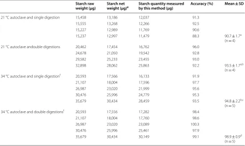
Table 1 Effects of autoclaving temperature and enzymatic digestion duration on the accuracy of starch measurement using this method
The letters a, b, c and d after mean ± SD indicate significant difference (p < 0.05, t-test) e The net weight excluded the weight of the water, protein and fiber in the corn starch
f The samples after autoclaving were split for single digestion and double digestions
Starch raw
weight (μg) Starch net weight (μg)e Starch quantity measured by this method (μg) Accuracy (%) Mean ± SD
121 °C autoclave and single digestion 15,458 13,186 12,037 91.3 15,555 13,268 12,266 92.5 15,227 12,989 11,769 90.6
15,237 12,997 11,479 88.3 90.7 ± 1.7a (n = 4) 121 °C autoclave andouble digestions 20,462 17,454 16,762 96.0
24,678 21,050 19,542 92.8 29,582 25,233 23,455 93.0
32,898 28,062 25,863 92.2 93.5 ± 1.7a,b (n = 4) 134 °C autoclave and single digestionf 20,593 17,566 16,133 91.9
21,107 18,004 17,596 97.7 26,987 23,020 21,999 95.6 30,476 25,996 24,779 95.3
35,679 30,434 28,459 93.5 94.8 ± 2.2b,c (n = 5) 134 °C autoclave and double digestionsf 20,593 17,556 17,282 98.4
21,107 18,004 17,760 98.6 26,987 23,020 23,089 100.3 30,476 25,996 25,461 97.9
The glucose product was measured using the dinitro-salicylic acid (DNS) method described in the previous “Glucose assay and calculation” section. The net weight of the corn starch (glucose in polymer) was determined by multiplying the quantity of the glucose product by 0.9.
Microalgal sample preparation—pigment extraction by using methanol‑tetrahydrofuran
Ten microliter of the day 5 microalgal culture (OD682
around 1.0) were harvested using swing bucket centrifu-gation (2600g, 3 min) at room temperature. The super-natant was discarded and the cells were transferred to a 2-mL screw cap microtube rapidly. After a brief high-speed centrifugation, the supernatant in the microtube was removed using a pipette and the cells were quickly frozen at − 15 °C in a mix of ice and crude sea salt. One microliter of cold methanol/tetrahydrofuran (v/v = 1/3) was added to the frozen cells. The cells were soaked in the solvent for 30 min in the ice-salt mix and agitated occasionally to extract the pigments. After centrifuging at 16,000g at 4 °C for 10 min, the solvent was discarded and the pellet was dried at 65 °C for 1 h. The dried pellet was repeatedly washed out using the sodium phosphate buffer (50 mM, pH 5.0) and the final volume was adjusted to 5 mL. A small amount of corn starch, weighed using the high accuracy analytical balance aforementioned and listed in Table 2, was added to each suspension serving as the internal control. This mix was autoclaved at 134 °C for 1 h. One mL of the autoclaved sample was smashed using a mini-beadbeater (BioSpec Products Inc., USA) to
fully release the starch from the cells, and 0.5 mL of the smashed sample was mixed with 0.5 mL of the sodium phosphate buffer then subjected to the double digestions aforementioned. After the double digestions, the sample was centrifuged again to precipitate any cell debris. Five hundred microliter of the supernatant was used for glu-cose measurement as described in the previous “Glucose assay and calculation” section.
Results
Estimate of the net weight of glucose polymers in the corn starch
As in the grains of crops, corn starch contains certain levels of water, protein and cellulosic fiber that affect the measurement of the net weight of starch (glucose polymer). Corn starch samples, stored in the lab refrig-erator, weighing more than 0.3 g were dried at 105 °C for 2 h in a ceramic crucible. The weight difference of each sample before and after the drying was measured using a high accuracy analytical balance. The water content in the starch was determined to be 13.5 ± 1.3% (mean ± SD, n = 3). The contents of protein and fiber in the corn starch were measured by the U.S. Department of Agri-culture (https ://ndb.nal.usda.gov/ndb/foods /show/30522 8), which together comprised 1.16% raw weight of corn starch. The amounts of other components such as lipids and minerals were very little as shown in our results and in the USDA database. They were thus ignored in the net weight calculation. Therefore, the net weight of starch (glucose polymer) was 85.3% of the raw weight of the corn starch used in this study.
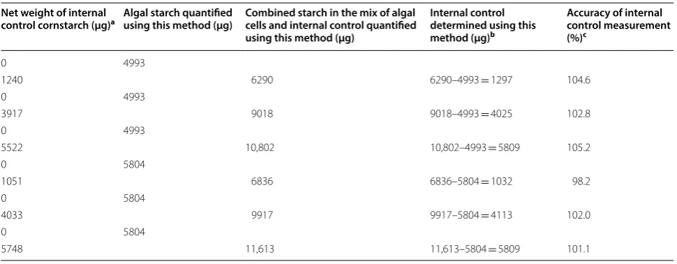
Table 2 Accuracy of measuring the internal control cornstarch mixed with algal samples using this method
a The net weight excluded the weight of the water, protein and fiber in the cornstarch
b The quantities were obtained by subtracting the amount of algal starch measured in the controls (without the cornstarch internal control) from the total starch
c Mean ± SD is 102.3 ± 2.5% (n = 6)
Net weight of internal
control cornstarch (μg)a Algal starch quantified using this method (μg) Combined starch in the mix of algal cells and internal control quantified using this method (μg)
Internal control determined using this method (μg)b
Accuracy of internal control measurement (%)c
0 4993
1240 6290 6290–4993 = 1297 104.6
0 4993
3917 9018 9018–4993 = 4025 102.8
0 4993
5522 10,802 10,802–4993 = 5809 105.2
0 5804
1051 6836 6836–5804 = 1032 98.2
0 5804
4033 9917 9917–5804 = 4113 102.0
0 5804
Effects of autoclaving temperature and duration of enzymatic digestion on starch degradation
Two parameters, autoclaving temperature and dura-tion of enzymatic digesdura-tion, can affect the completeness of starch digestion by glucoamylase. Starch granules are formed by compacted glucose polymers in the cells of green algae and plants. The compact structure hinders enzymatic reactions and thus it has to be disintegrated before the enzyme can completely digest the glucose polymers. The effect of autoclaving temperature on the disintegration of starch granules was examined. In addi-tion, the duration of the enzymatic digestion of dissolved glucose polymers is an important factor that determines the accuracy of starch measurement. This factor was also investigated in this study.
As shown in Table 1, the highest accuracy of the starch measurement using this procedure was achieved by autoclaving at 134 °C in conjunction with the glucoa-mylase double digestions (98.9 ± 0.9%, n = 5), followed by autoclaving at 134 °C coupled with single digestion (94.8 ± 2.2%, n = 5) and autoclaving at 121 °C with double digestions (93.5 ± 1.7%, n = 4. It should be noted that the difference between the two sets of measurements was not statistically significant), and lastly autoclaving at 121 °C with single digestion (90.7 ± 1.7%, n = 4).
Estimate of the accuracy of measuring endogenous starch in algal cells
Unlike processed corn starch, microalgal cells contain pigments that could impede the starch measurement which is based on spectrophotometry at 540 nm absorp-tion. Pigment extraction seems to be the best option to avoid this problem. The solvent methanol/tetrahydro-furan (v/v = 1/3) was employed to extract the pigments, and 30 min of extraction gave satisfactory results with the cells of the day 5 culture. After autoclaving at 134 °C and glucoamylase double digestions, the algal cells with or without the additional corn starch (the internal con-trols) were smashed and centrifuged to collect clear supernatant for the glucose assay. As shown in Table 2, the measurement accuracies of the additional corn starch (the internal controls) were close to 100% in the six tests using this procedure. The results suggest this procedure can achieve a high level of accuracy for the measurement of endogenous starch in green microalgal cells.
Discussion
This simple and accurate procedure that was built based on the corn starch internal controls for microalgal starch quantification is also low-cost, due to the use of the thermo-tolerant glucoamylase. This enzyme is able to hydrolyze α-1,6-glucosidic bonds in starch in addi-tion to α-1,4-glucosidic bonds (see the comments of the
International Union of Biochemistry of Molecular Biol-ogy, IUBMB about this group of enzymes in BRENDA database, and the product information of this enzyme issued by Tokyo Chemical Industry Company). The cost of the enzyme per assay (4 units/assay) in this study was less than USD 0.5 cent, a dramatic difference compared to USD 14.2 using the Sigma-Aldrich starch assay kit and Euro 2.6 using the Megazyme kit purchased in Taiwan. The mass of the internal controls was measured using a high accuracy analytical balance, which gave the readings very close to their true values. This provides the possibil-ity of gauging the accuracy of the results obtained using this quantification method.
In the study of Rose et al. (1991), the “accuracy” of the six starch quantification methods was compared, and up to 40% in variation was found in the results obtained by using those methods. As aforementioned, steps used in those six methods are still adopted recently (Branyikova et al. 2011; Dragone et al. 2011). The accuracy of starch quantifications nowadays is still a great concern. The six methods did not include internal controls or standards of purified starch in those assays. Therefore, the comparisons were actually about the precisions and the result varia-tions measured using those methods. The true values of the starch in their samples were not clear. Therefore, it is unlikely to determine which method was more accurate than the others among those six methods. In the two com-mercial starch assay kits produced by Sigma-Aldrich and Megazyme companies, the principles the two kits adopt are the same, which are (1) starch degradation to glucose using amylase/amyloglucosidase; (2) glucose conversion to 6-phosphosphate using hexokinase; (3) glucose-6-phosphate conversion to 6-phosphogluconate using glucose-6-phosphosphate dehydrogenase; and (4) meas-urement of the resulted NADH levels using OD340. The
two assays do include starch standards, but not internal controls, to calibrate the results from measuring unknown samples. However, the interferences from water contents and other non-glucose components in the starch standards were ignored in the protocols of the two kits. Besides, the three enzyme reactions are also a concern. Generally, more enzyme reactions give more bias in the quantifications.
Found in this study, the action of quick-freezing of the cells at − 15 °C right after harvest had positive influence on the accuracy of starch quantification. This might be due to the starch degradation by the endogenous micro-algal enzymes at room temperature when the cells were agitated. The measurement accuracy of the internal con-trols being a little over 100% on average in Table 2 was most likely due to the remaining pigments in the samples. Therefore, double solvent extractions might be necessary if the cell pellets of interest are still green after the first pigment extraction. Complete disintegration of starch granules and thorough glucose polymer digestion are crucial to achieve accurate measurement of starch. High temperature autoclaving at 134 °C and glucoamylase dou-ble digestions at 50 °C for more than 20 h yielded satisfac-tory results. High-speed centrifugation (16,000g at room temperature for 10 min) to precipitate the cell debris after the double digestions of the microalgal samples was required to obtain high accuracy of starch measurement.
Although it takes 2 days to quantify microalgal starch using this procedure, the work is not intensive since most time in the 2 days is used for autoclaving (about 4 h from the beginning to the end) and enzyme digestion (one night plus 7 h in the second day). This procedure will be a great tool for the studies of biochemical and genetic engi-neering that involve in starch biosynthesis or degradation in green microalgae.
Acknowledgements
The authors thank Prof. Keryea Soong for helpful discussion.
Authors’ contributions
TCY and C‑SC executed this study, interpreted the data, and reviewed this manuscript. C‑NNC conceived and directed this work, analyzed the data and prepared this manuscript. All authors read and approved the final manuscript.
Funding
This work was financially supported by the Grant MOST 106‑2221‑E‑110‑ 068‑MY2 from the Ministry of Science and Technology, Taiwan.
Availability of data and materials
All data generated or analyzed during the current study are included in this published article.
Ethics approval and consent to participate Not applicable.
Consent for publication Not applicable.
Competing interests
The author declare that they have no competing interests.
Received: 20 August 2019 Accepted: 5 October 2019
References
Berges JA, Franklin DJ, Harrison PJ (2001) Evolution of an artificial seawater medium: improvements in enriched seawater, artificial water over the last two decades. J Phycol 37:1138–1145
Bligh EG, Dyer WJ (1959) A rapid method of total lipid extraction and purifica‑ tion. Can J Biochem Physiol 37:911–917
Branyikova I, Marsalkova B, Doucha J, Branyik T, Bisova K, Zachleder V, Vitova M (2011) Microalgae—novel highly efficient starch producers. Biotechnol Bioeng 108:766–776
Chisti Y (2008) Biodiesel from microalgae beats bioethanol. Trends Biotechnol 26:126–131
Dragone G, Fernandes BD, Abreu AP, Vicente AA, Teixeira JA (2011) Nutrient limitation as a strategy for increasing starch accumulation in microalgae. Appl Energy 88:3331–3335
Folch J, Lees M, Sloane‑Stanley GH (1957) A simple method for the isola‑ tion and purification of total lipids from animal tissues. J Biol Chem 226:497–509
Juergens MT, Disbrow B, Shachar‑Hill Y (2016) The relationship of triacylglyc‑ erol and starch accumulation to carbon and energy flows during nutrient deprivation in Chlamydomonas reinhardtii. Plant Physiol 171:2445–2457 Kato Y, Ho SH, Vavricka CJ, Chang JS, Hasunuma T, Kondo A (2017) Evolutionary
engineering of salt resistance Chlamydomonas sp. strains reveals salinity stress‑activated starch‑to‑lipid biosynthesis switching. Bioresour Technol 245:1484–1490
Miller GL (1959) Use of dinitrosalicylic acid reagent for determination of reduc‑ ing sugar. Anal Chem 31:426–428
Pan YY, Wang ST, Chuang LT, Chang YW, Chen CNN (2011) Isolation of thermo‑ tolerant and high lipid content green microalgae: oil accumulation is predominantly controlled by photosysnthesis efficiency during stress treatments in Desmodesmus. Bioresour Technol 102:10510–10517 Rose R, Rose CL, Omi SK, Forry KR, Durall DM, Bigg WL (1991) Starch determina‑
tion by perchloric acid vs enzymes: evaluating the accuracy and precision of six colorimetric methods. J Agric Food Chem 39:2–11
Saqib AAN, Whitney PJ (2011) Differential behaviour of the dinitrosalicylic acid (DNS) reagent towards mono‑ and disaccharide sugars. Biomass Bioenergy 35:4748–4750
Siaut M, Cuine S, Cagnon C, Fessler B, Nguyen M, Carrier P, Beyly A, Beisson F, Triantaphylides C, Li‑Beisson Y, Peltier G (2011) Oil accumulation in the model green alga Chlamydomonas reinhardtii: characterization, vari‑ ability between common laboratory strains and relationship with starch reserves. BMC Biotechnol 11:7–15
Soh L, Montazeri M, Haznedaroglu BZ, Kelly C, Peccia J, Eckelman MJ, Zim‑ merman JB (2014) Evaluating microalgal integrated biorefinery schemes: emperical controlled growth studies and life cycle assessment. Bioresour Technol 151:19–27
Wang G, Michailides TJ, Bostock RM (1997) Improved detection of polygalac‑ turonase activity due to Mucor piriformis with a modified dinitrosalicylic acid reagent. Phytopathology 87:161–163
Publisher’s Note