High-grade spondylolytic spondylolisthesis
Peter C. Emary,
DC, MSc1Stefan A. Eberspaecher,
DC2John A. Taylor,
DC, DACBR31 Private Practice, Cambridge, ON
2 World Spine Care, Dominican Republic Clinic Supervisor, Patronato Cibao de Rehabilitación, Moca, Provincia Espaillat, República
Dominicana
3 Professor and Coordinator of Diagnostic Imaging, Chiropractic Department, D’Youville College, Buffalo, NY, World Spine Care Consultant
Corresponding author: Peter C. Emary
1 Private Practice, 201C Preston Parkway, Cambridge, ON N3H 5E8, Canada
e-mail: [email protected]
Consent: The patient has provided written consent to having her personal health information, including diagnostic images, published. The authors have no competing interests to declare.
© JCCA 2017
Case reports of high-grade spondylolisthesis have been rarely published in the chiropractic literature. Documented here is a case involving a 28-year-old woman who presented to the World Spine Care clinic in the Dominican Republic with minimal neuromusculoskeletal symptoms despite a grade 4 spondylolytic spondylolisthesis. The key imaging and etiological features of this clinical disorder are presented.
(JCCA. 2017;61(2):162-166)
k e y w o r d s: spondylolisthesis, spondylolysis, pars
interarticularis, chiropractic
Il y a eu peu d’études de cas de spondylolisthésis de haut degré publiées dans la littérature chiropratique. On consigne ici un cas concernant une femme de 28 ans qui s’est présentée à la clinique World Spine Care en République dominicaine avec des symptômes neuro-musculo-squelettiques minimaux malgré un spondylolisthésis spondylolytique de stade 4. Les principales caractéristiques d’imagerie et d’étiologie de ce trouble clinique sont présentées.
(JCCA. 2017;61(2):162-166)
m o t s c l é s : spondylolisthésis, spondylolyse, isthme
interarticulaire vertébral, chiropratique
Case Presentation
A 28-year-old female presented to the World Spine Care (WSC) clinic in Moca, Dominican Republic with a 10-year history of intermittent lower back pain (LBP) and
sitting was also provocative. Exercise at the gym, walk-ing, and performing yoga were described as palliative. Recent diagnostic imaging investigations had revealed a high-grade lumbosacral spondylolisthesis (Figures 1-3). The patient had consulted numerous surgeons and other medical specialists and was told that she would be un-able to have children because of her spondylolisthesis and would eventually be confined to a wheelchair if she did not have surgery. There were no signs or symptoms of cauda equina syndrome. The patient was in good general health and did not take medications. Because chiropractic treatment had provided relief in the past, she visited the WSC Moca clinic for a second opinion.
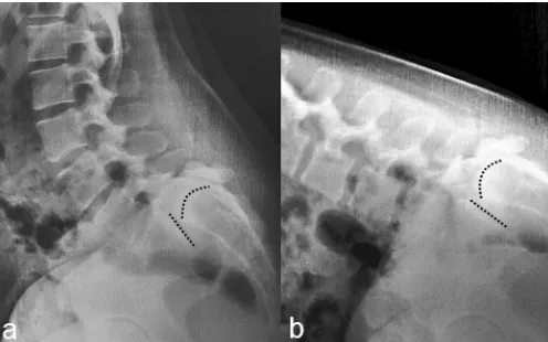
On examination, the patient was of normal weight and her gait was unremarkable. The lumbosacral lordosis was increased. Range of motion of the lumbar spine was full and pain-free, with the exception of mild lower back pain Figure 1. Lateral lumbar radiograph (a) and
mid-sagittal T2-weighted magnetic resonance (MR) image (b) reveal six lumbar-type vertebrae. The L6 vertebral body is trapezoidal shaped and has translated 80-90% anteriorly in relation to the S1 vertebral segment. A large gap is present in the region of the L6 pars interarticularis and has resulted in posterior translation of the spinous process and laminae of L6. The L6-S1 disc is obliterated and prominent buttressing of the anterior-inferior aspect of the L6 vertebral body is present. The L5-L6 disc appears well hydrated, but is narrowed and bulging posteriorly. The posterior elements of L5 approximate the posterior aspect of the sacral base resulting in prominent central stenosis at this level.
Figure 2. Anteroposterior lumbopelvic radiograph
reveals the characteristic “Bowline of Brailsford” or “Inverted Napolean Hat” sign. The arrows are pointing to the anterior cortex of the vertebral body and transverse processes of L6. This vertebral body is tipped forward significantly such that it resembles an axial projection.
Figure 3. Axial (b) T2-weighted MR image through the
on passive end-range extension. Bilateral Kemp’s test1
provoked mild LBP but the straight leg raise, slump, and femoral nerve stretch tests were negative. Neurologic examination of the lower limbs, including motor, reflex, sensory, vibratory, and Rhomberg’s testing, was normal except for absent Achilles tendon reflexes (graded as 0) bilaterally. Mid-thigh and calf muscle circumferences were symmetric bilaterally with no evidence of atrophy. A non-tender midline ‘step defect’ was palpated at the lumbosacral junction and mild pain was elicited with the application of posterior-to-anterior pressure over this re-gion. Palpation also revealed hypertonicity of the quad-ratus lumborum and lumbosacral erector spinae muscles, bilaterally. Milgram’s test1 (i.e. bilateral active straight
leg raise test, performed with the patient supine) severe-ly provoked the chief LBP complaint and elicited patient apprehension. Further radiographic imaging including dynamic views of the lumbar spine were obtained. The flexion radiographs revealed instability of the spondylo-listhesis (Figure 4). Based on clinical and imaging find-ings, a diagnosis of chronic unstable grade 4 spondylo-lytic spondylolisthesis was made.
Discussion
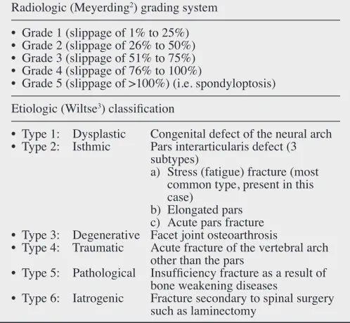
Table 1 lists the radiologic grading and etiological classi-fication systems for spondylolisthesis.2,3 The key imaging
features for high-grade spondylolytic spondylolisthesis are listed in Table 2. According to the Meyerding classi-fication2, high-grade spondylolistheses are characterized
by an anterior vertebral slippage of 50% or greater (i.e. grades 3, 4, and 5). The incidence of isthmic (spondy-lolytic) spondylolisthesis in the general adult population ranges between 3.7% and 8%;9 however, the
epidemi-ology of high-grade spondylolisthesis is unknown. To date, the evidence concerning the long-term prognosis and optimal (i.e. conservative versus surgical) manage-ment of patients with high-grade spondylolisthesis also remains insufficient and controversial.4-9 Moreover,
clin-ical symptoms in such patients often do not correlate with the degree of slip.10 Several authors advocate for surgical
intervention (e.g. fusion, with or without slip reduction) regardless of patient symptoms, in order to prevent fur-ther slippage and or symptom progression.7,8 Others have
suggested however that non-surgical management can be considered, particularly in asymptomatic or minimally symptomatic cases.5-8 Regarding natural history, the risk
of slip progression is greater in skeletally immature chil-dren and adolescents than it is in adults.8,10 As such
sur-gery is rarely indicated for this reason in adults.8
In the current case, the patient underwent a trial of con-servative therapy, including spinal manipulation (to the thoracic and upper lumbar spine), soft-tissue trigger-point therapy and Active Release Techniques® (to the
lumbo-sacral and thoracolumbar paraspinal muscles), and home-based spinal stabilization exercises (i.e. cat-camel mo-bilizations, bird-dogs, side-bridges, and crunches). The patient was also encouraged to continue with her weekly exercise activities (e.g. walking, gym exercise, and yoga). After nine visits (over five weeks) the patient had no func-tional limitations and her pain severity was reduced to a two out of 10. Furthermore, there were no adverse treat-ment effects during the course of her care.
Indications for surgery in patients with spondylolis-thesis are listed in Table 3. With a greater than 10% re-ported risk of complications including neurologic injury with surgical intervention6,8,11, some authors have
indicat-ed that non-surgical treatment may be suitable for patients in the absence of functional limitations or neurologic im-pairment.5-8 In the current case, the patient had clinical
Figure 4. Dynamic lateral lumbar radiographs obtained
and radiologic evidence of instability but no significant functional limitations, postural deformities, gait abnor-malities, or progressive neurologic signs or symptoms. Her LBP symptoms also responded favourably to a short-course of conservative treatment. She was advised to re-turn for chiropractic treatment as needed, or for re-evalu-ation should any neurologic signs or symptoms develop. As the patient was planning on getting married later in the year, the news that her spondylolisthesis would have no bearing on her ability to have children after all came as a welcome surprise.
Key Messages
• Published cases involving high-grade spondylolis-thesis are rare within the chiropractic literature • Indications for surgery in such patients include slip
progression, significant deformity and postural im-balance, neurologic deficit and/or cauda equina syndrome, and imaging confirmed radiculopathy unresponsive to conservative treatment
• Conservative therapy may be considered as a first line treatment option in patients with high-grade spondylolisthesis who present with minimal neuro-musculoskeletal symptoms
Table 2.
Key imaging features of high-grade spondylolytic (isthmic Type 2a) spondylolisthesis
Key imaging features
• Spondylolysis: pars interarticularis stress (fatigue) fracture that usually occurs during childhood or early adolescence • Most common at L5
• Anterior displacement of the vertebral body and corresponding posterior displacement of the neural arch resulting in
characteristic palpable “step defect” (see Figure 1) • Bowline of Brailsford on frontal radiograph (see Figure 2) • Trapezoidal shape of listhetic vertebral body and
corresponding “rounding” of the subjacent sacral base (see Figure 4) (These are signs of chronic, longstanding vertebral body displacement.)
• Vertebral body translation >4.5 mm observed on dynamic flexion-extension radiographs suggests radiologic instability (see Figure 4)
Table 3.
Indications for surgery in patients with spondylolisthesis Surgical indications8
• Slip progression
• High-grade spondylolisthesis with significant lumbosacral kyphotic deformity and sagittal imbalance
• Neurologic deficit
• Low back pain unresponsive to conservative treatment • Radicular pain and nerve root compression documented on
imaging studies unresponsive to conservative treatment
Table 1.
Radiologic grading and etiologic classification systems for spondylolisthesis
Radiologic (Meyerding2) grading system
• Grade 1 (slippage of 1% to 25%) • Grade 2 (slippage of 26% to 50%) • Grade 3 (slippage of 51% to 75%) • Grade 4 (slippage of 76% to 100%)
• Grade 5 (slippage of >100%) (i.e. spondyloptosis) Etiologic (Wiltse3) classification
• Type 1: Dysplastic Congenital defect of the neural arch • Type 2: Isthmic Pars interarticularis defect (3
subtypes)
a) Stress (fatigue) fracture (most common type, present in this case)
b) Elongated pars c) Acute pars fracture • Type 3: Degenerative Facet joint osteoarthrosis
• Type 4: Traumatic Acute fracture of the vertebral arch other than the pars
• Type 5: Pathological Insufficiency fracture as a result of bone weakening diseases
References
1. Evans RC. Illustrated Essentials in Orthopedic Physical Assessment. St. Louis: Mosby; 1994.
2. Meyerding HW. Spondylolisthesis. Surg Gynecol Obstet. 1932; 54:371-377.
3. Wiltse LL, Newman PH, Macnab I. Classification of spondylosis and spondylolisthesis. Clin Orthop Relat Res. 1976; 117:23-29.
4. DeWald CJ, Vartabedian JE, Rodts MF, Hammerberg KW. Evaluation and management of high-grade
spondylolisthesis in adults. Spine. 2005; 30(6 Suppl):S49-S59.
5. Bourassa-Moreau É, Mac-Thiong JM, Joncas J, Parent S, Labelle H. Quality of life of patients with high-grade spondylolisthesis: minimum 2-year follow-up after surgical and nonsurgical treatments. Spine J. 2013; 13(7):770-774.
6. Lundine KM, Lewis SJ, Al-Aubaidi Z, Alman B, Howard AW. Patient outcomes in the operative and nonoperative management of high-grade spondylolisthesis in children. J Pediatr Orthop. 2014; 34(5):483-489. 7. Xue X, Wei X, Li L. Surgical versus nonsurgical
treatment for high-grade spondylolisthesis in children and adolescents: a systematic review and meta-analysis. Medicine. 2016; 95(11):e3070.
8. Kasliwal MK, Smith JS, Kanter A, Chen CJ,
Mummaneni PV, Hart RA, Shaffrey CI. Management of high-grade spondylolisthesis. Neurosurg Clin N Am. 2013; 24(2):275-291.
9. Kreiner DS, Baisden J, Mazanec DJ, Patel RD, Bess RS, Burton D, et al. Guideline summary review: an evidence-based clinical guideline for the diagnosis and treatment of adult isthmic spondylolisthesis. Spine J. 2016; 16(12):1478-1485.
10. Haun DW, Kettner NW. Spondylolysis and
spondylolisthesis: a narrative review of etiology, diagnosis, and conservative management. J Chiropr Med. 2005; 4(4):206-217.