ORIGINAL ARTICLE
Sakae Horisawa · Yoh Sakuma · Shuichi Doi
Qualitative and quantitative PCR methods using species-specifi c primer for
detection and identifi cation of wood rot fungi
Abstract Species-specifi c oligonucleotide primers for detecting wood rot fungi, Gloeophyllum trabeum, Trametes versicolor, Coniophora puteana, and Serpula lacrymans, and the primer detecting a group of related fungi to G. sepiarium were developed. These primer sequences were picked up from the internal transcribed spacer region between small-subunit rDNA and large-subunit rDNA. The species selectivities of the developed primers were checked. Real-time polymerase chain reaction (PCR) was carried out using these highly specifi c primers to quantita-tively detect at least of 0.01 ng genome DNA of the target species. This quantitative PCR was also used to differenti-ate the target species DNA from mixed species DNA. A PCR-based technique using the species-specifi c primers would be applicable to multiple-sample assay in diagnosis of wood decay and to investigation of environmental fungal populations.
Key words Species-specifi c primer · Wood rot fungi · Quan-titative PCR · Wood decay
S. Horisawa (*)
Department of Environmental Systems Engineering, Faculty of Engineering, Kochi University of Technology, 185 Miyanokuchi, Kami, Kochi 782-8502, Japan
Tel. +81-887+57-2519; Fax +81-887-57-2520 e-mail: [email protected]
Y. Sakuma
Graduate School of Science and Engineering, Ehime University, Matsuyama 790-8577, Japan
S. Doi
Graduate School of Life and Environmental Sciences, University of Tsukuba, Tsukuba 305-8572, Ibaraki, Japan
Part of this article was presented at the International Symposium on Wood Science and Technology (IAWPC 2005), Yokohama, November 2005
Introduction
In the fi eld of wood preservation and in research on wood decay, it is important to identify the causal fungi of decay. In conventional methods, wood rot fungi have been identi-fi ed based on the morphology of the fruit bodies, spores, and mycelial growth conditions. However, the appraisers are required to have specifi c knowledge and experience to identify fungi using the conventional methods. As such, it is diffi cult to avoid occasional misidentifi cation of fungi. In particular, the identifi cation of wood rot fungi invading in wood becomes very diffi cult when no fruit body is obtained. In addition, the appraisers must detect wood rot fungi grown on the surface and distinguish it from other fungi such as mold.
Recently, several biomolecular techniques have been developed to detect and identify fungi.1,2 Protein assay such as sodium dodecyl sulfate-polyacrylamide gel electrophore-sis (SDS-PAGE) and matrix-aselectrophore-sisted laser desorption ion-ization time-of-fl ight mass spectrometry (MALDI-TOFMS) have been used to detect diversity among species and par-ticular proteins. An immunological method has been devel-oped as a sensitive diagnostic method and has been used in practical testing.3–6
Fourier transform infrared (FT-IR) spec-troscopy has been reported as a sensitive tool for detection of biomolecules or metabolites.7,8
DNA is a useful chemical form that can be used to detect and identify wood rot fungi in decayed wood or the environment because it is a relatively stable and amplifi able biomolecule. Random amplifi -cation of polymorphic DNA (RAPD) analysis9
and amplifi ed fragment length polymorphism (AFLP) analysis10,11
have been reported to produce genome DNA fi ngerprints for individual diversity. These DNA detection techniques are sensitive because the DNA is amplifi able by PCR.
For detecting basidiomycetes, small-subunit (SSU) and large-subunit (LSU) rDNA, internal transcribed spacer (ITS) region, intergenic spacer (IGS) region, and β-tubulin are employed due to their species-specifi c sequences as target genes.12–15
PCR using species-specifi c primers can be used to detect and identify wood rot fungi with great sensi-Received: October 6, 2008 / Accepted: December 3, 2008 / Published online: January 16, 2009
J Wood Sci (2009) 55:133–138 © The Japan Wood Research Society 2009
tivity. Suhara et al. and Schmidt and Moreth developed species-specifi c primers for detecting Phelebia brebispora, Serpula lacrymans, and Coniophora puteana.16–18
Quantita-tive PCR using species-specifi c primers is able to detect target species of fungi both qualitatively and quantita-tively.19–22
In the present study, species-specifi c primers for detecting Gloeophyllum sepiarium, Gloeophyllum trabeum, and Trametes versicolor, in addition to the reported species mentioned above, were developed. The quantitative capa-bilities of the species-specifi c primers were also examined.
Materials and methods
Fungal strains and DNA extraction
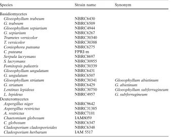
Coniophora puteana NBRC6275, C. puteana FPRI-m, Gloeophyllum sepiarium NBRC4944, G. sepiarium NBRC6267, Gloeophyllum trabeum NBRC6430, G. trabeum NBRC6509, Serpula lacrymans NBRC8697, S. lacrymans NBRC30955, Trametes versicolor NBRC30340, and T. ver-sicolor NBRC30388 were adopted as representative strains for making and checking species-specifi c primers. Fomitop-sis palustris, Gloeophyllum striatum, Gloeophyllum ungula-tum, Lentinus lepideus, Aspergillus restrictus, Chaetomium globosum, Cladosporium cladosporioides, and Cladospo-rium herbarum were employed as negative control strains (Table 1).
The DNA of each fungal strain was extracted by a modi-fi ed method previously reported.10
Mycelia of test strains were collected from their growing mycelial mat on potato dextrose agar (PDA) plates. To each fungus, approximately 0.1 g of mycelia was placed in a 2.0-ml microtube and 500 μl
of TE buffer [10 mM Tris-HCl, 0.1 mM ethylenediaminetet-raacetic acid (EDTA), pH 9.0] and 10 μl of mercaptoetha-nol were added. Sample mycelia were ground for 2 min with a 7-mm zirconia bead at a frequency of 25 Hz, using a mixer mill grinder (Tissuelyser, Qiagen, Valencia, CA, USA). Three hundred microliters of TE-saturated phenol and 100 μl of 10% SDS were added to each tube and incubated at 50°C for 30 min with reversing at 5-min intervals to keep the two phases thoroughly mixed. The tubes were centri-fuged at 11 000 g and 4°C for 10 min, and the supernatant was collected. DNA was extracted from the supernatants with a mixture of phenol, chloroform, and isoamyl alcohol (25 : 24 : 1). The DNA was precipitated with 2-propanol and sodium acetate and collected by centrifugation. The DNA pellets were rinsed with 70% ethanol followed by drying. The DNA was dissolved in 5 ml TE containing RNase and purifi ed on a column (Qiagen, Valencia, CA, USA).
Primer design and species-specifi c PCR
An ITS region was selected as a target sequence for species-specifi c PCR using a universal primer ITS 1 as a forward primer.23
Reverse primers were designed from a species-specifi c sequence in the ITS II region of each species, respectively (Fig. 1). The sequence of test strains and sequence information of objective and related species in the Genbank/DDBJ/EMBL database were aligned by ClustalW24
and species-specifi c sequences were searched by sight. The primers were designed with interrelation to the forward primer ITS 1 and the melting temperature (Tm) using primer-designing software Primer3.25
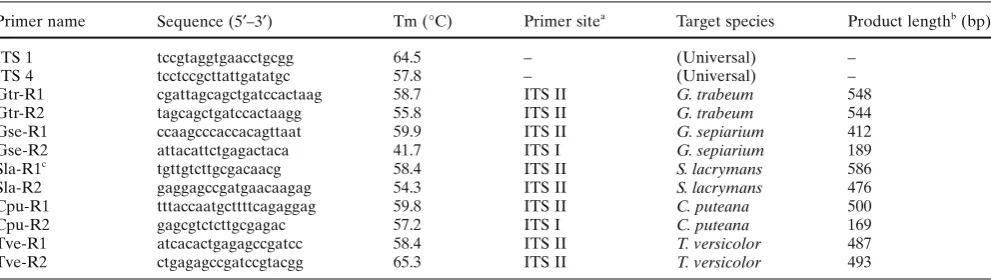
The primer sequences and prospective lengths of the amplicons are shown in Table 2.
Table 1. Tested strains
Species Strain name Synonym
Basidiomycetes
Gloeophyllum trabeum NBRC6430
G. trabeum NBRC6509
Gloeophyllum sepiarium NBRC4944
G. sepiarium NBRC6267
Trametes versicolor NBRC30340
T. versicolor NBRC30388
Coniophora puteana NBRC6275
C. puteana FPRI-m
Serpula lacrymans NBRC8697
S. lacrymans NBRC30955
Fomitopsis palustris NBRC30339 Gloeophyllum ungulatum NBRC6431
G. ungulatum NBRC6507
Gloeophyllum striatum NBRC30341 Gloeophyllum abietinum
G. striatum NBRC6429 G. abietinum
Lentinus lepideus NBRC30750 Gloeophyllum subferrugineum
L. lepideus NBRC4957 G. subferrugineum
Deuteromycetes
Aspergillus niger NBRC9642 Aspergillus restrictus NBRC31385
A. restrictus NBRC7101
Chaetomium globosum IAM8059
C. globosum NBRC6347
cycles of denaturization at 95°C for 5 s, annealing and extension at 60°C for 41 s. All the real-time PCR was carried out in triplicate by using three reaction wells for each tem-plate DNA. The threshold cycle (Ct) was determined auto-matically by the instrument.
Quantitative detection of target species by species-specifi c PCR
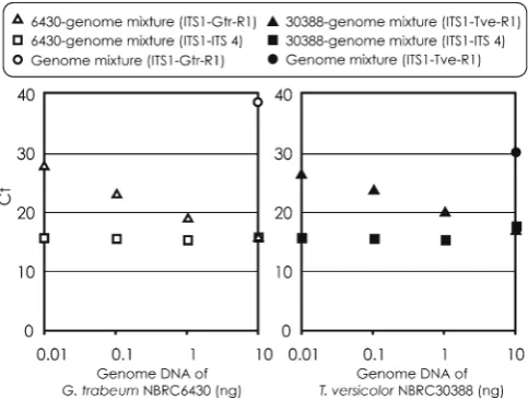
Quantitative PCR for detecting target species was con-ducted using species-specifi c primer Gtr-R1 or Tve-R1 from mixed-genome DNA. The mixed-genome DNA solution, except for target species, was prepared at a concentra-tion of 10 ng/μl, involving 1 ng/μl of each genome DNA (G. sepiarium NBRC6267 and NBRC4944, C. puteana NBRC6275 and FPRI-m, S. lacrymans NBRC8697 and NBRC30955, F. palustris NBRC30339, G. ungulatum NBRC6431, G. striatum NBRC30341, and L. lepideus NBRC30750). The real-time PCR for detecting G. trabeum was conducted using species-specifi c primer Gtr-R1 and a total of 10 ng DNA template, which was prepared from 0.01 ng, 0.1 ng, 1 ng, and 10 ng of G. trabeum NBRC6430 added to mixed-genome DNA, respectively. The PCR detecting T. versicolor was conducted using species-specifi c primer Tve-R1 and 10 ng of the mixed DNA solution added to 0.01 ng, 0.1 ng, 1 ng, and 10 ng DNA of T. versicolor NBRC30340, respectively. Other conditions of this real-time PCR were the same as those described above.
Results
Screening species-specifi c primer
The full length of ITS regions from the tested fungal strains in Table 1 were amplifi ed by PCR using the universal primers ITS 1 and ITS 423 to assess whether these primers were available, and a predicted length of PCR product was obtained for each strain (data not shown). Based on these results, the primer ITS 1 was employed as a forward primer for species-specifi c PCR in the present study. The specifi city
Fig. 1. Schematic diagram of the internal transcribed spacer (ITS)
region between small subunit (SSU) rDNA and large subunit (LSU) rDNA, which includes ITS I, ITS II, and 5.8S rDNA
Table 2. List of universal and species-specifi c primers
Primer name Sequence (5′–3′) Tm (°C) Primer sitea Target species Product lengthb (bp)
ITS 1 tccgtaggtgaacctgcgg 64.5 – (Universal) –
ITS 4 tcctccgcttattgatatgc 57.8 – (Universal) –
Gtr-R1 cgattagcagctgatccactaag 58.7 ITS II G. trabeum 548
Gtr-R2 tagcagctgatccactaagg 55.8 ITS II G. trabeum 544
Gse-R1 ccaagcccaccacagttaat 59.9 ITS II G. sepiarium 412
Gse-R2 attacattctgagactaca 41.7 ITS I G. sepiarium 189
Sla-R1c tgttgtcttgcgacaacg 58.4 ITS II S. lacrymans 586
Sla-R2 gaggagccgatgaacaagag 54.3 ITS II S. lacrymans 476
Cpu-R1 tttaccaatgcttttcagaggag 59.8 ITS II C. puteana 500
Cpu-R2 gagcgtctcttgcgagac 57.2 ITS I C. puteana 169
Tve-R1 atcacactgagagccgatcc 58.4 ITS II T. versicolor 487
Tve-R2 ctgagagccgatccgtacgg 65.3 ITS II T. versicolor 493
a See Fig. 1
b Predicted length by polymerase chain reaction using the primers of ITS 1 and species-specifi c primers c Sla-R1 was described by Schmidt and Moreth17
A PCR mixture was prepared from Takara Ex Taq (Takara Bio, Shiga, Japan) according to the manufacturer’s instructions. The mixture contained 10 ng of template DNA, each primer at a concentration of 0.5 pM, each dNTP at a concentration of 200 nM, 1.2 U/100 μl of DNA polymerase, and Expand reaction buffer with 1.5 mM MgCl2. PCR was performed with a GeneAmp 9700 thermal cycler (Applied Biosystems, Foster City, CA, USA) with the following parameters: an initial denaturization of 5 min at 95°C, 30 cycles of 30 s at 95°C for denaturization, 30 s at 48°C, 56°C, and 60°C for annealing, 80 s at 72°C for extension, and a fi nal extension of 7 min at 72°C. All PCR products were stored at 4°C until used. Aliquots of PCR products were examined by running them on 1.0% agarose gel.
Quantitative PCR using species-specifi c primer
of the developed species-specifi c primer was checked for the target genome DNA. Some of these results are pre-sented in Fig. 2. The primers that had high specifi cities to the target species were: Gtr-R1 and Gtr-R2 for Gloeophyl-lum trabeum; Tve-R1 and Tve-R2 for Trametes versicolor; Cpu-R1 and Cpu-R2 for Coniophora puteana; and Sla-R2 for Serpula lacrymans. These primers amplifi ed each portion of the ITS region, but only target species.
No primer having high specifi city for Gloeophyllum sepi-arium was produced. PCR using the Gse-R1 detected not only G. sepiarium but also Gloeophyllum ungulatum and Lentinus lepideus. Gse-R2 did not amplify any fragment from the tested strains at the annealing temperature of 60°C, but amplifi ed a part of the ITS region from G. sepia-rium, G. ungulatum, and L. lepideus at an annealing tem-perature of 56°C because of the low Tm of 47°C. No product was obtained by the primer Sla-R1.17
The reason for this result seems to be the differences in PCR conditions, includ-ing differences in the type of polymerase, buffer composi-tions, and thermal-cycle program effects on the reaction products. None of the primers developed for detecting wood rot fungi responded to any of the deuteromycetes tested.
Quantitative PCR
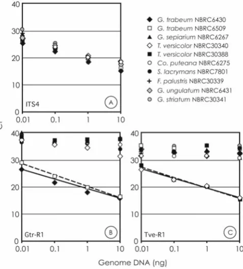
The real-time PCR using the universal primer and the species-specifi c primer was performed with various amounts of target genome DNA. The Ct values were plotted against the amount of genome (Fig. 3). The Ct value is defi ned as the actual PCR cycle when the fl uorescence signal increases above the background threshold. To construct a standard curve, genome DNA extracted from tested basidiomycete strains was serially diluted tenfold and was applied to quan-titative PCR using the universal primer pair, ITS 1 and ITS 4 (Fig. 3A). In this PCR, the detection limit was estimated to be more than 0.01 ng of genome DNA and the coeffi cient correlations were at least 0.96 in the standard curve of tested basidiomycete strains. A similar curve was obtained in the real-time PCR using the primers ITS 1 and Gtr-R1 from the genome DNA of G. trabeum NBRC6430 at amounts of 0.01–10 ng (Fig. 3B), and also the primer ITS 1 and Tve-R1 from T. versicolor NBRC30340 (Fig. 3C). However, the Gtr-R1 and the Tve-R1 primers did not amplify any products other than those of the target species.
The quantitative PCR assay to detect the target species from mixed-genome DNA was examined (Fig. 4). The Gtr-R1 and Tve-Gtr-R1 detected each target species quantitatively, and the curves of Ct against the template amounts were similar to the standard curve obtained in the PCR using the universal primer pair. The two species-specifi c primers did
Fig. 2. Specifi city of the primers designed for detecting the wood rot
fungi, Gloeophyllum trabeum, Gloeophyllum sepiarium, Trametes ver-sicolor, Coniophora puteana, and Serpula lacrymans. The numbers on the top indicate the template DNA used in each lane: 1, G. trabeum NBRC6430; 2, G. trabeum NBRC6509; 3, G. sepiarium NBRC6267; 4, G. sepiarium NBRC4944; 5, T. versicolor NBRC30340; 6, T. versicolor NBRC30388; 7, C. puteana NBRC6275; 8, C. puteana fpri-m; 9, S. lac-rymans NBRC8697; 10, S. laclac-rymans NBRC30955; 11, Fomitopsis palustris NBRC30339; 12, Gloeophyllum striatum NBRC6431; 13, G. striatum NBRC6507; 14, Gloeophyllum ungulatum NBRC30341; 15, G. ungulatum NBRC6429; 16, Lentinus lepideus NBRC30750; 17, L. lepi-deus NBRC4957; 18, Aspergillus niger NBRC9642. The primers used in each panel are shown on the left. The annealing temperatures are shown on the right
Fig. 3A–C. Standard curves obtained by quantitative polymerase
not amplify the regular products from the mixed DNA without the target. The template containing the target genome DNA and mixed DNA (10 ng in a total amount) presented the same Ct values in the PCR using the universal primer pair (ITS 1 and ITS 4) regardless of the amount of target genome DNA.
Discussion
The ITS region of eukaryotes, which is between SSU rDNA and LSU rDNA, is an intron and easily accumulates gene mutations. The ITS region is highly variable between species in both sequence and length; it therefore can be used for examination of the intraspecies and interspecies relation. The ITS sequence provides useful information for identify-ing organic species. In the present study, species-specifi c primers based on the ITS sequence were developed to clas-sify and identify the species of wood rot fungi, and the effectiveness of these primers was examined. Primers were developed to detect each fungal species, Coniophora puteana, Gloeophyllum trabeum, Serpula lacrymans, and Trametes versicolor, respectively.
The species-specifi c primer was designed as a reverse primer and the universal one was employed as a forward primer. The specifi c primers were picked from the ITS II region in order to obtain PCR products at lengths of around 300 bp for clear detection in agarose gel electrophoresis. The specifi city of the primer should be a most important consideration. Other sequences for discriminating taxo-nomic groups have the potential to make the species-specifi c primer.
The Gse-R1 primer was imperfect in detecting only the target species, although it was able to discriminate a group of some fungal species. A strategy that detects some group or genera of fungi is also effective in the diagnosis of wood decay. Identifi cation and classifi cation of S. lacrymans by means of ITS sequence variation has been discussed in pre-vious articles and the differences between the European and the Japanese strains were indicated.10,13,26
To design the species-specifi c primers, as much sequence information of the ITS from target and related species as possible should be acquired because intraspecifi c variation might be found in this region of some species.27,28
In addition, the PCR-based assay would show the dissimilar results due to that condition.
Combined use of the species-specifi c primers and real-time PCR technology allowed us to identify species rapidly and to estimate with sensitivity the quantity of wood rot fungi (minimum 10 ng). This technique is able to quantita-tively detect target fungal species from a mixed-genome DNA pool. It would be applicable to multiple-sample assay in diagnosis of wood decay and to investigation of environ-mental fungal populations.
Acknowledgments This work was partially supported by a
Grant-in-Aid for Young Scientists (B), 17780141, 2004. We thank the Bio-technology Center, Akita Prefectural University for DNA sequence analysis. We express our appreciation to Mr. Kensuke Hanata and Ms. Yukiko Hatakeyama of the Institute of Wood Technology, Akita Prefectural University, and Mr. Masaki Hirota of Kochi University of Technology for their technical assistance.
References
1. Palfreyman JW, Vigrow A, Button D, Herarty B, King B (1991) The use of molecular methods to identify wood decay organisms. Wood Protect 1:5–22
2. Palfreyman J, Vigrow A (1994) Molecular analysis of certain iso-lates of Serpula lacrymans. FEMS Microbiol Lett 117:281–286 3. Clausen C (1994) Dyed particle capture immunoassay for
detec-tion of incipient brown-rot decay. J Immunoassay 15:305–316 4. Palfreyman JW, Bruce A, Button D, Glanct H, Vigrow A, King B
(2001) Immunological method for the detection and characteriza-tion of wood decay basidiomycetes. Int Biodeterior Biodegrad 48:74–78
5. Clausen CA, Green F, Highley TL (1991) Early detection of brown-rot decay in southern yellow pine using immunodiagnostic procedures.Wood Sci Technol 26:1–8
6. Jellison J, Goodell B (1988) Immunological detection of decay in wood. Wood Sci Technol 22:293–297
7. Naumann A, Navarro-Gonzales M, Sudhakar P, Kues U, Polle A (2005) Fourier transformation infrared microscopy and imaging: detection of fungi in wood. Fungal Genet Biol 42:829–835 8. Erukhimovitch V, Pavlov V, Talyshinsky M, Souprun Y, Huleihel
M (2005) FTIR microscopy as a method for identifi cation of bacte-rial and fungal infections. J Pharm Biomed Anal 37:1105–1108 9. Horisawa S, Sakuma Y, Takata K, Doi S (2004) Identifi cation of
intra- and inter-species of the dry rot fungus Serpula lacrymans by PCR-RFLP and RAPD analysis. J Wood Sci 50:427–432
10. Terashima K, Matsumoto T, Hasebe K, Fukumasa-Nakai Y (2002) Genetic diversity and strain-typing in cultivated strains of Lentin-ula edodes (the shii-take mushroom) in Japan by AFLP analysis. Mycol Res 106:34–39
11. Maejer D, Mithen R, Lewis BG, Vos P, Oliver RP (1996) The use of AFLP fi ngerprinting for the detection of genetic variation in fungi. Mycol Res 100:1107–1111
Fig. 4. Quantitative detection by real-time PCR using the
12. Barry T, Colleran G, Glennon M, Dunican LK, Gannon F (1991) The 16s/23s ribosomal spacer region as a target for DNA probes to identify eubacteria. PCR Methods Appl 1:51–56
13. Tooley PW, Goley ED, Carras MM, Frederick RD, Kuldau GA (2001) Characterization of Claviceps species pathogenic on sorghum by sequence analysis of the β-tubulin gene intron 3 region and EF-1 α gene intron 4. Mycologoia 93:541–551
14. Roux J, Steenkamp ET, Marasas WFO, Wingfi eld MJ, Wingfi eld BD (2001) Characterization of Fusarium graminearum from Acacia and Eucalyptus using β-tubulin and histonegene sequences. Mycologia 93:704–711
15. Erwin, Takemoto S, Hwang W-J, Takeuchi M, Itoh T, Imamura Y (2008) Anatomical characterization of decayed wood in standing light red meranti and identifi cation of the fungi isolated from the decayed area. J Wood Sci 54:233–241
16. Suhara H, Maekawa N, Kubahashi T, Kondo R (2005) Specifi c detection of a basidiomycete, Phelebia brebispora accociated with butt rot of Chamaecyparis obtusa, by PCR-based analysis. J Wood Sci 51:83–88
17. Schmidt O, Moreth U (1998) Genetic studies on house rot fungi and a rapid diagnosis. Holz Roh Werkst 56:421–425
18. Schmidt O, Moreth U (2000) Species-specifi c PCR primers in the rDNA-ITS region as a diagnostic tool for Serpula lacrymans. Mycol Res 104:69–72
19. Woo TH, Patel BK, Cinco M, Smythe LD, Symonds ML, Norris MA, Dohnt MF (1998) Real-time homogeneous assay of rapid cycle polymerase chain reaction product for identifi cation of Leptonema illini. Anal Biochem 259:112–117
20. Kageyama A, Sakamoto M, Benno Y (2000) Rapid identifi cation and quantifi cation of Collinsella aerofaciens using PCR. FEMS Microbiol Lett 183:43–47
21. Lyons SR, Griffen AL, Leys EJ (2000) Quantitative real-time PCR for Porphyromonas gingivalis and total bacteria. J Clin Microbiol 38:2362–2365
22. Schweigkofl er W, O’Donnell K, Garbelotto M (2004) Detection and quantifi cation of airborne conidia of Fusarium circinatum, the causal agent of pine pitch canker, from two California sites by using a real-time PCR approach combined with a simple spore trapping method. Appl Environ Microbiol 70:3512–3520
23. White TJ, Burns T, Lee S, Taylor J (1990) Amplifi cation and direct sequencing of fungal ribosomal RNA genes for phylogenetics. In: Innis MA, Gelfand DH, Sninsky JJ, White TJ (eds) PCR protocols: a guide to methods and applications. Academic, San Diego, pp 315–332
24. Thompson JD, Higgins DG, Gibson TJ (1994) CLUSTAL W: improving the sensitivity of progressive multiple sequence align-ment through sequence weighting, position-specifi c gap penalties and weight matrix choice. Nucleic Acids Res 11:4673–4680 25. Rozen S, Skaletsky H (2000) Primer3 on the WWW for general
users and for biologist programmers. Methods Mol Biol 132: 365–386
26. Schmidt O, Moreth U (1999) Identifi cation of the dry rot fungus, Serpula lacrymans, and the wild merulius, S. himantioides, by amplifi ed ribosomal DNA restriction analysis (ARDEA). Holz-forschung 53:123–128
27. White NA, Dehal PK, Duncan JM, Williams NA, Gartland JS, Palfreyman JW, Cooke DEL (2006) Molecular analysis of intra-specifi c variation between building and “wild” isolates of Serpula lacrymans and their relatedness to S. himantioides. Mycol Res 105:447–452