Original Research Article.
The Prevalence of Mandibular Third Molar Angulation in Patients Who
Attended Arar Specialist Dental Centre: Panoramic Survey
Raed Aldahmeshi
1*, Abdulaziz Alshammri
2, Mohammed Alaenazi
2, Faisal Alrashdan
3,
Heatham Alasmari
41*Dentist, King Saud Medical City, Riyadh, Saudi Arabia. 2Dentist, Ar'ar Dental Center, Ar’ar, Saudi Arabia. 3Dentist, Star Dental Service, Riyadh, Saudi Arabia.
4Dentist,Ministry of Interior Medical Records, Riyadh, Saudi Arabia.
ABSTRACT
Aims: This study was carried out to investigate about the prevalence of mandibular third molar angulations and which specific type of angulation is more prevalent and the classification of impaction if present according to Pill's and Gregory classification and winter’s classification of impaction. Materials and Methods: The study was conducted on 2068 radiographic panoramic records for patients who attended to Ar’ar Specialist Dental Center with orthopantomograms collected from Digital Panoramic X-ray software. The orthopantomograms were examined and evaluated. After that the data were analyzed.
Results and Conclusion: According to data analyzed, from the 2068, total orthopantomograms, 1218 subjects had 3rdmolars, and 850 had not. According to Pill’s and Gregory’s classification, Class A was the most prevalent. On the other
hand, according to Winter’s classification, mesioangular angulation was Vertical more angulation, as well.
Key Words: Angulation, Third Molar, Impaction.
*Correspondence to:
Raed Aldahmeshi King Saud Medical city, Riyadh, Saudi Arabia. Article History:
Received: 25-11-2017, Revised: 15-12-2017, Accepted: 08-01-2018
Access this article online
Website:
www.ijmrp.com
Quick Response code
DOI:
10.21276/ijmrp.2018.4.1.024
INTRODUCTION
The development of third molars and their influence on the dental arches has long been of concern to the dental profession. The developmental path of third molars in human being is very irregular and the formation, calcification timing, position and course of eruption of these teeth show great variability. Frequently, third molars are impacted or congenitally missing.1
The eruption space for mandibular third molars is also affected by the direction of tooth eruption during the functional phase of eruption. Third molar will erupt if space is available and that its impaction is a manifestation of a tooth/tissue disharmony or crowding.1,2
There is considerable variation in the prevalence and distribution of impacted teeth in different regions of the jaw. Factors affecting the prevalence can be the age- group, timing of dental eruption, and the radiographic criteria for dental development and eruption.3
Different classifications for mandibular third molar angulation and impaction have been given such as Winter’s classification, Pell and Gregory’s classification, and other classifications for maxillary
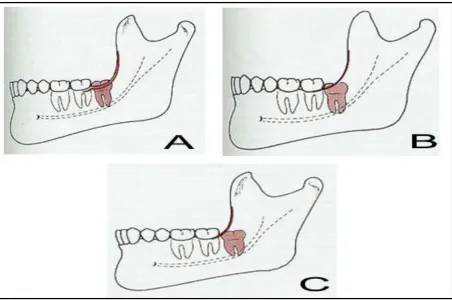
third molar impaction. Winter's classification is classified based on the inclination of the impacted tooth to the long axis of the second molar into mesioangular (38 in figure 1 A), distoangular (38 in figure 1B), vertical (38-48 in figure 1C) and horizontal (38 in figure 1D). This classification is used in this the study as it is simple and easily understandable.4
Figure 1: Winter’s classification of third molars: A) Mesioangular, B) Distoangular, C) Vertical, and D) Horizontal12.
Figure 2: Pell and Gregory’s classification of third molars: A) Class A, B) Class B, and C) Class C13
MATERIALS AND METHODS
The study was conducted on orthopantomograms for patients who attended to ArAr Specialist Dental Center using Digital Panoramic X-ray Software™. The sample consisted of 2068 patients, 1218 patients had at least one third molar, 666 of them were males and 552 were females, while 850 patients did not have third molars (either extracted or missing).
Then, the collected data were interpreted into graphs and charts using Google Drive Sheet™. The data were then analyzed using statistical package for social sciences (SPSS) version 16.0.
RESULTS
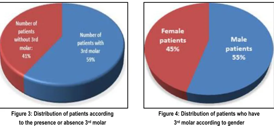
Among a total of 2068 orthopantomograms, 1218 patients (59%) had at least one mandibular third molar, and 850 patients (41%) did not have third molars (either extracted or missing) (Figure 3). Among the studied sample, 666 (55%) of those with third molar were males and 552 (45%) were females (Figure 4).
Among patients who had third molars (1218), 842 (69%) had two
third molars, whilst 376 (31%) had only one. The total examined third molars were 2060. These data are demonstrated in table 1. Among the patients who had two 3rd molars (842 patients), 458
(54.4%) were male patients and 384 (45.6%) were female patients (table 2).
As regards the patients who has one 3rd molar (376 patients), 208
of them were males (55.3%) and 168 (44.7 %) were females (Table 3).
Table 4 shows that there is no statistically significant difference between the number of 3rd molar in each patient in the sample in
relation to the gender, since p value = 0.765 (> 0.05).
Figure 3: Distribution of patients according to the presence or absence 3rd molar
Figure 4: Distribution of patients who have 3rd molar according to gender
Table 1: Distribution of patients according to the number of third molar teeth
Number of 3rd molar teeth Total
Patients having two 3rd molar 842
Patients having one 3rd molar 376
Number of examined 3rd molar 2060
Table 2: Distribution of patients who have two 3rd molar according to gender
Patients who have two 3rd molar teeth Number
Male patients 458
Female patients 384
Table 3: Distribution of patients who have one 3rd molar according to gender
Patients who have one 3rd molar tooth Number
Male patients 208
Female patients 168
Table 4: The relations between the number of 3rd molar and gender of patients
Gender Two 3rd molar One 3rd molar X² OR 95 CI P
Male 458 208 0.09 1.038 0.81 – 31.325 0.765
Female 384 168
As regards the classification of angulation and impaction, patients were classified according to both Pell and Gregory classification and Winter’s classification. The results of these classification were as follows:
1: According to Pell and Gregory classification:
the studied sample (38.8%) were classified in this class, with 423 molars male patients (52.9%) and 377 molars female patients (47.1%). The rest 298 patients (14.5%) were classified as Class C with males constituting 50.7% (151molars) and females constituting 49.3% (147 molars). It was found that there was a statistically significant difference in Classes (A-B-C) between both
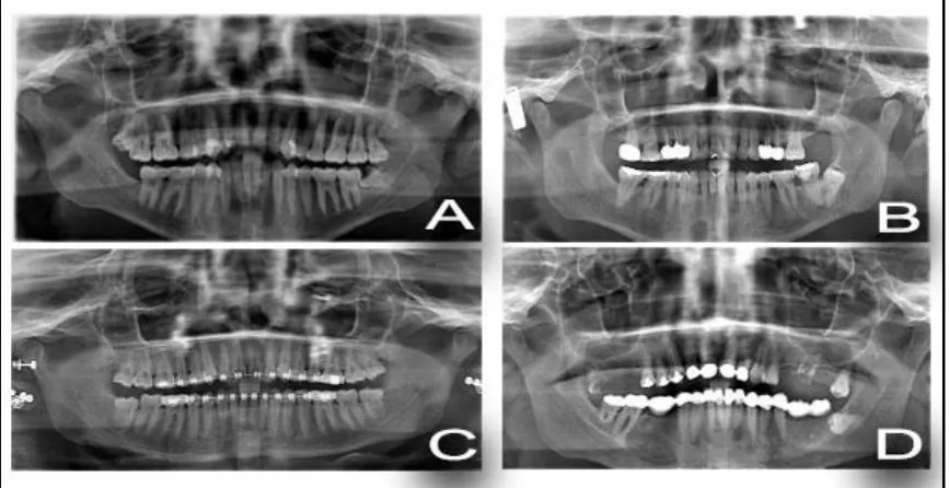
genders in favor of female which has the highest mean (1.7255); the T test was 2.915 with a P-value of 0.004 (< 0.05). For testing this, T-independent samples test was applied and the results are depicted in table 5. Figure 5 demonstrates examples of patients with class A, B, and C third molars according to Pell and Gregory’s classification.
Figure 5: Patients’ radiographs showing class A (a), class B (b), and class C (c) third molars
according to Pell and Gregory’s classification.
Figure 6: Patients’ radiographs showing mesioangular (a), distoangular (b), verical (c) and horizontal (d) third molars according to Winter’s classification.
Table 5: Differences among patients according to Pell and Gregory classification
Classes N Mean (S.D) T P-value
Male 1124 1.6335 (.70172) 2.915 0.004
Female 918 1.7255 (.71958)
Table 6: Difference among patients according to Winter’s classification
Winter’s Classification N Mean (S.D) T P-value
Male 1134 1.9788 (1.11615) 4.695 0.000
2: According to Winter’s classification: 976 out of 2060 (47.4%) had mesioangular 3rd molars (609 molars in males
(62.4%) and 367 molars in females (37.6%)), 786 (38.1%) had vertical 3rd molar (361 molars in male patients (45.9%) and 425
molars in female patients (54.1%)), 122 (5.9%) had distoangular 3rd molars (58 molars in male patients (47.5%) and 64 molars
female patients (52.5%)), and 176 (8.5%) had horizontal 3rd
molars (112 molars in male patients (63.6%) and 64 molars in female patients (36.4%)). It was found that there was also a statistically significant difference in Winter’s Classification between both genders in favor of female which has the highest mean (2.2048); and a T test of 4.695 with a P-value of 0.000 (< 0.05) as shown in table 6. Figure 6 demonstrates patients with different types of angulation according to Winter’s classification.
DISCUSSION
The current study was carried out to initiate a background about the angulations of mandibular 3rdmolars and how that is prevalent and to make comparisons between variant types of angulations in males and females and with other studies.
According to Winter’s classification, the most prevalent angulation that was found in this study was mesioangular angulation which poses 47.7% of the sample and that results come along with other studies such as a study conducted by Santhosh Kumar et al in 2015. 3 They found that 69 % of angulations were mesioangular.6
In addition, researchers in another study that was conducted by Kramer and Williams6, concluded that the mesioangular
angulation comprises 75% of impactions.
Vertical angulation was found to be the second prevalent angulation in this study. And that is variable from a study to another. According to Suneel Kumar Punjabi et. Al7, they found
vertical angulation to be the most prevalent type of angulation in a sample of 500 patients. The same is for another study for Santhosh Kumar et al3, they found that vertical angulation is the
third prevalent type. Mesioangula and vertical cases are considered to be risk factors for pericoronitis according to Sasano T, Venta et al8, Knutsson et al9 and Punwutikorn et al.10
Distoangular and horizontal angulation, however, is considered to be less prevalent than the previous types. In a clinical study by Ajrish George11, he found that there were only 2 distoangular
cases and 5 horizontal cases in a sample of 26 patients. In another study by Punjabi et. Al7, the prevalence of distoangular
angulation was 11.2% and horizontal angulation was 16.6%.
CONCLUSION
Among the studied sample, patients with two 3rdmolars were more than those with one 3rd molar. According to Pill’s and
Gregory’s classification, Class A was the most prevalent. On the other hand, according to Winter’s classification, Vertical angulation was more than mesioangular angulation.
REFERENCES
1. Saysel MY, Meral GD, Kocadereli I, Taşar F. The effects of first premolar extractions on third molar angulations. Angle Orthod. 2005;75(5):719-722.
doi:10.1043/0003-3219(2005)75[719:TEOFPE]2.0.CO;2.
2. Ahmed I, Gul-e-Erum, Kumar N. Mandibular third molar angulation in extraction and non extraction orthodontic cases. J Ayub Med Coll Abbottabad. 2011;23(3):32-35.
3. Santhosh Kumar MP, Aysha S. Angulations of impacted mandibular third molar: A radiographic study in saveetha dental college. J Pharm Sci Res. 2015;7(11):981-983.
4. Alattar MM, Baughman R a, Collett WK. A survey of panoramic radiographs for evaluation of normal and pathologic findings. Oral Surg Oral Med Oral Pathol. 1980; 50: 472 - 478. doi:http://dx.doi.org/10.1016/S0030-4220(80)80017-X.
5. Spiotto MT, Juodzbalys G, Daugela P. Mandibular Third Molar Impaction: Review of Literature and a Proposal of a Classification. J Oral Maxillofac Res. 2013;4(2). doi:10.5037/jomr.2013.4201. 6. Kramer RM, Williams AC. The incidence of impacted teeth. A survey at Harlem Hospital. Oral Surgery, Oral Med Oral Pathol. 1970;29(2):237-241. doi:10.1016/0030-4220(70)90091-5. 7. Punjabi SK, Khoso NA, Butt AM, Channar KA. Third molar impaction: Evaluation of the symptoms and pattern of impaction of Mandibular third molar teeth. J Liaquat Univ Med Heal Sci. 2013;12(1):26-29.
8. Sasano T, Kuribara N, Iikubo M, et al. Influence of angular position and degree of impaction of third molars on development of symptoms: long-term follow-up under good oral hygiene conditions. Tohoku J Exp Med. 2003;200(2):75-83. doi:10.1620/tjem.200.75.
9. Ventä I, Turtola L, Murtomaa H, Ylipaavalniemi P. Third molars as an acute problem in Finnish university students. Oral Surgery, Oral Med Oral Pathol. 1993;76(2):135-140. doi:10.1016/0030-4220(93)90192-7.
10. Knutsson K, Brehmer B, Lysell L, Rohlin M. Pathoses associated with mandibular third molars subjected to removal. Oral Surg Oral Med Oral Pathol Oral Radiol Endod. 1996;82(1):10-17. doi:10.1016/S1079-2104(96)80371-4.
11. Ajrish George S. The prevalence of impacted third molars and their associated pathologies in adult patients with age groupo 25-60. J Pharm Sci Res. 2015;7(10):871-872.
12. Adaki SR, Yashodadevi BK, Sujatha S, Santana N, Rakesh N, Adaki R. Incidence of cystic changes in impacted lower third molar. Indian J Dent Res. 2013;24(2):183-187. doi:10.4103/0970-9290.116674.
13. Hashemipour MA, Tahmasbi-Arashlow M, Fahimi-Hanzaei F. Incidence of impacted mandibular and maxillary third molars: A radiographic study in a southeast iran population. Med Oral Patol Oral Cir Bucal. 2013;18(1). doi:10.4317/medoral.18028.
[
Source of Support: Nil. Conflict of Interest: None Declared.
Copyright: © the author(s) and publisher. IJMRP is an official publication of Ibn Sina Academy of Medieval Medicine & Sciences, registered in 2001 under Indian Trusts Act, 1882. This is an open access article distributed under the terms of the Creative Commons Attribution Non-commercial License, which permits unrestricted non-commercial use, distribution, and reproduction in any medium, provided the original work is properly cited.
Cite this article as: Raed Aldahmeshi, Abdulaziz Alshammri, Mohammed Alaenazi, Faisal Alrashdan, Heatham Alasmari. The Prevalence of Mandibular Third Molar Angulation in Patients Who Attended Arar Specialist Dental Centre: Panoramic Survey. Int J Med Res Prof. 2018 Jan; 4(1):122-26.