Original Research Article.
Management of Benign Spinal Cord Tumours: A Prospective Study
Sandip R. Solanki
1, Ankur Bhupendrakumar Pachani
2*, Jaimin K. Shah
3, Raj V. Agarbattiwala
1,
Brijesh A. Panchal
1, Keyur H. Prajapati
1, Parth D. Lalakia
41Senior Resident, 2*Assistant Professor, 3Associate Professor and Head,
Department Of Neurosurgery, B. J. Medical College and Civil Hospital, Ahmedabad, Gujarat, India.
4Observer Pre Medical School Student, Rutgers University, Camden, New Jersey, USA.
ABSTRACT
Introduction: Spinal cord tumors account for about 15% of central nervous system neoplasm. According to their location, spinal tumors are conveniently classified as extradural and intradural, although some can be both inside and outside the dura. Intradural tumors can be intramedullary (intramedullary spinal cord tumor [IMSCT]) or extramedullary (intradural extramedullary [IDEM]).
Methods and Materials: This study was designed as a prospective cross-sectional observational study of a cohort of 71 patients who underwent surgery in the Tertiary Care Hospital, Ahemedabad, from August 2013 to December 2016 for ―Benign spinal cord tumors.
Results: Our study shows incidence of benign spinal tumor was maximum in age group of 21 to 30 year. Patients presented with weakness as the most common complain followed by walking difficulty. Most common location of tumor is in dorsal region. Most common tumors are nerve sheath tumors. Most common location of tumor is in Intradural Extramedullary (IDEM) and then Intradural Intramedullary (IDIM).
Conclusion: Tumour type and biology plays an important role, with benign IDEM tumours displaying the best prognosis for long term survival after complete excision. The pretreatment
neurological status of the patient is important in determining outcome after treatment. Delayed presentation is the main reason for poor preoperative neurologic status, leading to poor outcome in the management of spinal tumors. A high level of suspicion and acknowledging the classical symptoms of cord compression are the most important factors in shortening the time to diagnosis of spinal tumors.
Keywords: Spinal Cord Tumours, Intramedullary, Extramedullary, Intradural.
*Correspondence to:
Dr. Ankur Bhupendrakumar Pachani,
Assistant Professor, Department Of Neurosurgery, B. J. Medical College and Civil Hospital,
Ahmedabad, Gujarat, India. Article History:
Received: 16-07-2017, Revised: 04-08-2017, Accepted: 22-08-2017
Access this article online
Website:
www.ijmrp.com
Quick Response code
DOI:
10.21276/ijmrp.2017.3.5.069
INTRODUCTION
Spinal cord tumors account for about 15% of central nervous system neoplasm. There are few organs in the human body in which neoplastic disease occurs in a more benign form, and the results of surgery are more brilliant than in the spinal cord and its membranes. At the same time, there is no organ in which total restoration of function following the removal of the neoplasm is so completely dependent on an early diagnosis.1
Benign and malignant neoplasms can arise from intraspinal structures such as meninges, spinal cord, nerve roots, blood vessels and other tissues. These are 10 times less frequent than intracranial tumors with majority of them being benign. According to their location, spinal tumors are conveniently classified as extradural and intradural, although some can be both inside and outside the dura. Intradural tumors can be intramedullary (intramedullary spinal cord tumor [IMSCT]) or extramedullary (intradural extramedullary [IDEM]).2
Space occupying lesions in the spinal canal cause compression of the structures with resultant neurological deficits. Rapidly growing lesions cause severe loss of function as there is no time for the spinal cord to adjust itself. The presence of a tumor interferes with the normal movements of the cord, which occur during movements of the spinal column. Such impairment contributes to cord damage.
Spinal tumors are rare and potentially devastating lesions that threaten the patient’s mobility or even life. Despite their rarity, every neurosurgeon in clinical practice has to deal with them regularly. With modern imaging, microsurgical technique and improved understanding of spinal biomechanics and modern instrumentation system, the fate of complete paraplegia can be avoided if therapy is instituted in time.In this study, we analyzed 71 cases of benign spinal cord tumors which was surgically managed by us, to see the trend of spinal tumors in our institute. The cases were evaluated with regard to the pathological diagnosis, preoperative medical history, clinical symptoms, Radiology, surgical treatment, outcome and complication. Observations have been made from the above study and compared with those available in literature.Patients studied were those who were admitted directly as well as those referred from the departments of medicine, pediatrics and the department of neurology.
AIMS AND OBJECTIVES
In this prospective and observational study of 71 patient who Underwent surgery in the Tertiary Care Hospital, Ahmedabad, from August 2013 to December 2016 for benign spinal cord tumors. Following aims and objectives were kept as the bases for this study:
1. To study various clinical presentation and radiological correlation of benign spinal tumors.
2. To identify pathological nature of these tumors.
3. To study various surgical approaches pertaining to the management of these tumours.
4. To study morbidity and mortality in the surgical management of these tumours.
5. To study the recovery and improvement in follow up of patients operated for ―Benign spinal tumors.
MATERIALS AND METHODS
This study was designed as a prospective cross-sectional observational study of a cohort of 71 patients who underwent surgery in the Tertiary Care Hospital, Ahmedabad, from August 2013 to December 2016 for benign spinal cord tumors. These patients were direct admissions from the OPD of the department as well as those referred from the departments of pediatrics, medicine, and neurology and pediatric surgery.
Inclusion Criteria
▪ Intradural extramedullary beningn spinal tumors.
▪ Extradural benign spinal tumors extending into intradural compartment.
▪ Intradural Intramedullary benign congenital tumor. Exclusion Criteria
▪ Intramedulary malignant tumor like astrocytoma and ependymoma
▪ Vascular malformations
▪ Infective pathologies including tuberculoma, hydatid cyst etc
▪ Primary bony spinal tumor.
▪ Tumor like conditions like eiosinophillic granuloma, Histiocytosis X, aneurysmal bone cyst.
OBSERVATIONS AND RESULTS
In this study, (Table 1) shows incidence of ― Benign spinal tumor was maximum in age group of 21 to 30 year with 21.12 % , 41-50
year age with 19.71 % and 31-40 year age with 15.49 % while minimum in older age group more than 70 with 1.40 % , 51-60 year age with 8.45 % and 61-70 year age with 9.85 %. Benign spinal tumor was also frequent in pediatric age group in first and second decade.
In our series, sex incidence of “Benign spinal tumors” in male is 49.29% and 50.70 % in female which was almost equal.
In our series of “ Benign spinal tumors” , patients presented with weakness (74.64 %) as the most common complain followed by walking difficulty (71.83%), back pain (67.60%), tingling (32.39%), numbness (33.80%), radiating pain (36.61%), limb pain (9.85%), spine deformity(2.81%), back swelling (1.4%).
In our study the most common bowel-bladder symptom was urinary retention which was present in 11.26 % of patients, followed by constipation in 8.45 % and urinary and fecal incontinence in 4.22%.
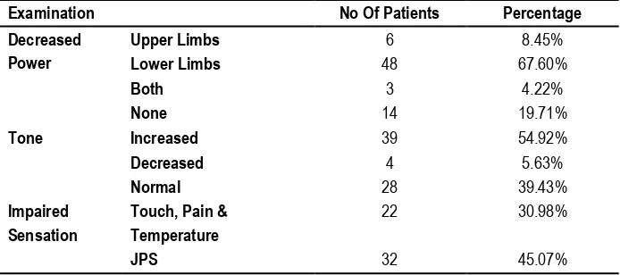
Out of the 71 patients in our study 67.60 % had weakness in lower limbs, 8.45 % had weakness in upper limbs, 4.22 % had weakness in both and 19.71 % patients had no weakness. 54.92 % of our patients presents with hypertonia, 5.63 % with hypotonia and 39.43% with normal tone. 30.98 % of patients presents with impaired touch, pain and temperature sensation and 45.07 % with impaired joint and position sensation.
In our study of “Benign spinal tumors”, most common location of tumor is in dorsal region (57.74%). Next common location is cervical region (16.90%), followed by lumbar region (14.08%). Some tumor extends in junction area of nearby regions like Dorsolumbar (8.45%), Cervicodorsal (2.81%), but no tumor found in Lumbosacral and sacral region.
In our study of benign spinal tumors‖, most common location of tumor is in intradural extramedullary (IDEM) and then itradural intramedullary (IDIM).Least common site for tumor is extradural extramedullary (EDEM).
In our study majority of the tumors are located posterolateral, (53.52%). Next most common location is anterolateral (14.08) and within the cord (14.08) followed by posterior (8.45%), anterior (5.63%) and others (4.22%).
In our study effect on spinal cord by Benign spinal tumor shows displacement of the cord in 23.94 % of patient, compression in 45.07 % of patients, enlargement of the cord in 14.08 % and no effect in 16.90 % of patients. In our series two patients has associated anomaly of spine, showing dorsal kyphoscoliosis. In our study complete tumor removal was achieved in 94.36 % of patients, while in 5.63% near total removal was done. Out of the total 71 patients, in 66 patients dura was closed primarily, while in 5 patients fascia lata graft was used for dural repair.
In our study 4.22% of patients develops CSF collection in wound which was treated with repeated aspirations and antibiotics, none of the patients required re-exploration. 1 patient develops local wound hematoma, which was treated conservatively. 1 patient develops pulmonary infection secondary to leak of neuroenteric cyst content into chest. In 2.81 % of the patients in our study there was transient deterioration of power which gradually improved over time and one of them having deterioration of power due to spine instability by lysthesis after 3month of post op.
cyst and neuroenteric cyst are 2.81% and dermoid, hemangioblastoma, lipoma, teratoma, meningeal cyst and simple cyst are 1.40%. In our series of 71 patients of “Benign spinal
tumors” 85.91 % of patients improved neurologically while 2.81 % patients deteriorated neurologically and 11.26 % of patients remain same as preoperative status of neurology.
Table 1: Age Distribution
S.N. Age Group (Yr) Total No. Patient Percentage %
1. 1-10 7 9.85
2. 11-20 10 14.08
3. 21-30 15 21.12
4. 31-40 11 15.49
5. 41-50 14 19.71
6. 51-60 6 8.45
7. 61-70 7 9.85
8. ≥ 71 1 1.40
Table 2: Sex Distribution
Sex Total No. Patient Percentage %
Male 35 49.29
Female 36 50.70
Table 3: Clinical Symptoms
Symptom No. of Patients Percentage (%)
Back pain 48 67.60
Radiating pain 26 36.61
Limb pain 7 9.85
Numbness 24 33.80
Tingling 23 32.39
Weakness 53 74.64
Walking difficulty 51 71.83
Back swelling 1 1.40
Spine deformity 2 2.81
Table 4: Bowel and Bladder Involvement
Symptom No. Of Patients Percentage (%)
Urinary Symptoms
Retention 8 11.26
Incontinence 3 4.22
Bowel Symptoms
Constipation 6 8.45
Incontinence 3 4.22
Table 5: Neurological Examination
Examination No Of Patients Percentage
Decreased Power
Upper Limbs 6 8.45%
Lower Limbs 48 67.60%
Both 3 4.22%
None 14 19.71%
Tone Increased 39 54.92%
Decreased 4 5.63%
Normal 28 39.43%
Impaired Sensation
Touch, Pain & Temperature
22 30.98%
Table 6: Radiological Location of Tumor (Vertical)
LOCATION Total Percentage (%)
Cervical 12 16.90%
Cervicodorsal 2 2.81%
Dorsal 41 57.74%
Dorsolumbar 6 8.45%
Lumbar 10 14.08%
Lumbosacral 0 0%
Sacral 0 0%
Total 71 100%
Table 7: Location of Tumor
LOCATION EDEM (%) IDEM (%) IDIM (%)
Cervical 1 1.40 9 12.67 2 2.81
Cervicodorsal 0 0 1 1.40 1 1.40
Dorsal 6 8.45 33 46.47 2 2.81
Dorsolumbar 0 0 4 5.63 2 2.81
Lumbar 0 0 7 9.85 3 4.22
Lumbosacral 0 0 0 0 0 0
Sacral 0 0 0 0 0 0
Total 7 9.85 54 76.05 10 14.08
Table 8: Radiological Location of Tumor (Horizontal)
Location Number of Patients Percentage (%)
Anterior 4 5.63
Anterolateral 10 14.08
Posterolateral 38 53.52
Posterior 6 8.45
Within The Cord 10 14.08
Others 3 4.22
Table 9: Effect on the Cord by Tumors
S.N. Feature Total No. Patient Percentage (%)
1 Compressed 32 45.07
2 Displaced 17 23.94
3 Enlarged 10 14.08
4 None 12 16.90
Table 10: Associated Anomaly of Spine
Patient Percentage
Total No. of Patients 71 100
Kyphoscoliosis 2 2.81%
None 69 97.18%
Table 11: Extent of Tumor Removal
Tumor Removal Total Percentage (%)
Complete 67 94.36%
Near total 4 5.63%
Total 71 100
Table 12: Dura Closure
CLOSURE TOTAL PERCENTAGE (%)
PRIMARY 66 92.95 %
Table 13: Postoperative Complication
Complication No. Of Patients Percentage
CSF Collection 3 4.22%
Wound Hematoma 1 1.40%
Pulmonary Infection 1 1.40%
Deterioration Of Power 2 2.81%
Spine Instability 1 1.40%
Table 14: Histopathological Incidence
S.N Diagnosis Patients %
1 Meningioma 24 33.80%
2 Nerve Sheath Tumor
Schwannoma 23 8
31 43.66%
Neurofibroma
3 Arachnoidcyst 2 2.81%
4 Neuroenteric/Enterogeneous Cyst 2 2..81%
5 Epidermoid 6 8.45%
6 Dermoid 1 1.40%
7 Hemangioblastoma 1 1.40%
8 Lipoma 1 1.40%
9 Teratoma 1 1.40%
10 Meningeal Cyst 1 1.40%
11 Simple Cyst 1 1.40%
Table 15: Follow Up For Weakness
Status Of Weakness No. Of Patients Percentage
Improved 61 85.91%
Deteriorated 2 2.81%
Same 8 11.26%
DISCUSSION
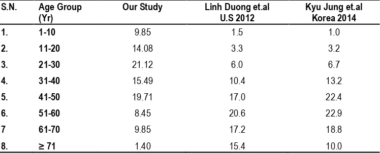
In this study, (Table 1) shows incidence of benign spinal tumor was maximum in age group of 21 to 30 year with 21.12 %, 41-50 year age with 19.71 % and 31-40 year age with 15.49 % while minimum in older age group more than 70 with 1.40%, 51-60 year age with 8.45% and 61-70 year age with 9.85%. Benign spinal tumor was also frequent in pediatric age group in first and second decade. We have compared our study age group with study of Linh Duong et.al4 and Kyu Jung et.al.5
Tumors of the central nervous system are common in the pediatric population and constitute the second most prevalent tumor type of childhood. Within this group, spinal cord tumors are a relatively rare diagnosis and account for 1% to 10% of all pediatric central
nervous system tumors.6-8Spinal cord tumors and masses are a
rare diagnosis in the pediatric population. The types of tumors seen in children tend to be different then the adult types. In evaluating the trends seen in this population, we had a large percentage of developmental tumors (dermoid, epidermoid, and teratomas) which differs from other, similar reports on tumors in this population.8-11 This does, however, counter a report by
Townsend et al in a small case series of 10 patients.12
Our study (Table 2) shows sex incidence of ―Benign spinal tumors‖ in male is 49.29% and in female is 50.70 % which was almost equal. . We have compared our study sex group with study of Linh Duong et al4 and Kyu Jung et al.5
S.N. Age Group (Yr)
Our Study Linh Duong et.al U.S 2012
Kyu Jung et.al Korea 2014
1. 1-10 9.85 1.5 1.0
2. 11-20 14.08 3.3 3.2
3. 21-30 21.12 6.0 6.7
4. 31-40 15.49 10.4 13.2
5. 41-50 19.71 17.0 22.4
6. 51-60 8.45 20.6 22.9
7 61-70 9.85 17.2 18.8
SEX OUR STUDY Linh Duong et.al U.S 2012
Kyu Jung et.al Korea 2014
MALE 49.29 38.1 42.2
FEMALE 50.70 61.9 57.8
The literature indicates that in western populations, the primary spinal tumors occur more frequently in females, whereas Asian studies show a slight male preponderance.13,14We had a male to
female ratio of 1:1 among our patients. Similar male to female ratio has been reported by other studies from India.15,16 Our study
(Table 3) shows In series of ― Benign spinal tumors‖, patients presented with weakness (74.64 %) as the most common complain followed by walking difficulty (71.83%), back pain (67.60%), tingling (32.39%), numbness (33.80%), radiating pain (36.61%), limb pain (9.85%), spine deformity (2.81%), back swelling (1.4%). We have compared our study clinical symptoms with study of Rajnish kumar et al.17Our study (Table 4) shows the most common bowel-bladder symptom was urinary retention which was present in 11.26 % of patients, followed by constipation in 8.45 % and urinary and fecal incontinence in 4.22%. We have compared our study bladder and bowel involvement with study of Rajnish kumar et.al17 North India 2015.
Our study (Table 5) shows Out of the 71 patients in this study 67.60 % had weakness in lower limbs, 8.45 % had weakness in upper limbs, 4.22 % had weakness in both and 19.71 % patients had no weakness. 54.92 % of our patients presents with hypertonia, 5.63 % with hypotonia and 39.43% with normal tone. 30.98 % of patients presents with impaired touch, pain and temperature sensation and 45.07 % with impaired joint and position sensation. We have compared our study clinical examination with study of Rajnish kumar et.al17 North India 2015. Our study (Table 6) shows most common location of tumor is in dorsal region (57.74%). Next common location is cervical region (16.90%), followed by lumbar region (14.08%). Some tumor extends in junction area of nearby regions like Dorso-lumbar (8.45%), Cervicodorsal (2.81%), but no tumor found in Lumbosacral and sacral region. We have compared our study of tumor location with study of Rajnish kumar et.al17 North India 2015.
SYMPTOM OUR STUDY Rajnish kumar et.al
North India 2015
Pain 67.60 41.44
Parasthesias 33.80 18
Motor weakness 74.64 70.27
Walking difficulty 71.83 70.27
Spine deformity 2.81 1.8
Symptom Our Study Rajnish kumar et.al
North India 2015
Bladder 15.49 34.23
Bowel 12.67 8.1
Examination Our Study Rajnish kumar et.al
North India 2015
Decreased Power 74.64 % 70.27 %
Tone Increased 54.92 % 70.27 %
Decreased 5.63 % 9.1 %
Impaired Sensation
45.07 % 49.54 %
LOCATION Our study Rajnish kumar et.al
North India 2015
Cervical 16.90% 18.91
Cervicodorsal 2.81% 9.1
Dorsal 57.74% 34.23
Dorsolumbar 8.45% 13.51
Lumbar 14.08% 10.81
Lumbosacral 0% 5.40
Our study (Table 7) shows most common location of tumor is in Intradural Extramedullary (IDEM) and then Intradural Intramedullary (IDIM). Least common site for tumor is Extradural Extramedullary (EDEM). We have compared our study of tumor location with study of Rajnish kumar et.al17 North India 2015 and Kenichi Hirano et.al18 Japan 2012. Our study results are almost same with japan study.
Primary spinal cord tumors account for 4–10% of all central nervous system tumors and are characterized based on their location as intramedullary (IDIM), IDEM, and extradural.19 Totally, 2/3 of all spinal tumors are said to be IDEM and 10% IDIM Spinal cord tumor (SCT)20 but we had nearly equal incidence of IDEM (76.05 %) and IDIM SCTs (14.08%) in our series. This difference
may be due to tertiary referral at our institute or this may represent the epidemiological trend of a developing country. Our study (Table 8) shows majority of the tumors are located posterolateral, (53.52%). Next most common location is anterolateral (14.08) and within the cord (14.08) followed by posterior (8.45%), anterior (5.63%) and others (4.22%). Our study (Table 9) shows effect on spinal cord by benign spinal tumor causes displacement of the cord in 23.94 % of patient, compression in 45.07 % of patients, enlargement of the cord in 14.08 % and no effect in 16.90 % of patients. Our study (Table 10) shows two patients has associated anomaly of spine, showing dorsal kyphoscoliosis (2.81%). We have compared our study with study of Rajnish kumar et.al17 North India 2015 in which it is about 1.8%.
Study EDEM (%) IDEM (%) IDIM (%)
Our Study 9.80 76.05 14.08
Rajnish kumar et.al North India 2015
27.02 36.03 36.93
Kenichi Hirano et.al Japan 2012
4.0 77.6 18.3
Our study (Table 11) shows complete tumor removal was achieved in 94.36 % of patients, while in 5.63% near total removal was done. A posterior approach using standard microsurgical techniques was performed in all cases, and this was irrespective of the location of a tumor. We did not require instrumentation in any case. We have compared our study of tumor location with study of Rajnish kumar et.al17 North India 2015.
Tumor Removal
Our Study Rajnish kumar et.al North India 2015
Complete 94.36% 51.35
Neartotal 5.63% 19.81
Subtotal 0% 27.02
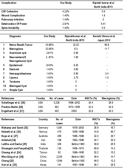
Our study (Table 12) shows Out of the total 71 patients, in 66 patients dura was closed primarily, while in 5 patients fascia lata graft was used for dural repair. Out of which two (4.22%) duroplasty was having CSF leak in which one is FLG and one is primary dural repair. Both of which was treated by conservative management like repeated aspiration, antibiotic and tab Diamox. In study of Bokhari et al21 2016 Karachi incidence of CSF leak was 2.36 %. Our study (Table 13) shows about 4.22% of patients develops CSF collection in wound which was treated with repeated aspirations and antibiotics, none of the patients required re-exploration. 1 patient develops local wound hematoma, which was treated conservatively. 1 patient develops pulmonary infection secondary to leak of neuroenteric cyst content into chest. In 2.81 % of the patients in our study there was transient deterioration of power which gradually improved over time and one of them having deterioration of power due to spine instability by lysthesis after 3month of Postoperative. We have compared our study Complication with study of Rajnish kumar et.al17 North India 2015 in given below table. Our study (Table 14) shows most common tumors are nerve sheath tumors, accounts for 43.66% of the total tumors. Next most common tumor is meningioma, which accounts for 33.80% of the tumors and epidermoid is 8.45%. Other least
common tumour are arachnoid cyst and neuroenteric cyst are 2.81% and dermoid, hemangioblastoma, lipoma, teratoma, meningeal cyst and simple cyst are 1.40%. We have compared our study of tumor Histopathology with study of Rajnishkumar et.al17 North India 2015 and Kenichi Hirano et.al18 Japan 2012. The incidence of primary spinal cord tumors in various series is listed in below Table. Compared with reports from other parts of the world, there are evident differences in the frequencies of nerve sheath cell tumors (NSCTs: schwannomas and neurofibromas) and meningiomas. In Asian countries,22-25including what we found
in the current study, the frequency of NSCTs is higher than that of meningiomas. This tendency is even stronger in eastern Asia. 22-24,26 On the other hand, the incidence of meningiomas in
non-Asian countries (USA, Europe, Australia), is equal to or higher than that of NSCTs27-31 with the exception of only one report.32In
the series from Germany,32 authors counted multiple tumors in one
patient (probably, neurofibromatosis or schwannomatosis). Therefore, NSCTs outnumbered meningiomas in their series. Neuroepithelial tumors seem to occur less frequently in Asian countries than in non-Asian countries. The frequency of vascular tumors seems to vary less among countries studied. Our results confirm previous reports22,24,26 which demonstrate different
frequencies of NSCTs, meningiomas, and neuroepithelial tumors between Asian countries and other parts of the world.
Meningiomas account for 25-46% of all primary intraspinal neoplasms and spinal meningiomas are only 7.5-12.5% of all meningiomas because most meningiomas are found in the brain.36
Spinal meningiomas are mostly located in the thoracic vertebra and they are more common in females, which is presumably due to the influence of female hormones.36 In this study, 33.8%
were performed without additional procedures because all the tumors were easily separated from the dura mater and no recurrence was observed at the last follow-up.
As mentioned above, there are dissimilarities in the relative frequencies of spinal cord tumors among various reports in above table. While some of these discrepancies are due to differences in the actual rate of occurrence of these tumors, others might result from different ways in which data are obtained. For various reasons, the comparison of data from different sources in various
communities might not reveal real differences among the communities. Moreover, the diversity in the types of tumors
included, and the classification used in different series further complicates the collation of statistics. However, despite dissimilarities in the materials and methods, comparing these series might reveal important points on the relative frequency of spinal cord tumors in different parts of the world, suggesting the possible roles of environmental, genetic, and hormonal factors in the etiology of spinal cord tumors. For surgeons, it is very useful to recognize what type of tumor is likely to be encountered, based not only on preoperative imaging characteristics such as magnetic resonance imaging (MRI), but also on the reported incidence of the various tumors in each country or region.
Complication Our Study Rajnish kumar et.al
North India 2015
CSF Collection 4.22% 0.9
Wound Complication 1.40% 1.8
Pulmonary Infection 1.40% 0
Deterioration Of Power 2.81% 5.41
Spine Instability 1.40% 0.9
Diagnosis Our Study Rajnishkumar et.al
North India 2015
Kenichi Hirano et.al Japan 2012
1 Nerve Sheath Tumor 43.66% 22.52 60.6
2 Meningioma 33.80% 6.3 11.7
3 Arachnoid cyst 2.81% 0.90 -
4 Neuroenteric/E Neurogeneous Cyst
2..81% 1.80 -
5 Epidermoid 8.45% 6.30 -
6 Dermoid 1.40% 3.60 -
7 Hemangioblastoma 1.40% 0.90 3.4
8 Lipoma 1.40% 2.70 1.5
9 Teratoma 1.40% 0 -
10 Meningeal Cyst 1.40% 0 -
11 Simple Cyst 1.40% 0 -
References Country No. of cases Date NSCTs (%) Meningioma (%)
Schellinger et al. [30] USA 3,226 1998–2002 24.4 28.9
Preston-Martin [28] USA 462 1972–1985 22.3 42.9
Engelhard et al. [31] USA 430 2000 22.6 24.2
References Country No. of
cases
Date NSCTs
(%)
Meningioma (%)
Klekamp and Samii [32] Germany 1,081 1978–2003 26.9 16.7
Helseth et al. [33] Norway 415 1955–1986 10.8 46.7
Kaye et al. [27] Australia 266 1986–1988 32.3 29.7
Ardehali [34] Iran 108 1962–1986 40.7 33.3
Lalitha and Dastur [25] India 326 Before 1980 39.9 25.5
Shuangsh and Panyatha[23] Thailand 120 1956–1973 65.8 14.2
Cheang et al. [24] Taiwan 92 1988–1995 52.2 15.2
Wen-Qing et al. [26] China 2,245 Before 1982 49.4 14.7
Cheng [22] China 1,549 Before 1982 49.5 13.2
There were potential limitations in the current study. Since this was a non-population-based study, and we dealt only with surgical cases, we could not determine the actual frequency of spinal cord tumors occurring within the population.
That is, we did not include asymptomatic cases or cases in which the spinal cord tumors were diagnosed with imaging and treated conservatively. We learned the relative frequencies of different spinal cord tumors which were treated surgically with this study. A population-based study on primary spinal cord tumors should be planned in India, similar to broad-based studies from other countries.
Our study (Table 15) shows 71 patients of ― Benign spinal tumors‖ 85.91 % of patients improved neurologically while 2.81 % patients deteriorated neurologically and 11.26 % of patients remain same as preoperative status of neurology. We defined "good outcome" as the improvement in patient's preoperative modified McCormick score, at the time of last follow-up. Those who had an improvement of ≥2 grades were labeled as having "significant improvement". The patients who either remained same or showed a deterioration of modified McCormick score were considered "poor outcome". We have compared our study outcome with study of Rajnish kumar et.al17 North India 2015.
OUTCOME OUR
STUDY
Rajnish kumar et.al North India 2015(17)
IMPROVED 85.91% 79.27
DETERIORATED 2.81% 5.41
SAME 11.26% 15.31
A delay in diagnosis or management of patients with mass lesions causing compression of neuronal tracts in the spinal cord may result in residual deficits and poor outcome.
Dutch investigators27collected a series of 108 patients with both
intradural and extradural tumors. They found that 35% of patients were diagnosed more than 2 years after the onset of symptoms. Delayed presentation is one of the main factors leading to poor neurological grade at time of surgery/intervention. This affects the postoperative outcome.
The average preoperative duration of symptoms in our series was 15.17 months, with a range from 15 days to 10 years and a significant number of our patients were in poor neurological status at presentation (71.83%).
The long-standing compressive pathologies cause profound and irreversible neuronal degeneration due to destructive changes such as ischemic necrosis and neuronal loss in gray matter as well as demylenation in white matter and in posterior and lateral white columns.28
The various series of spinal tumors had a good functional outcome in 15- 90% of patients.32-37 In our series, a total of 85.91% patients were mobile at last follow-up regardless of prognostic factors.
This compares favorably with another series. Similar observations also have been made by other authors. We recommend that surgical intervention if indicated should be considered for spinal tumors regardless of the prognostic factors. Even patients with poor neurological grade preoperatively may improve in their functional status.
SUMMARY
▪ Our study shows incidence of ― Benign spinal tumor‖ was maximum in age group of 21 to 30 year with 21.12 % , 41-50 year age with 19.71 % and 31-40 year age with 15.49 % while minimum in older age group more than 70 with 1.40 %, 51-60 year age with 8.45 % and 61-70 year age with 9.85 %. Benign spinal tumor was also frequent in pediatric age group in first and second decade. In our study male is 49.29% and in female is 50.70 % which was almost equal.
▪ Patients presented with weakness (74.64 %) as the most common complain followed by walking difficulty (71.83%). Most common bowel- bladder symptom was urinary retention which was present in 11.26 % of patients, followed by constipation in 8.45 % and urinary and fecal incontinence in 4.22%.Out of the 71 patients in this study 67.60 % had weakness in lower limbs, 8.45 % had weakness in upper limbs, 4.22 % had weakness in both and 19.71 % patients had no weakness. 54.92 % of our patients presents with hypertonia. 30.98 % of patients presents with impaired touch, pain and temperature sensation and 45.07 % with impaired joint and position sensation.
▪ Our study shows most common location of tumor is in dorsal region (57.74%). Next common location is cervical region (16.90%), followed by lumbar region (14.08%).
▪ Our study shows most common location of tumor is in Intradural Extramedullary (IDEM) and then Intradural Intramedullary (IDIM). Least common site for tumor is Extradural Extramedullary (EDEM) with majority of the tumors are located posterolateral, (53.52%). Benign spinal tumor causes displacement of the cord in 23.94 % of patient, compression in 45.07 % of patients, enlargement of the cord in 14.08 % and no effect in 16.90 % of patients.
▪ Our study shows complete tumor removal was achieved in 94.36 % of patients, while in 5.63% near total removal was done. Out of the total 71 patients, in 66 patients dura was closed primarily, while in 5 patients fascia lata graft was used for dural repair. Out of which two (4.22%) duroplasty was having CSF leak in which one is FLG and one is primary dural repair. Both of which was treated by conservative management like repeated aspiration, antibiotic and tab Diamox. Other complications are local wound hematoma, pulmonary infection secondary to leak of neuroenteric cyst content into chest. In 2.81 % of the patients in our study there was transient deterioration of power which gradually improved over time and one of them having deterioration of power due to spine instability by lysthesis after 03 months of postoperative.
▪ Our study shows most common tumors are nerve sheath tumors, accounts for 43.66% of the total tumors. Next most common tumor is meningioma, which accounts for 33.80% of the tumors and epidermoid is 8.45%.Other least common tumour are arachnoid cyst and neuroenteric cyst are 2.81% and dermoid, hemangioblastoma, lipoma, teratoma, meningeal cyst and simple cyst are 1.40%.
CONCLUSION
This study of ― Benign spinal cord tumors‖ describing the demographic characteristics, histopathological features, anatomical location, and vertebral level of these tumors treated surgically. Similar to other reports from Asian countries, there is an equal male/female ratio for all benign spinal cord tumors in this study. There is also a higher proportion of NSCTs, and a lower proportion of meningiomas and neuroepithelial tumors as compared to NSCTs. Data in the current study represent the characteristics of primary spinal cord tumors in Asian countries. Tumour type and biology plays an important role, with benign IDEM tumours displaying the best prognosis for long term survival after complete excision. The pretreatment neurological status of the patient is important in determining outcome after treatment. Delayed presentation is the main reason for poor preoperative neurologic status, leading to poor outcome in the management of spinal tumors. A high level of suspicion and acknowledging the classical symptoms of cord compression are the most important factors in shortening the time to diagnosis of spinal tumors.
REFERENCES
1. Spurling RG, Mayfield FH. Neoplasms of the spinal cord-A review of forty-two surgical cases. JAMA.1936;107:924–9.
2. Ramamurthi R, Rao SM. Clinical features and diagnosis. In: Ramamurthy B, Tandon PN, editors. Text Book of Neurosurgery. 3rd ed. New Delhi: Jaypee Medical Publishers; 2012. p. 1181.
3. Williams R, Foote M, Deverall H. Surgical treatment of 264 primary spinal tumors. Global Spine J.2012;2:249-66.
4. Linh Duong et.al, Descriptive Epidemiology of Malignant and Nonmalignant Primary Spinal Cord, Spinal Meninges, and Cauda Equina Tumors, United States, 2004–2007.
5. Kyu Jung et al, Incidence of Primary Spinal Cord, Spinal Meninges, and Cauda Equina Tumors in Korea, 2006-2010.
6. Hardison HH, Packer RJ et al. Outcome of children with primary intramedullary spinal cord tumors. Childs Nerv Syst. 1987;3:89–92. 7. Stiller CA, Nectoux J. International incidence of childhood brain and spinal tumours. Int J Epidemiol.1994;23:458–464.
8. Nadkarni TD, Rekate HL. Pediatric intramedullary spinal cord tumors. Critical review of the literature.ChildsNervSyst.1999;15:17 28. 9. Constantini S, Epstein F. Pediatric intraspinal tumors. In: Choux M, Di Rocco E, Hockley A, Walker M, editors. Pediatric Neurosurgery. London, UK: Churchill Livingstone; 1999. pp. 601–602.
10. Greenwood J., Jr. Surgical removal of intramedullary tumors. J Neurosurg. 1967;26:276–282.
11. Malis LI. Intramedullary spinal cord tumors. Clincal Neurosurg. 1978;25:512–539.
12. Townsend N, Handler M et al. Intramedullary spinal cord astrocytomas in children.Pediatr Blood Cancer. 2004;43:629–632 13. Hirano K, Imagama S, Sato K, Kato F, Yukawa Y, Yoshihara H, et al. Primary spinal cord tumors: Review of 678 surgically treated patients in Japan. A multicenter study. Eur Spine J. 2012;21:2019–26. 14. Wen-qing H, Shi-ju Z et al. Statistical analysis of central nervous system tumors in China. J Neurosurg. 1982;56:555–64.
15. Bansal S, Ailawadhi P et al. Ten years’ experience in the management of spinal intramedullary tumors in a single institution. J Clin Neurosci. 2013;20:292–8.
16. Chandy MJ, Babu S. Management of intramedullary spinal cord tumours: Review of 68 patients. Neurol India. 1999;47:224–8. 17. Rajnish Kumar Arora et.al. Spinal tumors: Trends from Northern India. Asian Journal of Neurosurgery. Oct-Dec 2015; 10(4)291. 18. Kenichi Hirano et al. Primary spinal cord tumors: review of 678 surgically treated patients in Japan. A multicenter study Eur Spine J.
2012 Oct; 21(10): 2019–2026.
19. Segal D, Lidar Z, Corn A, Constantini S. Delay in diagnosis of primary intradural spinal cord tumors.Surg Neurol Int. 2012;3:52. 20. Chamberlain MC, Tredway TL. Adult primary intradural spinal cord tumors: A review. Curr Neurol Neurosci Rep. 2011;11:320–8. 21. Bokhari et al 2016 karachi, Journal of the College of Physicians and Surgeons Pakistan 2016, Place and Duration of Study: Neurosurgery Department, JPMC, Karachi, from March 2011 to February 2014.
22. Cheng MK. Spinal cord tumors in the People’s Republic of China: a statistical review. Neurosurgery.1982;10:22–24.
23. Shuangsh S, Panyatha R. Neural neoplasms in Thailand—study of 2,897 cases. Neurology.1974;24:1127–1134.
24. Cheang CM, Hwang SL, Hwong SL. An analysis of intraspinal tumors in south Taiwan. Kaohsiung J Med Sci. 1997;13:229–236. 25. Lalitha VS, Dastur DK. Neoplasms of the central nervous system— histological types in 2237 cases. Indian J Cancer. 1980;17:102–106.
26. Wen-qing H, Shi-ju Z et al. Statistical analysis of central nervous system tumors in China. J Neurosurg. 1982;56:555–564.
27. Kaye AH, Giles GG, Gonzales M. Primary central nervous system tumours in Australia: a profile of clinical practice from the Australian Brain Tumour Register. Aust N Z J Surg. 1993;63:33–38.
28. Preston-Martin S. Descriptive epidemiology of primary tumors of the spinal cord and spinal meninges in Los Angeles County, 1972-1985. Neuroepidemiology. 1990;9:106–111.
29. Helseth A, Mork SJ. Primary intraspinal neoplasms in Norway, 1955 to 1986. A population-based survey of 467 patients. J Neurosurg. 1989;71:842–845.
30. Schellinger KA et al. Descriptive epidemiology of primary spinal cord tumors. J Neurooncol. 2008;87:173– 179.
31. Engelhard HH et al. Clinical presentation, histology, and treatment in 430 patients with primary tumors of the spinal cord, spinal meninges, or cauda equina. J Neurosurg Spine. 2010;13:67–77. 32. Klekamp J, Samii M. Surgery of spinal tumors. Heidelberg: Springer; 2006.
33. Helseth A, Mork SJ, Johansen A, Tretli S. Neoplasms of the central nervous system in Norway. IV. A population-based epidemiological study of meningiomas. APMIS. 1989;97:646–654. 34. Ardehali MR. Relative incidence of spinal canal tumors. Clin Neurol Neurosurg. 1990;92:237–243.
35. Suh YL, Koo H, Kim TS et al. Tumors of the central nervous system in Korea—a multicenter study of 3221 cases. J Neurooncol. 2002; 56: 251–259.
36. Solero CL, Fornari M, Giombini S, et al. Spinal meningiomas: review of 174 operated cases.Neurosurgery. 1989;25(2):153–160. 37. Saito T et al. A novel technique for surgical resection of spinal meningioma. Spine. 2001;26(16):1805– 1808.
[
Source of Support: Nil. Conflict of Interest: None Declared.
Copyright: © the author(s) and publisher. IJMRP is an official publication ofIbn Sina Academy of Medieval Medicine & Sciences, registered in 2001 under Indian Trusts Act, 1882. This is an open access article distributed under the terms of the Creative Commons Attribution Non-commercial License, which permits unrestricted non-commercial use, distribution, and reproduction in any medium, provided the original work is properly cited.