CODEN (USA)-IJPRUR,e-ISSN: 2348-6465
Original Article
Comparative Account of Microbial Load Assessment in a
University Cafeteria
Lovely Gupta, Mohini Agarwal, Kumud Bala*
Amity Institute of Biotechnology, AUUP, Noida, India.
A R T I C L E I N F O A B S T R A C T
_______________________________________________________________________________
1. INTRODUCTION
Proper supply of safe, complete and healthy food is
essential for the health and well - being of the humans
(Adak et al., 2005).2Consumption of contaminated or
unsafe foods may result in illnessand can lead to food
borne diseases (WHO,2000; Bryan, 1997). Food
hygiene is essentially aimed at producing food which is
International Journal of Pharma Research and Health Sciences
Available online atwww.pharmahealthsciences.net
Received: 05 Oct 2015 Accepted: 30 Oct 2015
The physical environment of a cafeteria may influence some factors such as hygiene and food preparation, and both factors can play a crucial role in the transmission of infectious disease among students, hostellers and staff using the cafeteria. Food contact surfaces are a major concern for food service facilities in the cafeteria for controlling the spread of various kinds of pathogenic infections.
The Microbiological analysis of main cafeteria of a University, in Noida was taken for the study. Random sampling was done taking sample from food storage area, food preparation area, washing area, serving area and eating area of the cafeteria. Samples were later, assessed using standard microbiological methods.
Various genera of bacteria and fungus were isolated and identified, they were mainly Gram positive, Gram negative bacteria such as Staphylococcus aureus, E. coli,Streptococcus along with Aspergillus, Penicilium and yeast colonies were also observed. These are some of the infectious agents which cause diarrhoea, typhoid, abdominal pain, microbial food poisoning etc. for which no clinical or laboratory findings are provided.
In general, it has been observed that the level of personal hygiene of the food handlers in food establishment was unsatisfactory due to poor sanitation and wrong practices. This can be enhanced by regular cleaning & monitoring of the cafeteria by the staff, process owner/authorities as per food safety practices. Regular training/workshops on personal hygiene will increase the awareness and good practices in cafeteria.
Keywords: personal hygiene, bacterial load, cafeteria microbial load assessment, food handler.
Corresponding author *
Dr Kumud Bala
3rdFloor, Room No-312, Amity Institute of Biotechnology, Sector-125, Noida, Uttar Pradesh, India.
safe for human consumption and is required for good
health (Scheule, 2001).25
Biological contaminants such as bacteria, viruses,
fungi, protozoa and helminthes constitute the major
cause of concern ranging from mild infection to life
threatening illness or both. Diseases such as cholera,
campylobacterosisE. coli gastroenteris, salmonelosis,
shigellosis, typhoid fever, brucellosis, amoebiasis are
being reported due to unhygienic food preparation and
storage conditions in most of the developing countries.
(Edema, 2005).11
Lacking awareness and personal hygiene amongst food
handlers is one of the most commonly reported
practices contributing to food – borne illness. Dirty
hand of the workers and work surface hygiene also
adds to the problem (WHO,2000; Bryan, 1997). The
risk of food borne illness due to contact with hands or
surface depends on level of contamination and
transmission of the disease causing vector during food
preparation and storage till its consumption.
The presence and absence of pathogenic
microorganisms in food materials, food preparation
surfaces, equipments and utensils has led to a high
degree of chronic illness (Egonu and Alan, 2000).Food
safety need to be ensured during preparation,
production, processing, storage, distribution and
preparation of food to minimize the contamination and
to maintain its safety for human consumption (Edemaet
al.,2005).11 Effective cleaning is of prime importance
since it does not only remove gross contamination but
also residues that could support the subsequent survival
and growth of microorganisms (Bean et al.,1990).3
Few reasons which predominates the contamination of
the food and outbreak of food borne diseases are
subsequently identified as unsafe sources,
contaminated raw food items, improper food storage,
and poor personal hygiene during food preparation,
inadequate cooling and reheating of food items and a
prolonged time lapse between preparation and
consumption of food items (Linda du and Irma, 2005).
In large scale cooking, specially, in cafeteria,
restaurants, hotels and dhabas, food passes through
many hands, thereby increasing the chances of food
contamination due to improper handling which might
endanger the health of consumers (Omaye,2004).21
In the year 1998, Zhao et al., 28 reported that
contamination from food handlers usually results due
to inadequately washed hands, improper food
preparation techniques, incorrect cleaning procedures
of food preparation surfaces, chopping boards and
tables. Bacteria have been reported to survive on
chopping-boards for more than three hours, especially
when boards are not properly cleaned (Zhao et al.,
1998; Salo et al., 2000)24, 28
In addition, Salo and colleagues (2000) reported that
wet items such as dishcloths, hand towels, Apron, and
sponges, as well as sink drain areas with leaking pipes,
uncovered drains, garbage, leftovers might also serve
as continuous reservoirs that harbor potentially harmful
microorganisms, which may end up settling on kitchen
surfaces (Zhao et al., 1998; Salo et al., 2000). 24, 28
Improper food hygiene practices and unclean surfaces
have been associated with opportunistic pathogenic
microorganisms such as Staphylococcus aureus
(Andargie et al., 2008; Garcia, 2007).2, 14 Improperly
cleaned surfaces along with deficient food handling
practices have led to an increase in microbiological
hazards in food preparation areas (Nkhebenyane,
2010).20
2. MATERIAL AND METHODS
2.1. Study Area
As per the guideline of FSME (Food Safety
Management System) of the University, study was
carried out in theUniversity cafeteria in Noida, Uttar
Pradesh. Various samples were collected from
of the study was during the month of March 2014 till
March 2015
2.2. Sample Collection
Random Sampling was done. Sterilized swab sticks
were used to collect the samples. The samples were
taken from the hands of food handlers, tables, apron,
utensils (storage, cooking), washed utensils (includes
spoons, plates, trays, bowls), juicer, fridge, food
storage area, food making area, washing area, serving
area and eating area. Swab sticks were dipped in 1ml
of double distilled water tubes and brought to the
laboratory for further examination.
2.3 Sample Processing
50 µL of sample were plated onto Luria Bertuni agar
(LA) for bacterial count and on Potato Dextrose agar
(PDA) for fungal count using spread plate method. LA
and PDA plates were incubated at 37°C and 25°C for
24 - 72hrs respectively.
2.4 Identification of Microbial Isolates
After incubation period, the bacterial and fungal
colonies were counted; the morphological
characteristics were observed. Later the gram staining
procedure was performed for the identification of the
gram positive and gram negative strains of
Bacteria.Fungal/ Bacterial/Yeast isolates were also
observed under the microscope. Biochemical test were
performedsuch as oxidase, catalase, coagulase, indole,
urease, citrate, sugar utilization as described by Speck
(1986) 26 and Cheesebrough (2004) to identify the
bacterial species.
3. RESULT
The microbial quality assessment of hands of food
handlers, tables, apron, utensils (includes storage,
cooking), washed utensils (includes spoons, plates,
trays, bowls), juicer, fridge, food storage area, food
making area, washing area, serving area and eating
area were examined using standard method. The total
bacterial and fungal counts were done on Luria Bertuni
agar (LA) and Potato Dextrose Agar (PDA)
respectively. Occurrence of microbial isolates of
specimens obtained from students’ cafeteria in the
University is presented in Table 1& 2. The results
reveal that, on PDA plates the average bacterial
colonies present were 3.78×10³ CFU/ml,mainly in
fridge, wash basin and spoon. The highest bacterial
colonies were ranging from 10 -25×103
CFU/mlincluding chopping vegetables, worker’s hands
and juicer. Whereas the least bacterial colonies were
ranging from 0.2 -7.2×10³ CFU/ml including tray,
plates and eating table.21 fungal colonies including
Aspergillus andPenicilliumwere present in worker’s
hand, worker’s clothes, salad chopper, cooked food
storing area, eating table, etc. and one sample (fridge)
showed pink coloured yeast colonies. On LA plates,
plates the average bacterial colonies present were
2.44×10⁵CFU/ml, mainly in salad chopper, worker’s
hand, fridge, juicer, wash basin, spoon and eating
tables. The highest bacterial colonies were 1×10⁸
CFU/ml including roti area, chopping vegetables,
cooked food storing area, washed cooker, storage
containers and utensils. Whereas the least bacterial
colonies were ranging from 2×10²-9.4×10³ CFU/ml
including worker’s clothes, washed utensils, tray,
serving area and billing counter.Two samples of
washed utensils also showed pink colored yeast
colonies. Preliminary analysis of microbes was done
with the help of biochemical test(Table3)
4. DISCUSSION
In the present study, nine genera of bacteria were
isolated and identified. They were identified as E.coli,
Clostridium sp, Micrococcus sp, Staphylococcus sp,
Streptococcus sp, Pseudomonas sp, Shigellasp,
Bacillus sp, Salmonella sp by comparing their
morphology and biochemical characteristic(Table 3)
The presence of organism such as E. coli, Salmonella
sp, Clostidiumsp and other organisms in this study is of
special concern and perhaps the greatest danger
associated with the water for food processing and
drinking purpose (Lynch et al., 2003). 18 Qualitative
hand swab results showed that a high fraction of the
personnel’s hand were contaminated by E. coli,
shigellasp, micrococcus sp even though the source of
those contaminants was not determined they are highly
indicative of inadequate hand sanitation (Collins et al.,
1989; Brown et al., 2000). 5, 9 However the workers
serving food were not found contaminated as they were
wearing gloves. Large number of the Staphylococcus
sp and Streptococcus sp were isolated although they
are normal commensal on human which reflect
improper hygiene practice such as pocking nose with
fingers (Collins 2001). It was observed that there was
no hand sanitizer/soap available for the workers to
clean their hand after using the toilet or handling
food/raw foods. It has also been observed that the
common practice after washing is to dry their hands in
their apron, garment which could probably serve as
source of further contamination, which has been
reported by Moyo and Baudi (2004). 19 From these
assessments, the food handlers personal hygiene
standard and food handling practices were
unsatisfactory, the tables and plates used for eating
could also be a source of spread of food borne diseases
unless corrective sanitary measures are put in place.
In this study, the presence of Staphylococcus sp, E.
coli, Pseudomonas sp, Bacillus sp in the plates shows
the existing poor sanitary qualities of food utensils,
ineffective washing techniques, improper handling and
storage of clean utensil. Repeated usage of water for
cleaning utensils and their hand increases the severity
of the infection. Clothes and mops used for wiping and
drying plates and tables are also improperly cleaned.
The presence of Bacillus, fungi such as Aspergillus,
Penicilliumspp in the foods could be due to the fact
that they are spore formers. These heat-resistant spores
may have survived processing while vegetative cells
were eliminated. Contamination of foods could have
resulted from inappropriate processing, incomplete
heating, or secondary contamination via contact with
contaminated hand, equipments and utensils (Oranusi
et al., 2013).22
Yeasts, including Candida
albicans, Rhodotorula rubra, Torulopsis andTrichospor
oncutaneum, have been found living in between
people's toes as part of their skin flora (Oyeka and
Ugwu 2002). Yeasts are found to be present in the gut
flora of mammals and some insects such as flies,
cockroaches, thus the worker’s hand and bad hygienic
condition of the cafeteria can lead to food
contamination. Yeasts are able to grow in foods with a
low pH (5.0 or lower) and in the presence of sugars,
organic acids, and other easily metabolized carbon
sources which results in food spoilage as reported by
Kurtzman (2006).15
Fig. 1a and 1b: Media plates and microscopic image are showing
Aspergillus colonies isolated from worker’s clothes (chopping area).
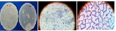
Fig. 2a and 2b: Plates are showing bacterial and yeast colonies isolated from fridge; microscopic view of isolated yeast.
Fig. 4a, 4b and 4c-LA plate is showing gram positive bacterial colonies and PDA plate,Aspergillus colony with their microscopic view isolated from clean eating table
Fig. 5a, 5b and 5c- plates are showing gram positive bacterial and yeast colonies with its microscopic view isolated from washed bowl
Fig. 6a, 6b and 6c- Media plates are showing gram negative bacterial and yeast colonies with its microscopic view isolated from washed utensils.
Fig. 7a, 7b and 7c- LA plate is showing gram negative bacterial colonies and PDA plate penicillium colony with their microscopic view isolated from papad tray.
Food handlers with skin lesion, respiratory infection,
eyes and nose discharge could have served as the
source of Staphylococcus aureus on the plate. As
Staphylococcus aureus lives and flourishes in the
human nose, eyes, skin and throat, the likely hood of
recontamination of cleaned plate by infected food
handlers is quite high. This has also been observed that
the workers are not aware of these infectious agents
and their harmful effects. Many researchers all over the
world have reported bacterial and fungal infection of
the food in restaurants, hotels and cafeteria. However,
no concrete plan of action has been found in place to
handle such contamination at public place. There is an
urgent need to maintain the workers’ health chart along
with the immunization detail by the cafeteria owners so
that the infective people could not be employed at such
places.
Globally some of the reported research such as Collins
and Brown in the year 2000 has shown that bacterial
count of a food reflect the hygienic and unhygienic
condition of the food outlets and the food handlers.
Bryan in 19977has emphasized on the importance of
training and awareness about the infectious diseases
and its mode of action in the human so that each
worker feels the responsibility of minimizing it at the
worksite.Study of Abdullahi et al., 1in the year 2004
has reported that the most of the harmful disease
causing bacteria are found in the hand of worker such
as Staphylococcus, E. coli, Pseudomonas ,
Klebsiellaetcso, proper awareness will definitely help
in the minimization of the these agents.
As we see that the contamination is within the premises
of food preparation and processing so precautionary
measures such as hand sanitizers, hand dryer, clean
garments, aprons will help to further minimize the
contamination. Moyo and Baudi, in the year 2004 has
also emphasized on the clean working environment.
Feglo and Sakyi, 201213has reported that most
ready-to-eat foods in Kumasi (Ghana) were contaminated
with enteric bacteria and other potential food poisoning
organisms with bacterial counts higher than the
acceptable levels. Utensils and equipment used in food
preparation and processing need to be properly cleaned
to stop the cross contamination. Disinfecting
equipment, hands, surface, and utensils have been
advocated by Linda et al., in the year 2005. Storage
area is more prone to spore producing bacilli and lactic
acid bacterium as mentioned by Lucyna et al., 2013.17
5. CONCLUSION
Conclusively, it should be noted that the working
surface or any surface which comes under direct
contact with food shall not contain more than 100
total microbial count of the hand should be considered
as negligible. Process owners has to take the
responsibility of providing personal hygiene/sanitation
training to the staff and should also bear the moral
responsibility of developing tactics to motivate food
handlers to practice food hygiene and implement a
regular screening of food outlet for microbial load.
0 50 100 150 N o. of C ol oni es Axis Title Total Microbes Bacteria Fungus Yeast
GraphA 1: Comparative analysis of microbes in highly contaminated samples after 24 hrs, 48hrs of incubation on LA plates and 72hrs of incubation on PDA plates.
0 200 N o. o f C ol on ie s Bacteria Bacteria
Graph A 2: Comparative analysis of Bacterial colonies in highly contaminated samples after 24hrs of incubation on LA plates.
0 10 N o. o f C ol on ie s Fungus Fungus
Graph A 3: Comparative analysis colonies of Fungus in highly contaminated samples after 72hrs of incubation on PDA plates.
0 5 Sal ad … W or ke r's … W or ke r's … Co ok ed … Fr id ge 2 Pap ad tr ay Bi lli ng … D irt y… Cl ean … Co ok ed … Ju ic er w as h bas in sp oo n W as he d… W as he d… Ch op pi ng … N o. o f C ol on ie s Yeast Yeast
Graph A4: Comparative analysis of Yeast colony in highly contaminated samples after 48hrs of incubation on PDA Plates
6. ACKNOWLEDGEMENT
Authors acknowledge Amity University for providing
necessary facilities and infrastructure to perform the
tests.
Table 1: Comparative account of Fungal load from the sample collected area on PDA plate.
S. no. Sample Area
Sample size
No. of colonies grown Physical characteristics Lactophenol cotton blue stain Bacterial count (Cfu/ml) Fungal count
1 Roti area 50µl 3.6×10³ 0 2 Noodles
area
50µl 0.2×10³ 0
3 Chopping vegetables
2*
50µl 15×103 1 White color with black
spores.
Aspergillus*
4 Washed cooker
50µl 0.2×10³ 0
5 Salad
chopper*
50µl 10×103
2 White color with black
spores.
Aspergillus*
6 Worker’s
hand*
50µl 20×103 5 White color with black spores. Aspergillus* (chopping) 7 Worker’s clothes*
50µl 0 8 White color
with black spores.
Aspergillus*
(chopping)
8 Rice area* 50µl 2.2×10³ 1 White color with black
spores.
Aspergillus*
9 Fridge 1 50µl 6.6×10³ 0
10 Fridge 2* 50µl 7.2×10³ 0 Pink color Yeast* 11 Juicer 50µl 25×103
0 12 Wash basin 50µl 3.8×10³
0
13 Spoon 50µl 2.6×10³ 0 (washing
area)
14 Papad tray* 50µl 0.2×10³ 1 Green colored colonies
Penicillin*
15 Billing counter*
50µl 0.6×10³ 1 Aspergillus*
16 Dirty eating table*
50µl 0.2×10³
1 Aspergillus*
17 Clean eating table*
50µl 1.8×10³ 1 Aspergillus*
18 Washed plates (serve
to students)
50µl 0.2×10³ 0
19 Front serving area
50µl 0.2×10³ 0
20 Apron 50µl 0.2×10³ 0
21 Roti
container
50µl 0.4×10³ 0
22 Cooked food storing
area*
50µl 1×10³
1 White colored colonies with black spores.
Aspergillus*
23 Jalebi tray 50µl 3×10³ 0
24 Washed bowl
50µl 14×103 0
25 Washed utensils (in
washing area)
50µl 0.4×10³ 0
26 Washing slab containing
utensils
50µl 6×10³ 0
Table 2: Comparative account of Bacterial load from the sample collected area on LA plates.
S. no. Sample Area Sample size No. of colonies grown Physical characteristics Gram stain Bacterial count (Cfu/ml)
1 Roti area 50µl 1×10⁸ Cream coloured lawn
G+ve
2 Roti area 2 50µl 1×10⁸
White isolated colonies
G-ve
3 Noodles area 50µl 1×10⁸ Cream coloured lawn
G+ve
4 Chopping
vegetables 1
50µl 1×10⁸
Cream lawn with some isolated
colonies
G-ve
5 Chopping
vegetables 2
50µl 1×10⁸ Cream coloured lawn
G+ve
6 Storage utensil container
50µl 1×10⁸
Cream coloured lawn
G+ve
7 Large storage container
50µl 1×10⁸ Bacterial lawn G-ve
8 Washed
cooker
50µl 1×10⁸ 2 Bacterial lawns G+ve
9 Storage area utensils
50µl 1×10⁸
Cream coloured lawn
G+ve
10 Salad
chopper*
50µl 2.2×10⁴ Many Cream coloured colonies
G+ve
11 Worker’s hand
(chopping)
50µl 1×10⁶
2 types of Bacterial lawns
G+ve
12 Worker’s
clothes (chopping)*
50µl 2.4×10³
Yellowish colony
13 Rice area 50µl 1.6×10³ White coloured colonies
G+ve
14 Fridge 1 50µl 7×10⁴
White coloured colonies
G+ve
15 Fridge 2 50µl 4×10²
White coloured colonies
G-ve,G+ve
16 Juicer* 50µl 1×10⁶ Bacterial lawn
with isolated colonies
G+ve
17 Wash basin* 50µl 4×10⁴ Brown and cream coloured colonies
G+ve
18 Spoon 50µl 1.4×10⁴
Brown and cream colonies
G+ve
(washing area)*
19 Papad tray* 50µl 4.8×10³ White and cream colonies
G-ve
20 Billing counter*
50µl 2×10²
White coloured mycelium 21 Dirty eating
table*
50µl 1.1×10⁴ Brown and cream coloured colonies
G+ve
22 Clean eating table*
50µl 1.4×10⁴ Brown and cream coloured colonies
G+ve
23 Washed plates (serve to students)
50µl 6×10²
White coloured colonies
G+ve
24 Front serving area
50µl 4.4×10³
Cream coloured colonies
G+ve
25 Apron 50µl 1.4×10³ Cream coloured
colonies
G+ve, G-ve
26 Roti container 50µl 8×10²
White coloured colony
G-ve, G+ve
27 Cooked food storing area
50µl 1×10⁸ Cream coloured lawn
G+ve, G-ve
28 Jalebi tray 50µl 1×10⁸ Cream coloured lawn
G-ve, G+ve
29 Washed bowl* 50µl 2×10³ White coloured isolated colonies
G+ve, G-ve yeast*
30 Washed
utensils (in washing
area)*
50µl 9.4×10³
White and brown coloured isolated
colonies
G-ve, yeast*, G+ve
31 Washing slab containing
utensils
50µl 1×10³ White colonies G-ve, G+ve
Note:* shows higher concentration of Bactrial contamination in the specified area. Mean Bacterial count= 2.44×10⁵and Standard deviation= 2.32×10⁵
Table 3: Biochemical Identification of bacterial colonies
S.No. Suspected G ra m Sta ini n g O x id a se C a ta la se C o a g ul a se C itr a te U re a se Indo le G luc o se La cto se Suc ro se M a nni to l M o ti li ty microorganism
1 E.Coli - - - + + + + + +
2 Salmonella sp. - - - - + - - + - d + +
3 Shigella sp. - - - + + - d
-4 Clostridium sp. + - - - + + + +
5 Pseudomonas
sp.
- + + - + - - + - - + +
6 Streptococcus
sp.
+ - - - + d d +
-7 Staphylococcus
sp.
+ - + + - - - + - - +
-8 Bacillus sp. + + - - - d + +
9 Micrococcus sp. + - + - + + - - -
-*d-differential result
7. REFERENCES
1. Abdullahi, I.O., Umoh, V.J., Ameh, J.B.,
&Galadima, M. Hazards Associated with Killishi
Preparation in Zaria, Nigeria. Nigerian Journal of
Microbiology 2004; 18(1-2): 339-345.
2. Adak, G.K., Meakins, S.M., Yip, H., Lopman,
B.A., &O'Brien, S.J. Disease risks from foods,
England and Wales, 1996– 2000. Emerging
Infectious Diseases 2005; 11(3): 365–372.
3. Andargie, G., Kassu, A., Moges, F., Tiruneh,
M.,&Huruy, K. Prevalence of bacteria and
intestinal parasites among food-handlers in Gondar
town, northwest Ethiopia.
Journal of Health, Population and Nutrition 2008;
26: 451-455.
4. Bean, N., Griffin, M., Guilding, J.S.,& Ivey C.
Food Borne Diseases Outbreak – Year Summary
1983. Journal of Food Protection 1990; 53-711.
5. Brown, A. Understanding Food; Principles and
Preparation. Belmont, C. A. Wadsworth –
Thompson Learning. 2000.
6. Brown, M.H., Gill, C.O., Hollingsworth, J. N.,
the safety of beef. International Journal of Food
Microbiology 2000; 62:7–16.
7. Bryan, F.L. Risk of Practices, Procedures and
Processes that Lead to Outbreaks of Food -
Borne-illness. Journal Food Protection 1997; 51(8): 663–
673.
8. Chesbrough, M. District Laboratory Practice in
Tropical Countries, Cambridge University press,
United Kindom. 435.2004.
9. Collins, C.H., Lynes, P.M., &Grange, J.M.
Microbial methods, Butter worth and Co
Publishers Ltd, 6th edition.1989.
10. Collins, J.E. Impact of changing consumer
Lifestyles on the emergence of food borne
pathogens. Emerging Infectious Diseases 2001;
3(4):1- 13.
11. Edema, M., Atayese, A.O., &Idowu. Microbial
Quality of Food Wave Processed Foods in Book of
Abstract of the 29th Annual Conferences and
General Meeting on Microbes as Agents of
Sustainable Development, Organized by Nigerian
Society For Food Microbiology (NSM), UNAAB,
Abeokuta from 6–10th November. 2005.
12. Enogu, E.C., &Allan, M.C. The Temperature
Related Effect on the Repressive Activities of
Bacillus cereus in Associative Growth in Custard
Medium. Nigerian Food Journal 2000;14(1): 6368.
13. Feglo, P. &Sakyi, K.Bacterial contamination of
street vending food in Kumasi, Ghana.Journal of
Medical and Biomedical Sciences 2012; 1(1): 1-8.
14. Garcia, L.S.Diagnostic Medical Parasitology, (5th
ed), ASM Press, Washington, DC.2007.
15. Kurtzman, C.P. Detection, identification and
enumeration methods for spoilage yeasts. In:
Blackburn, C. de. W, editor. Food spoilage
microorganisms. Cambridge, England: Woodhead
Publishing 2006; 28–54.
16. Linda du, T., &Irma, V. Food practices associated
with increased risk of bacterial food-borne disease
of female students in self-catering residences at the
Cape Peninsula University of Technology. Journal
of Family Ecology and Consumer Sciences 2005;
33.
17. Lucyna, K., Zadernowska, A.&Wierzchowska,
W.C. Microbiological contamination of dried and
lyophilized garlic as a potential source of food
spoilage. Journal of Food Science and Technology
2013; 1-6.
18. Lynch, R.A., Elledge, B.L., Griffith, C.C.,
&Boatright, D.T. A Comparison of food safuty
knowledge anong Restaurant Managger s by
Source of training and Experience in Oklahoma.
Journal of Environmental Health 2003; 66(2):9-14.
19. Moyo, D.Z.,&Baudi. A Bacteriological
Assessment of the Cleaning and Disinfection
Efficacy at the Midland State University Canteen,
Zimbabwe. Pakistan Journal of Biological and
Agricultural Sciences2004; 7(11): 1996–2001.
20. Nkhebenyane, J.S. Microbial hazards associated
with food preparation in central South African
HIV/AIDS hospices. M.Tech. dissertation. Central
University of Technology, Free State,
Bloemfontein.2010.
21. Omaye, S.T. “Food and Nutritional Toxicology,”
CRC Press LLC, Boca Raton, 2004; 163-173.
22. Oranusi, S.U., Oguoma, O.I., &Agusi, E.
Microbiological quality assessment of foods sold
in student’s cafeterias. Global Research Journal of
Microbiology 2013; 3(1):1–7.
23. Oyeka, C.A., &Ugwu, L.O. "Fungal flora of
human toe webs". Mycoses 2002; 45 (11–12):
488–491.
24. Salo, S., Lane, A., Alanko, T., Sjoberg, A.M.,
&Wirtanen, G. Validation at the microbiological
swabbing in surface hygiene control: A Nordic
collaborative study. Journal of AOAC International
2000; 83:1357-1365.
25. Scheule, B., &Sneed, J. From farm to fork: critical
control points for food safety.
Journal of Nutrition in Recipe & Menu
Development 2001; 3(1): 3-27
26. Speck, M.L.Compedium of methods for the
Microbiological Examination of Foods. American
Public Health Association, Washington DC.
200-500.1986.
27. World Health Organization. Food borne diseases; a
focus for health education. 53rdWorld Health
Assembly. Geneva.2000.
28. Zhao, P., Zhao, T., Doyle, P.M., Rubino, J.R.,
&Meng, J. Development of a model for evaluation
of microbial cross-contamination in the kitchen.
Journal of Food Protection 1998; 61: 960-963.
Conflict of Interest: None