S Y S T E M A T I C R E V I E W
Open Access
Comparison of intramedullary nailing and
plate fixation in distal tibial fractures with
metaphyseal damage: a meta-analysis of
randomized controlled trials
Liangcong Hu
1†, Yuan Xiong
1†, Bobin Mi
1, Adriana C. Panayi
2, Wu Zhou
1, Yi Liu
1, Jing Liu
1, Hang Xue
1,
Chengcheng Yan
1, Abudula Abududilibaier
1, Lang Chen
1and Guohui Liu
1*Abstract
Background:Distal metadiaphyseal tibial fractures are commonly seen lower limb fractures. Intramedullary nail fixation (IMN) and plate internal fixation (PL) are the two mainstay treatments for tibial fractures, but agreement on the best internal fixation for distal tibial fractures is still controversial. This meta-analysis was designed to compare the success of IMN and PL fixations in the treatment of distal metadiaphyseal tibial fractures, in terms of complications and functional recovery.
Methods:A systematic research of the literature was conducted to identify relevant articles that were published in PubMed, MEDLINE, Embase, the Cochrane Library, SpringerLink, Clinical Trials.gov, and OVID from the database inception to August 2018. All studies comparing the complication rate and functional improvement of I2MN and PL were included. Data on the 12 main outcomes were collected and analyzed using the Review Manager 5.3.
Results:Eleven studies were included in the current meta-analysis. A significant difference in malunion (RR = 1.76,
95%CI 1.21–2.57,P= 0.003), superficial infection (RR = 0.29, 95%CI 0.13–0.63,P= 0.002), FFI (MD = 0.09, 95%CI 0.01–
0.17,P= 0.02), and knee pain (RR = 3.85, 95%CI 2.07–7.16,P< 0.0001) was noted between the IMN group and PL group.
No significant difference was seen in the operation time (MD =−10.46, 95%CI −21.69–0.77, P= 0.07), radiation
time (MD = 7.95, 95%CI −6.65–22.55, P= 0.29), union time (MD =−0.21, 95%Cl −0.82–0.40, P= 0.49.), nonunion
(RR = 2.17,95%CI 0.79–5.99, P= 0.15), deep infection (RR = 0.85, 95%CI 0.35–2.06, P= 0.72), delay union (RR = 0.
92, 95%CI 0.45–1.87, P= 0.82), AOFAS (MD 1.26, 95%Cl −1.19–3.70, P= 0.31), and Disability Rating Index in 6 or
12 months (MD =−3.75, 95%CI −9.32–1.81, P= 0.19, MD =−17.11, 95%CI −59.37–25.16, P= 0.43, respectively).
Conclusions: Although no significant difference was seen between IMN and PL fixation with regards to the operation time, radiation time, nonunion, deep infection delay union, union time, AOFAS, and Disability Rating Index, significant differences were seen in occurrence of malunion, superficial infection, FFI, and knee pain. Based on this evidence, IMN appears to be a superior choice for functional improvement of the ankle and reduction of postoperative wound superficial infection. PL internal fixation seems to be more advantageous in achieving anatomical reduction and decreasing knee pain.
Keywords:Distal tibial fracture, Distal metaphyseal fractures, Intramedullary nail, Plate
* Correspondence:[email protected]
†Liangcong Hu and Yuan Xiong contributed equally to this work. 1Department of Orthopedics, Union Hospital, Tongji Medical College,
Huazhong University of Science and Technology, 1277, Jiefang Avenue, Wuhan 430022, China
Full list of author information is available at the end of the article
Introduction
The optimal type of internal fixation for treatment of a dis-tal radius fracture is still under debate. The tibia is an im-portant weight bearing bone in the lower limb, which articulates proximally with the femur at the knee and distally with the talus at the ankle. Fractures of the distal tibial metaphysis, diaphysis, and adjacent diaphysis are commonly seen in road traffic accidents or sports injuries. These metadiaphyseal fractures are distinct in terms of their
management from articular impaction “pilon” type
fractures and middle third diaphyseal injuries [1]. The
over-all incidence of tibial fractures is 51.7 per 100,000 a year, and the incidence of diaphyseal and distal tibia fractures is
15.7 and 9.1 respectively per 100,000 a year [2]. Common
definitions of distal tibial fractures include distal
extra-articular tibial fractures which are located between 4 and 12 cm from the tibial plafond (AO 42A1 and 43A1). Further subdivisions are made on the basis of the morph-ology and degree of comminution of the fracture: 43-A1 are non-comminuted extra-articular fractures, 43-A2 are wedge fractures, and 43-A3 are comminuted extra-articular fractures. Simple extension of the fracture into the joint without depression of the joint surface are classified as 43-B1 and are often treated in the same way as 43-A
fractures [3–5].
Use of IMN for fracture fixation has been shown that there is limited interference of the device with the soft tis-sue around the fracture, but the technique of placement is difficult and the learning curve is long. In addition, it has been shown to be linked to complications such as
malu-nion and knee pain after surgery [6–9]. Common surgical
procedures for internal fixation of the plate include open reduction and internal fixation and bridge fixation (MIPPO). Open reduction and internal fixation is an ana-tomical reduction under direct vision, but it is very dis-turbing to the soft tissue surrounding the fracture. In severe soft tissue injury cases, it is often necessary to ex-tend the preoperative preparation time to optimize soft tissue recovery. Compared with open reduction and in-ternal fixation, MIPPO technology requires fixation with a steel plate, but it also indirect bridge fixation. It is more likely to lead to soft tissue injury than open reduction, but also has a higher rate of fracture malformation and
in-creased local soft tissue pressure possibility [10–13].
The aim of this systematic review and meta-analysis was to compare the efficacy of two fixation methods, plate fixation, and intramedullary nail fixation, in the treatment of distal metadiaphyseal tibial fractures with or without articular involvement.
Materials and methods Search strategy
The databases searched were PubMed, MEDLINE, Embase, the Cochrane Library, SpringerLink, Clinical Trials.gov, and
OVID from inception to August 2018. The following search terms were used: distal tibial fracture; intramedullary nail; plate; internal fixation.
Data selection
To evaluate inclusion eligibility, two investigators inde-pendently screened the title and abstracts of all articles. Any disagreements were resolved with discussion between the authors. A third researcher was the adjudi-cator when there was disagreement between the two in-vestigators. The included studies had to meet the following criteria: (1) must be designed as RCTs; (2)
participants must be at least 16 years old; (3) the arti-cles compare intramedullary nail fixation and plate fixation.
Data extraction
Two authors independently extracted the following data from each eligible study: study design, type of study population, age, number of participants, and interven-tions. Any discrepancies in data extraction were resolved by a third investigator.
Quality and risk of bias assessments
The modified Jadad scale was used to assess the
methodo-logical quality of each study. A score of≥4 indicated high
quality. The Cochrane Handbook for Reviews of Interven-tions (RevMan Version 5.3) was used to assess the risk of bias. Two independent authors subjectively reviewed all
articles and assigned a value of“high,” “low,”or“unclear”
based on the following items: selection bias; performance bias; detection bias; attrition bias; reporting bias and other bias. Any disagreements were resolved with discussion to reach a consensus. If a consensus could not be reached a third investigator was consulted.
Statistical analysis
The RevMan software was used to analyze the numerical data from the included studies. For binary data, the risk ratios (RR), and 95% confidence intervals (CI) were
assessed (ɑ= 0.05 for the inspection standards). For
con-tinuous data, means and standard deviations (SD) were pooled to a weighted mean difference (WMD) and a 95% confidence internal (CI) in the meta-analysis.
Het-erogeneity was tested using the I2statistic. Studies with
anI2statistic of 25 to 50% were considered to have low
heterogeneity, those with an I2 statistic of 50 to 75%
were considered to have moderate heterogeneity and
those with anI2statistic > 75% were considered to have
high heterogeneity. When theI2statistic was > 50%,
sen-sitivity analyses were performed to identify potential sources of heterogeneity. Statistical significance was
in-dicated by aPvalue < 0.05.
Results
Description of studies and demographic characteristics A total of 889 articles were identified as potentially
rele-vant studies (Fig.1). A total of 687 full publications were
screened based on title and abstracts followed by
re-moval of duplicates (n= 202). Twenty full manuscripts
were assessed and a further 9 trials were excluded, leav-ing 11 trials eligible to be included in the meta-analysis.
The demographic characteristics are summarized in
Tables1 and2. All studies compared postoperative
com-plication rates, postoperative joint function recovery, fracture healing time, delayed fracture healing, wound in-fection, soft tissue irritation, and postoperative outcomes in the treatment of distal radius fractures in the INM and PL groups. The incidence of knee pain was also extracted from the studies.
Risk of bias in included studies
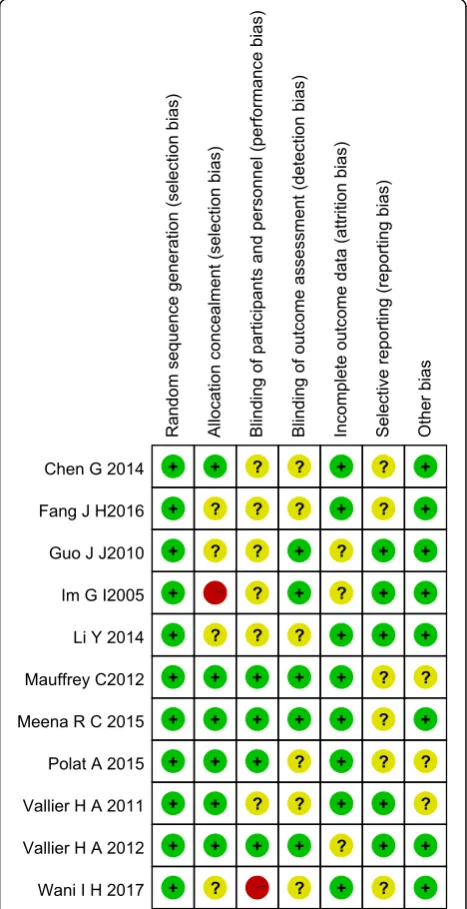
Assessment of risk of bias is presented in Fig.2. All
tri-als included in this study are randomized trial designs
[12,13]. One trial [14] did not provide detailed
informa-tion of random sequence generainforma-tion, and one trial [15]
did not describe the method of concealing group alloca-tion. Blinding of participants and personnel (perform-ance bias) was unclear and incomplete outcome data
(attrition bias) was high risk in two trials [15,16]. Three
trials [15–17] lost patients to follow-up.
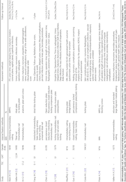
Table 1The characteristics of included studies
Study Year Country Patients(n) Age(Y) Study
Design
Fracture type
Quality Score
IMN PL IMN PL
Wani IH [13] 2017 India 30 30 36.4 ± 9.7 38.4 ± 8.7 RCT OTA42 A1-3 5
Vallier HA [12] 2011 USA 56 48 38.1 38.5 RCT OTA 42 6
Im GI [15] 2005 Korea 34 30 42(19,65) 40(17,60) RCT A1-3,C1 5
Fang JH [18] 2016 China 28 28 35.0 ± 9.2 38.6 ± 7.5 RCT OTA 42 6
Chen G [19] 2014 China 60 120 53.0 ± 8.1 25.53 ± 8.73 RCT AO 42A-B 4
Li Y [20] 2014 China 46 46 44(18-78) 43(18-79) RCT OTA42 6
Mauffrey C [21] 2012 UK 12 12 50(39-60) 33(24-43) RCT EAFDT 6
Guo JJ [16] 2010 China 44 41 44.2(27-70) 44.4(23-69) RCT OTA43A1-3 6
Costa ML [22] 2017 UK 161 160 44.3 ± 16.3 45.8 ± 16.3 RCT EAFDT 6
Polat A [14] 2015 Turkey 10 15 34.0 ± 9.7 36.4 ± 10.7 RCT OA42/43A1 6
Vallier HA [17] 2012 USA 45 41 41.0 37.8 RCT OTA42 5
Operation, radiation, and union time Operation time
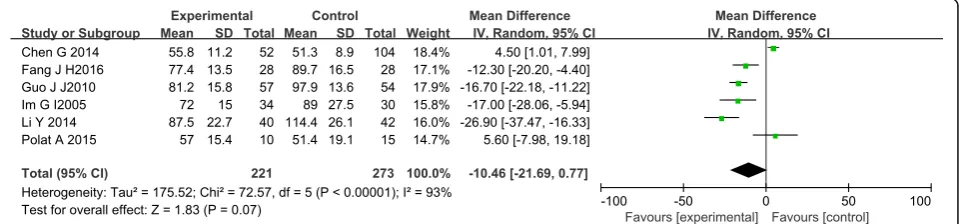
Six studies [14–16,18–20] with a total of 494 patients in
both groups provided data on operative time. Heterogen-eity in these studies was large, and the random-effects
model was used (I2= 93%). The meta-analysis showed no
significant difference in operative time between the IMN
group compared with PL group (MD =−10.46, 95%CI−
21.69 to 0.77,P= 0.07) (Fig.3).
Radiation time
Appropriate data on radiation time were available in 3
articles [14, 16, 19] with a total of 366 patients. Polat et
al. [14] recorded the radiation time in milliseconds,
whereas the other two study records in minutes, so the comparison could only be done once the time was con-verted to minutes. There was significant heterogeneity among the studies requiring analysis with a
random-ef-fects model (I2= 100%). Meta-analysis showed no
signifi-cant difference in radiation time between the IMN
group and the PL group (MD = 7.95, 95%CI −6.65 to
22.55,P= 0.29) (Fig.4).
Union time
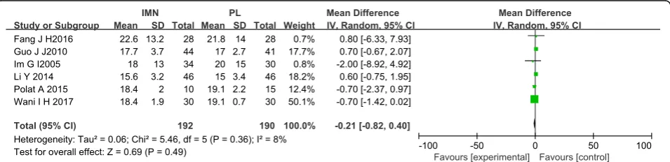
Six studies [13–16, 18, 20] reported data on union time
in the IMN group compared with the PL group. No sig-nificant difference in union time was noted between the
IMN group and the PL group (MD =−0.21, 95%Cl −
0.82 to 0.40,P= 0.49) (Fig.5).
Complication Nonunion
Seven studies [12–16, 18, 20] provided data on
union. There was no significant difference in the non-union rate between the IMN group and the PL group
(RR = 2.17, 95%CI 0.79 to 5.99,P= 0.15) (Fig.6).
Deep infection
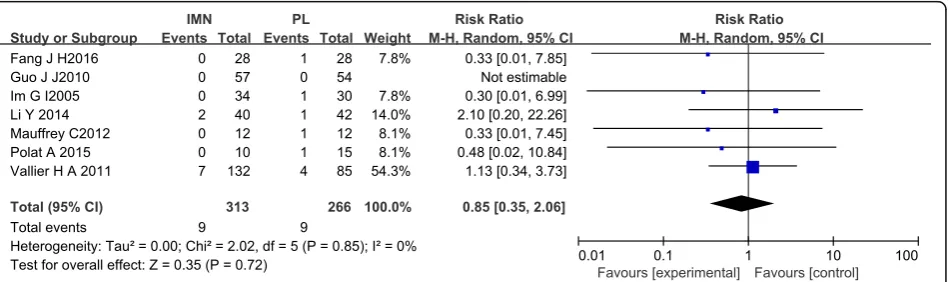
Six studies [12,14–16,18,20,21] with a total of 579
pa-tients in both groups reported deep infection. There was
no heterogeneity among these studies (I2= 0%). Data
were pooled using the random-effects model and the meta-analysis indicated that there was no significant dif-ference in deep infection occurrence between IMN and
PL groups. (RR = 0.85, 95%CI 0.35 to 2.06, P= 0.72)
(Fig.7).
Malunion
Seven studies [12,14,15,17–20] with a total of 676
pa-tients in both groups reported malunion. Mild
hetero-geneity existed among these studies (I2= 4%). Data were
pooled by fixed-effects analysis and the meta-analysis indicated that the IMN group had significantly higher
Fig. 2Risk of bias summary
malunion versus the PL group. (RR = 1.76, 95%CI 1.21 to
2.57,P= 0.003) (Fig.8).
Knee pain
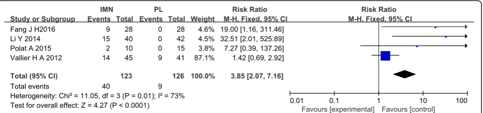
Four studies [14, 17, 18, 20] of 249 patients in both
groups reported on knee pain. Heterogeneity was
sub-stantial heterogeneity among studies (I2= 53%). Data
were pooled using a fixed-effects analysis, and the meta-analysis indicated that the IMN group had significantly higher knee pain versus the PL group (RR = 3.85, 95%CI
2.07 to 7.16,P< 0.0001) (Fig.9).
Superficial infection
Eight studies [13–16, 18–21] of 577 patients in both
groups reported on superficial infection. There was no
heterogeneity among the studies (I2= 16%). Data were
pooled using a fixed-effects analysis, and the meta-analysis indicated that the IMN group had significantly lower superficial infection rates compared to the PL group
(RR = 0.29, 95%CI 0.13 to 0.63, P= 0.002) (Fig. 10).
Delay union
Five studies [15, 16, 18, 20, 21] of 337 patients in both
groups provided data on delayed union. There was no
heterogeneity among studies (I2= 0%). Data were pooled
using the fixed-effects model, and the meta-analysis in-dicated no significant difference between the two groups
(RR = 0.92,95%CI 0.45 to 1.87,P= 0.82) (Fig.11).
Objective score
American Orthopaedic Foot and Ankle Surgery (AOFAS) score at the 6-month follow-up
Three studies [16,18,19] of 297 patients in both groups
provided data on the AOFAS at 6 months follow-up. A significant difference in the AOFAS score was noted between the PRP group and the HA group (MD 1.26,
95%Cl −1.19−3.70, P= 0.31). However, this result
should be interpreted with caution due to presence of
low or insignificant statistical heterogeneity (χ2= 0.88,
I2
= 0%) (Fig.12).
Foot Function Index
Four studies [13,14,17,19] comprising of 297 patients in
both groups provided date on the Foot Function Index. Low heterogeneity among the studies indicated use of the
fixed-effects model (I2= 0%). The meta-analysis found a
significant difference in the Foot Function Index between the IMN group compared to the PL group (MD = 0.09,
95%CI 0.01 to 0.17,P= 0.02) (Fig.13).
Disability rating index
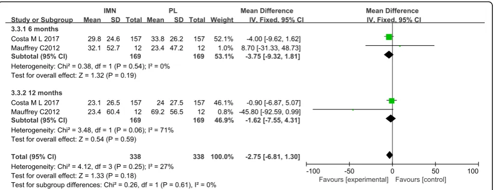
Two studies [21, 22] focusing on 338 patients in both
groups, reported on the Disability Rating Index. Low heterogeneity among the studies at the 6 month time
point required adoption of the fixed-effects model (I2=
0%). The meta-analysis showed no significant difference in the Disability Rating Index between the IMN group
compared with the PL group (MD =−3.75, 95%CI −
9.32 to 1.81, P= 0.19) (Fig. 14). High heterogeneity
Fig. 4Forest plot of radiation time in the IMN group compared with the PL group
among the studies at the 12 month time point required
adoption of the random-effects model (I2= 71%).
Meta-analysis showed no significant difference in the Disability Rating Index between the IMN group and the
PL group (MD =−17.11, 95%CI −59.37 to 25.16, P=
0.43). Low heterogeneity among the studies in the sub-group analysis indicated use of the fixed-effects model
(I2= 27%) and no statistically significant difference was
noted (MD =−2.92,95%CI −8.71 to 2.87, P= 0.32)
(Fig.14).
Discussion
The earliest PubMed record describing the use of intrame-dullary nails in the treatment of fractures was a 1946 paper
by Otoole reporting the treatment of femoral fractures [23];
use was later reported in the treatment of tibiofibular
frac-tures [24], and subsequently widely applied in limb
length-ening. Even in irregular bones such as clavicle fractures, the bone is characterized by highest protection of periosteal blood supply and lowest soft tissue irritation at the fracture end, thus providing a favorable soft tissue environment for fracture healing. IMN can be either reamed or non-reamed, where reaming is more beneficial for reduction. Animal
studies have confirmed that there is no significant increase in blood perfusion and osteophyte strength at the fracture
end when comparing reamed with unreamed IMN [25].
However, stress occlusion of the fracture end can lead to destruction of local fracture integrity and lead to negative clinical outcomes, occurrences which are expected to de-crease with improvement of instrument design techniques
and surgery [26]. After the fracture end is reset, the locking
nail is locked and pressurized, and the fracture end micro-motion is provided to promote fracture healing, however, if fixed too firmly, the alignment may be poor, the
rotation is deformed, or the nail broken [27]. In contrast,
open reduction and internal fixation is direct reduction and fixation, so contralateral alignment of the fracture end is su-perior to IMN fixation. MIPPO is similar to IMN fixation but does not require alignment of the fracture end under direct vision. In addition, it does not require strong fixation and emphasizes soft tissue and blood around the fracture. Limiting the disruption of the blood supply ensures ad-equate perfusion and maintenance of the biological
envir-onment at the fracture ends [28–30]. In addition, treatment
of distal radius fractures includes external fixation, external fixation combined with limited open reduction and internal
Fig. 6Forest plot of nonunion in the IMN group compared with the PL group
fixation, intramedullary nail and steel plate fixation, but these are not included in the scope of this meta-analysis.
To our knowledge, this is the first meta-analysis look-ing at the most recent randomized controlled trials com-paring the efficacy of IMN and PL in distal tibial fractures and distal metadiaphyseal tibial fractures. This meta-analysis was based on 11 RCTs that included 526 patients treated with IMN and 571 patients treated with PL fixation. The main outcomes investigated were union time, operation time, nonunion, and delay union, super-ficial infection, deep infection, malunion, and knee pain. In our meta-analysis, when IMN is compared with the PL group, there was no significant difference in AOFAS, delayed union time, deep infection, operation time, and radiation time. However, the IMN group was superior to the PL group in terms of the FFI, knee pain, and malu-nion. In terms of superficial infection occurrence, the PL group was significantly higher than the IMN group. These results highlight that IMN and PL are effective fixation methods for distal tibial fractures, with no sig-nificant differences in fracture healing time and opera-tive time. But the INM group was shown to recover better than the PL group, and the superficial infection rate was lower. However, the malunion and postopera-tive knee pain in the IMN group were higher than those
in the PL group. The operation time, fracture healing time, and intraoperative fluoroscopy time showed no statistically significant difference.
Prior studies [13, 31–33] and several meta-analyses
[34–37] investigating the efficacy of IMN have shown
that IMN has no superiority over PL when the two methods are compared in well-designed double-blinded trials. The reported beneficial effects of IMN in most tri-als may have been due to insufficient blinding methods. Our results showed that there was a statistically signifi-cant difference in malunion between the IMN and the PL group, with a higher rate of malunion in the IMN group. This is consistent with the results of a prior meta-analysis where no significant difference in oper-ation time and radioper-ation time between INM and PL
group was seen (P= 0.07, P= 0.29, respectively) [34].
This is not consistent with our conclusions. In addition, the limited number of RCTs and patients may also affect these results.
As is the case of many meta-analyses, our study is not devoid of limitations. First, the current meta-analysis fo-cuses only on papers that have been previously pub-lished. Inclusion of unpublished research may have increased heterogeneity and changed the current results. Second, the frequency of follow-up varied between
Fig. 8Forest plot of malunion in the IMN group compared with the PL group
Fig. 12Forest plot of AOFAS score in the IMN group compared with the PL group
Fig. 10Forest plot of superficial infection in the IMN group compared with the PL group
Fig. 11Forest plot of delay union in the IMN group compared with the PL group
studies, with five studies following up for more than 18 months, five studies limited to a year, and one study fol-lowing up for only 6 months. This variation may affect heterogeneity and hence the results. Furthermore, the type of incision and plate were not consistent across the different studies. There exist several different types of surgical incisions in the use of steel plate fixation, four studies used the MIPPO technique, whereas the rest used open reduction and internal fixation. In terms of type of steel plate, two studies used non-locking steel plates, five used compression-type steel plates, one used an anatomical steel plate, two used a bridge steel plate, and one used ordinary steel plates. Further rigorously designed RCTs, with larger sample sizes, are necessary to better compare the efficacy of IMN and PL fixation.
Conclusion
The results of this meta-analysis highly support that both the IMN and PL internal fixation methods are ef-fective for the treatment of distal tibial fractures with metaphyseal involvement. However, knee pain has been recorded following use of IMN, malunion is more fre-quent with IMN fixation, and the risk of superficial wound infection is higher in PL internal fixation than IMN use. In terms of FFI scores, IMN internal fixation appears to be superior to PL. Further studies investigat-ing the use of IMN internal fixation for knee pain after distal tibial fractures are required. Furthermore, the rate of fracture deformity is higher with IMN fixation, while treatment and prevention of superficial wound infections after PL surgery increases the ankle joint function.
Abbreviations
–:Indicates not reported by the study; AOFAS: The American Orthopaedic Foot and Ankle Surgery; DRI: Disability Rating Index; DTF: Distal tibial fracture; EAFDT: Extra-articular fracture of distal tibia fracture; EQ-5D-3 L: The EuroQol Health-Related Quality-of-Life 3-level score; F/M: Female/male; FFI: Foot
Function Index; IMN: Intramedullary nail; KSS: The Knee Society’s Knee Scoring System; MFA: Musculoskeletal Function Assessment; MIPO: Minimally invasive plate osteosynthesis; OMAS: The Olerud and Molander Ankle Score; OTA: Orthopaedic Trauma Association; GAT: Gustilo and Anderson type; PL: Plate; PTP: Proximal tibial plating; TSC: Tscherne classification
Acknowledgements
This work was accomplished with the help of the library of Huazhong University of Science and Technology.
Funding
Projects on national natural science foundation (81772345).
Availability of data and materials
Please contact author for data requests.
Authors’contributions
LH and YX contributed equally to this paper. LH and YX participated in the design of the study. YL and BM carried out the data curation. JL and HX performed the statistical analysis. JL and LH carried out the investigation. BM, JL, and AA carried out the project administration. YL and YX operated the software. YL carried out the supervision. LH and YL carried out the validation. LH, YX, CCY, and LC conceived of the study, participated in its design and coordination, and helped to draft the manuscript. GL and PC participated in the sequence alignment and drafted the manuscript. All authors read and approved the final manuscript.
Ethics approval and consent to participate
Not applicable.
Consent for publication
Not applicable.
Competing interests
The authors declare that they have no competing interests.
Publisher’s Note
Springer Nature remains neutral with regard to jurisdictional claims in published maps and institutional affiliations.
Author details
1
Department of Orthopedics, Union Hospital, Tongji Medical College, Huazhong University of Science and Technology, 1277, Jiefang Avenue, Wuhan 430022, China.2Department of Plastic Surgery, Brigham and
Women’s Hospital, Harvard Medical School, Boston 02152, USA.
Received: 10 November 2018 Accepted: 11 December 2018
References
1. Bedi A, Le TT, Karunakar MA. Surgical treatment of nonarticular distal tibia fractures [J]. J Am Acad Orthop Surg. 2006;14(7):406.
2. Wennergren D, Bergdahl C, Ekelund J. Epidemiology and incidence of tibia fractures in the Swedish fracture register [J]. Injury. 2018;11:
S0020138318304893.
3. Marsh J, Slongo T, Broderick J, et al. Fracture and dislocation classification compendium - 2007: Orthopaedic trauma association classification, database and outcomes committee [J]. J Orthop Trauma. 2007;21(10 Suppl): 1–133.
4. Newman SD, Mauffrey CP, Krikler S. Distal metadiaphyseal tibial fractures [J]. Injury-international Journal of the Care of the Injured. 2011;42(10):975–84. 5. Robinson CM, Mclauchlan GJ, Mclean IP, et al. Distal metaphyseal fractures
of the tibia with minimal involvement of the ankle. Classification and treatment by locked intramedullary nailing [J]. J Bone Joint Surg Br. 1995; 77(5):781–7.
6. Busse JW, Morton E, Lacchetti C, et al. Current management of tibial shaft fractures: a survey of 450 Canadian orthopedic trauma surgeons [J]. Acta Orthop. 2008;79(5):689–94.
7. Bode G, Strohm PC, Südkamp NP, et al. Tibial shaft fractures - management and treatment options. A review of the current literature [J]. Acta Chir Orthop Traumatol Cechoslov. 2012;79(6):499.
8. Seyhan M, Unay K, Sener N. Intramedullary nailing versus percutaneous locked plating of distal extra-articular tibial fractures: a retrospective study [J]. Eur J Orthop Surg Traumatol. 2013;23(5):595–601.
9. VidovićD, MatejčićA, Ivica M, et al. Minimally-invasive plate osteosynthesis in distal tibial fractures: results and complications. Injury-international Journal of the Care of the Injured. 2015;46:S96–S9.
10. Richard RD, Kubiak E, Horwitz DS. Techniques for the surgical treatment of distal tibia fractures [J]. Orthop Clin N Am. 2014;45(3):295–312.
11. Gupta RK, Rohilla RK, Sangwan K, et al. Locking plate fixation in distal metaphyseal tibial fractures: series of 79 patients [J]. Int Orthop. 2010;34(8): 1285–90.
12. Vallier HA, Beth Ann C, Patterson BM. Randomized, prospective comparison of plate versus intramedullary nail fixation for distal tibia shaft fractures [J]. J Orthop Trauma. 2011;25(12):736–41.
13. Wani IH, Ul GN, Yaseen M, et al. Operative Management of Distal Tibial Extra-articular Fractures - intramedullary nail versus minimally invasive percutaneous plate Osteosynthesis [J]. Ortop Traumatol Rehabil. 2017:537–41. 14. Polat A, Kose O, Canbora K, et al. Intramedullary nailing versus minimally
invasive plate osteosynthesis for distal extra-articular tibial fractures: a prospective randomized clinical trial [J]. J Orthop Sci. 2015;20(4):695–701. 15. Gun-il I, Suk-Kee T. Distal metaphyseal fractures of tibia: a prospective
randomized trial of closed reduction and intramedullary nail versus open reduction and plate and screws fixation [J]. J Trauma. 2005;59(5):1219. 16. Guo JJ, Tang N, Yang HL, et al. A prospective, randomised trial comparing
closed intramedullary nailing with percutaneous plating in the treatment of distal metaphyseal fractures of the tibia [J]. Ned Tijdschr Traumatol. 2010; 18(6):185–6.
17. Vallier HA, Beth Ann C, Patterson BM. Factors influencing functional outcomes after distal tibia shaft fractures [J]. J Orthop Trauma. 2012;26(3): 178–83.
18. Fang JH, Wu YS, Guo XS, et al. Comparison of 3 minimally invasive methods for distal tibia fractures [J]. Orthopedics. 2016;39(4):e627.
19. Chen G, Qian MQ, Zhu GX, et al. Percutaneous closed reduction locking compression plate, percutaneous closed reduction interlocking intramedullary nail and open reduction plate in the treatment of tibial fracture: comparison of biostability [J]. Zhongguo Zuzhi Gongcheng Yanjiu. 2014;18(44):7151–6.
20. Li Y, Jiang X, Guo Q, et al. Treatment of distal tibial shaft fractures by three different surgical methods: a randomized, prospective study [J]. Int Orthop. 2014;38(6):1261–7.
21. Mauffrey C, Mcguinness K, Parsons N, et al. A randomised pilot trial of
“locking plate”fixation versus intramedullary nailing for extra-articular fractures of the distal tibia [J]. J Bone Joint Surg Bri. 2012;94(5):704. 22. Costa ML, Achten J, Griffin J, et al. Effect of locking plate fixation vs
intramedullary nail fixation on 6-month disability among adults with
displaced fracture of the distal tibia: the UK FixDT randomized clinical trial [J]. Jama. 2017;318(18):1767–76.
23. O’toole M. Fracture of shaft of the femur treated with Küntscher intramedullary nail [J]. Nurs Times. 1946;42(34):640.
24. SPRIET. [nailing of fractures of the lower third of the femur and of the tibia by the low approach; introduction of a nail for tibial and condylar malleoli] [J]. Mem Acad Chir. 1952;78(8–9):259–62.
25. Schemitsch EH, Kowalski MJ, Swiontkowski MF, et al. Comparison of the effect of reamed and unreamed locked intramedullary nailing on blood flow in the callus and strength of union following fracture of the sheep tibia [J]. J Orthop Res. 1995;13(3):382–9.
26. Filardi V. The healing stages of an intramedullary implanted tibia: a stress strain comparative analysis of the calcification process [J]. J Orthop. 2015; 12(Suppl 1):S51–61.
27. Lefaivre KA, Pierre G, Holman C, et al. Long-term follow-up of tibial shaft fractures treated with intramedullary nailing [J]. J Orthop Trauma. 2008;22(8): 525–9.
28. Jens F, Geoffroy V, Frank V, et al. Percutaneous plate fixation of fractures of the distal tibia [J]. Acta Orthop Belg. 2004;70(2):148–54.
29. Borrelli J Jr, Prickett W, Song E, et al. Extraosseous blood supply of the tibia and the effects of different plating techniques: a human cadaveric study [J]. J Orthop Trauma. 2002;16(10):691.
30. Farouk O, Krettek C, Miclau T, et al. Minimally invasive plate osteosynthesis and vascularity: preliminary results of a cadaver injection study [J]. Injury-international Journal of the Care of the Injured. 1997;28(Suppl 1):A7. 31. Chen F, Huang X, Ya Y, et al. Finite element analysis of intramedullary
nailing and double locking plate for treating extra-articular proximal tibial fractures [J]. J Orthop Surg Res. 2018;13(1):12.
32. Shen J, Xu J, Tang MJ, et al. Extra-articular distal tibia facture (AO-43A): a retrospective study comparing modified MIPPO with IMN [J]. Injury-international Journal of the Care of the Injured. 2016;47(10):2352–9. 33. Qi Y, Jie N, Li-bin P, et al. Locked plating with minimally invasive
percutaneous plate osteosynthesis versus intramedullary nailing of distal extra-articular tibial fracture: a retrospective study. Zhonghua Yi Xue Za Zhi. 2013;93(47):3748–51.
34. Guo C, Ma J, Ma X, et al. Comparing intramedullary nailing and plate fixation for treating distal tibail fractures: A meta-analysis of randomized controlled trials [J]. Int J Surg. 2018;53:5–11.
35. Mao Z, Wang G, Zhang L, et al. Intramedullary nailing versus plating for distal tibia fractures without articular involvement: a meta-analysis [J]. J Orthop Surg Res. 2015;10(1):1–12 10,1(2015-06-16).
36. Yu J, Li L, Wang T, et al. Intramedullary nail versus plate treatments for distal tibial fractures: A meta-analysis [J]. Int J Surg. 2015;16(Pt A):60–8.