Name-calling in the hippocampus (and beyond): coming to terms
with neuron types and properties
D. J. Hamilton.D. W. Wheeler.C. M. White.C. L. Rees. A. O. Komendantov.M. Bergamino.G. A. Ascoli
Received: 11 March 2016 / Accepted: 24 May 2016 / Published online: 9 June 2016 The Author(s) 2016. This article is published with open access at Springerlink.com
Abstract Widely spread naming inconsistencies in neuro-science pose a vexing obstacle to effective communication within and across areas of expertise. This problem is par-ticularly acute when identifying neuron types and their properties. Hippocampome.org is a web-accessible neu-roinformatics resource that organizes existing data about essential properties of all known neuron types in the rodent hippocampal formation. Hippocampome.org links evidence supporting the assignment of a property to a type with direct pointers to quotes and figures. Mining this knowledge from peer-reviewed reports reveals the troubling extent of ter-minological ambiguity and undefined terms. Examples span simple cases of using multiple synonyms and acronyms for the same molecular biomarkers (or other property) to more complex cases of neuronal naming. New publications often use different terms without mapping them to previous terms. As a result, neurons of the same type are assigned disparate names, while neurons of different types are bestowed the same name. Furthermore, non-unique prop-erties are frequently used as names, and several neuron types are not named at all. In order to alleviate this nomenclature confusion regarding hippocampal neuron types and properties, we introduce a new functionality of
Hippocampome.org: a fully searchable, curated catalog of human and machine-readable definitions, each linked to the corresponding neuron and property terms. Furthermore, we extend our robust approach to providing each neuron type with an informative name and unique identifier by mapping all encountered synonyms and homonyms.
Keywords HippocampusNeuronTypeProperty Nomenclature
1 Introduction
From its beginning, neuroscience has been tied to ad hoc neuron naming, which is subject to the whims of researchers with diverse interests. It has always been the inclination of neuroscientists to name neurons based on certain observed properties. Already in the 1800s, researchers leveraged ongoing progress in optical microscopy and newly discov-ered staining techniques to identify neuron types and their morphological features. Historical examples include Betz’ naming of ‘‘giant pyramids’’ [1] and Cajal’s description of ‘‘psychic cells’’ (nowadays known as pyramidal neurons) as characterized by ‘‘…a dendritic shaft and tuft directed toward the cerebral surface [and] the existence of collateral spines on the dendritic processes…’’ [2]. Thousands of reports describing neurons and their characteristics have been published since, and several dozens of distinct types of neurons had been already recognized before the turn of the millennium in each of several prominent neural systems, such as among the ‘‘GABAergic non-principal cells’’ of the hippocampus [3].
The often subjective and arbitrary naming of neurons led to a cluttered literature landscape in which breakdowns in D. J. HamiltonD. W. WheelerC. M. White
C. L. ReesA. O. KomendantovM. Bergamino G. A. Ascoli (&)
Center for Neural Informatics, Structure, & Plasticity, Molecular Neuroscience Dept., Krasnow Institute for Advanced Study, MS2A1, George Mason University, Fairfax, VA 22030-4444, USA
e-mail: [email protected]
Present Address: M. Bergamino
Laureate Institute for Brain Research, 6655 S Yale Ave, Tulsa, OK 74136, USA
communication can hinder the understanding of the struc-ture and function of the brain. A comprehensive solution would require establishing a broadly applicable and widely accepted classification scheme defining neuron types based on their properties. However, despite early efforts focused on identifying key neuronal properties with precise termi-nology [4], to this date there is a high level of disorgani-zation when it comes to reporting neuronal property information. Although community efforts exist for the expert curation of neuroanatomical terms pertaining to brain regions [5] and grass root scholarly collation of neuroscience terminology [6], the continuously increasing pace of data acquisition is paradoxically yielding an ever more fractured lexicon, creating serious impediment to progress.
We have previously proposed an ontological approach to defining neurons based on necessary and sufficient part-relation-value triple-store techniques [7]. In the absence of comprehensive data and unbiased sampling, however, it may be impossible to select a priori the appropriate defining properties [8]. Using too few or too many con-straints results in under-defining or over-defining a neuron type. The former case (‘‘over-lumping’’) leads to a few large groups of neurons that share very few properties; the latter (‘‘over-splitting’’) leads to myriad types of doubtful interpretation. To complicate this matter further, the con-tinuous gradation of key properties may require a shift to fuzzy classification approaches [9].
A recent empirical assessment of inter-investigator agreement on morphological classes of neocortical interneurons demonstrated a variable level of consensus across neuron types and properties [10]. One of the most reliable identifiers of neuron types is the presence or absence of axons and dendrites within well-defined neu-roanatomical boundaries. Spotlighting this, Hippocam-pome.org [11] recently established unambiguous definitions of neuron types primarily based on axonal and dendritic distributions across all the main subregions and layers of the hippocampal formation. This classification approach yielded an initial catalog of 122 neuron types identified from the scientific literature. It is important to stress that the classification criteria employed by Hip-pocampome.org operate independently of previously used names.
In this framework, a neuron type is initially identified by its (putative) neurotransmitter and the presence of axons and dendrites in the distinct layers of dentate gyrus, CA3, CA2, CA1, subiculum, and entorhinal cortex. Each type is further characterized by available information on bio-marker expression and electrophysiological features. This relatively simple characterization allows dense curation of the published literature through text mining and annotation. The resulting information is instantiated as a
machine-readable electronic relational knowledge base that is pub-licly and freely available, facilitating web accessibility and computational analytics. With critical properties compiled in an easily accessible portal, Hippocampome.org provides a unique opportunity to establish a consistent set of defi-nitions and a naming protocol that could be expanded to other cortical areas, aiding research and scientific communication.
The remaining of this report is organized as the fol-lowing. The next section provides illustrative examples of the terminological confusion regarding neuron types and properties from the hippocampal literature. The following section outlines the three steps toward a solution: first, we describe the design of a database to define, store, browse, search, and retrieve human-interpretable but machine-readable definitions of neuron types based on their prop-erties, as recently implemented at Hippocampome.org. Second, we introduce a newly deployed functionality that maps all relevant property terms to corresponding con-cepts, linking their occurrence in the published evidence to community-accepted definitions. Third, we offer a formal definition of the resulting neuron types and detail the process to assign each of them with a unique common name. The last section closes the paper with concluding remarks.
2 A neuronal ‘‘Tower of Babel’’
The nomenclatures of neuron types and of their features are both vexed with ambiguities, resulting in a ‘‘many-to-many’’ mapping between neurons and names as well as inconsistent definitions of properties. We illustrate below representative examples of the most common scenarios from the hippocampal literature.
When neurons are described in a publication, they are typically named in isolation, out of context with respect to the rest of the brain circuit and the literature. Sometimes neuron types or individual neurons are indicated solely by a non-descriptive label (e.g., ‘‘Type I’’ cells [12] or ‘‘cell #7’’ [13], and occasionally they are not named at all. When proper terms are used, it may still be difficult to discern whether a word is meant to be a name or merely a description, as when referring to ‘‘multipolar cells’’ [12,14]. The result is often a baffling web of associations between names and neuron types.
distributed in the CA1 oriens and radiatum layers (though also extending into the subiculum), but dendrites limited to oriens [16]. Unfortunately, neurons with these distinct characteristics had already been bestowed the different name of ‘‘CA1 trilaminar cells’’ in an earlier article [17]. Nevertheless, the label ‘‘CA1 trilaminar cell’’ was also used to describe yet another neuron type that had a similar axonal distribution, but dendrites invading lacunosum-moleculare [16]. But the confusion does not end here, as other labs independently referred to this latter morphology as either ‘‘CA1 Schaffer-associated’’ [18] or ‘‘CA1 apical dendrite innervating’’ [19].
We should note that these are not exceptional instances, but absolutely frequent occurrences, as depicted by several additional examples in Fig.1 [20–29]. There are also multiple cases of the same referencing article calling a morphologically defined type by different synonyms, such as ‘‘perforant path-associated’’ and ‘‘CA1 R-LM’’ referring to neurons with axons and dendrites in CA1 stratum lacunosum-moleculare and dendrites in radiatum [18] (Fig.2a). At the same time, these are not sterile spelling quibbles, because the specific laminar pattern of dendrites and axons defines the potential connectivity of the circuit and therefore the computational functions of neurons.
The confusion is not limited to neuron types but also affects the nomenclature of neuronal features, including morphological, electrophysiological, and molecular termi-nology. Qualitative phraseology is especially common in reporting morphological properties. An examination of the evidence collated in Hippocampome.org pertaining to the relative abundance of axons in an anatomical location of interest reveals ample use of terms such as ‘‘most,’’ ‘‘ma-jority,’’ and ‘‘usually.’’ Furthermore, categorical terms are often employed to indicate continuous spatial distributions, as in ‘‘superficial/deep layer X,’’ ‘‘proximal/distal area Y,’’ and ‘‘septal/temporal region Z.’’ A clear consensus of how such terms should be adopted and interpreted, and what terms are to be avoided, reduces ambiguity. Hippocam-pome.org proposes a set of protocols for the description of neurites and their locations (hippocampome.org/full-interp).
The electrophysiological lexicon suffers not only from ambiguous descriptors but also from inconsistent defini-tions of the parameters themselves. For example, some investigators measure action potential amplitude from the resting membrane potential to the peak of the spike [30]. A complementary subset of studies, however, calculates action potential amplitude relative to the spike threshold
Fig. 1 Relationships between cited names [3,15–29] and neuron types. Thisbipartite graphhighlights the naming confusion that is typical
potential [31]. The relationship between the minimum and the steady-state membrane potentials resulting from a hyperpolarizing current is similarly ambiguous. The sag ratio quantifies the relative difference between the peak hyperpolarization and steady-state hyperpolarization [32]. Alternatively, the sag percentage reports the fractional change in membrane potential from peak to steady state relative to the steady state [33]. Figure2b schematically shows the differences between these parameter definitions. Plainly, the use of identical or similar names for terms with different electrophysiological meanings can lead to the propagation of confusion and, worse, incorrect interpreta-tions of data that are incorporated into the literature mov-ing forward.
Molecular biomarkers bear an overabundance of syn-onyms, homsyn-onyms, hypsyn-onyms, hypernyms, and abbrevia-tions. There is movement toward standardizing the naming of proteins, but it is debatable whether the efforts are alleviating or augmenting confusion. For instance, the entire family of mammalian neuronal transporters has been given the official name of ‘‘solute carrier family [X] member [Y].’’ The new names confer that the proteins are transporters, but provide little information beyond that. As an example, some authors now refer to vesicular glu-tamate transporter 2 (Gene ID: 84487, ncbi.nlm.nih.gov/ protein/NP_445879.1) by the abbreviation Slc17a6, short for the official full name ‘‘solute carrier family 17, member
6,’’ while others keep the familiar vGluT2. If these two alternatives were not enough, the marker is also known by the symbols Dnpi and Vgl [34–37] (Fig.2c).
One of the worst cases of molecular biomarker termi-nology confusion in neuroscience involves glutamate receptors. Metabotropic glutamate receptors (mGluRs) are not to be confused with three classes of ionotropic recep-tors (GluRs): AMPA, kainate, and NMDA, sometimes referred to as AMPARs, KAs, and the NRs [38]. In the promising new naming schema for glutamate receptors, metabotropic receptors retain use of mGluR, while AMPA receptors use GluA, kainate GluK, and NMDA GluN [39]. It is yet to be seen how widely used either of these sche-mata will be. Alas, even if the entire research community compactly embraced them today, the problem of linking new information with previous publications would remain.
3 Resolving the neuron-type crossword puzzle
The solution to both the naming dilemma and property-based neuronal classification lies in establishing and con-sistently applying an unambiguous, clearly defined, unique nomenclature with links to antecedent synonyms. With property terms, scholarly resources can serve as broadly accepted references and dictionaries, such as the Medical Subject Headings (MeSH) by the US National Library of
Fig. 2 Examples of confusing nomenclature.aMorphological terms
[18, 47]. b Physiological properties. Neuronal responses to suprathreshold depolarizing (top) and hyperpolarizing (bottom) cur-rent injections. Greenand red labels show different definitions of electrophysiological parameters (action potential amplitude and sag
ratio).Vrestresting membrane potential,Vthreshthreshold potential, Vmin minimum of membrane potential drop, Vss steady-state membrane potential under long-lasting hyperpolarizing stimulation,
Medicine [40] and NeuroLex by the National Institutes of Health-contracted Neuroscience Information Framework [41]. However, using such services requires turning atten-tion away from the material with the confusing or unknown term, navigating external web site(s), finding and pro-cessing the definition(s), then refocusing attention to the original material. A terms portal integrated into the original material would greatly simplify the process.
3.1 Data schema for property-based classification of hippocampal neurons
To solve the neuronal naming problem, the neuroscience community would ideally adopt a robust approach to classification. Using the distributions of axons and den-drites across identifiable anatomical areas is advantageous for a number of reasons. Axonal and dendritic patterning is fundamental to all neurons, yet sufficiently information-rich to allow grouping at a useful level of abstraction on the spectrum from considering all neurons the same (as would be the case if spike integrator were the chosen property) and each individual neuron unique (as would result if using exact matches of the neurite arbors). In addition, neurite patterns are more stable and less dependent upon experi-mental conditions than molecular markers and electro-physiology, respectively. Lastly, as demonstrated below, this approach naturally provides the means of creating unique, concise, informative names of neuron types.
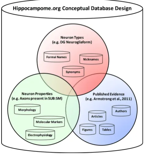
We designed an open-source online system enabling machine-readable information accessibility. Knowledge about each Hippocampome.org neuron type, including the names, synonyms, properties, and evidence, is stored in a relational database sourcing a user-friendly web-accessible interface. Figure3 depicts the conceptual organization of the database based on three general categories: neuron types, neuron properties, and published evidence. Links between data and relations are captured in separate relation tables, to both increase flexibility and reduce complexity, thereby facilitating continuous development and long-term maintainability.
Converting information published for human consump-tion into machine-readable form dictates system level decisions to minimize the energy cost of processing. We chose a three-step workflow. The first step is for researchers (doctoral students, postdocs, and faculty) to identify and study relevant articles, gleaning salient infor-mation and encoding it into spreadsheets. The second step involves python code to ingest these spreadsheets into data tables, populating along the way relation tables. The third step consists of rendering the resulting structured data in web pages dynamically leveraging the database. Perform-ing the most time consumPerform-ing tasks up front (steps one and two) allows for fast web-based lookup access by the
end-user community. The data/relation table design adds a layer of complexity to the database, but simplifies the resultant query implementation complexity, considerably speeding up real-time interactive retrieval.
3.2 Neuron term machine-readable definition identifier
In order to facilitate the collation of machine-readable definitions of relevant terms, we designed and implemented a novel functionality of Hippocampome.org for online assistance in disambiguating neuron property nomenclature (Fig.4). This new resource (Hippocampome.org) inte-grates key neuron term descriptors into a curated catalog of web-accessible human- and machine-readable definitions. Users can browse, search, and filter terms from drop-down menus augmented with autocomplete-as-you-type function. After selecting one or more terms, the portal returns the mapped concept with mouse/cursor-layover display of all available synonyms and the context in which they appear, along with a list of available definitions and direct hyper-links to the corresponding source providers. Users can also search for specific keywords of interest within the defini-tions. Furthermore, when browsing Hippocampome.org and all cited evidence within, terms with available defini-tions are now highlighted: users can display a definition
Fig. 3 Hippocampome.org conceptual design. The database groups
pop-up with mouse/cursor-layover or directly click on the term for linking out to the corresponding entry from the providing resource.
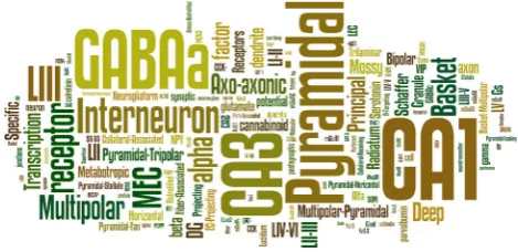
The first challenge in deploying this novel functionality was to identify the set of terms requiring machine-readable definitions. This research leveraged two primary sources of terms: Petilla [4] and the article excerpts cited as evidence in Hippocampome.org [11]. The Neuron Registry [7] constituted a third minor source of terms. The Petilla ter-minology consists of a finite list of (*232) published terms. Hippocampome.org, in contrast, contributes a less neatly bounded set of terms exceeding 10 K discrete tokens (as estimated by the wordle.net utility, Fig.5). To parse these tokens into a manageable set, we filtered the Hip-pocampome.org terms at each extreme of the occurrence count spectrum. This preprocessing step eliminated words with very large ([1000) occurrence counts, including uninformative strings such as ‘‘a,’’ ‘‘the,’’ and ‘‘of,’’ as well as words with very small (\100) occurrence counts, rep-resenting rare and typically uninteresting terms like ‘‘out-side-out’’ and ‘‘sheetlike.’’ Lastly we hand-curated the remaining set of approximately 700 terms to remove non-scientifically relevant words yielding a final corpus of 490 evidence-derived terms. An additional 782 terms corre-sponded to neuron names, anatomical regions, biomarkers,
and electrophysiological parameters stored in Hippocam-pome.org. In all, due to minor overlaps among the above lists, this collation accounted for 1478 distinct terms.
To find machine-readable definitions we devised a pre-ferred portal/repository approach. For general neurobio-logical terms, we first searched Neurolex.org, MeSH browser (nlm.nih.gov/mesh), the Bioportal services from the National Center for Biomedical Ontology [42], and the US Public Health Service CRISP database [43]. The terms from Hippocampome.org evidence primarily refer to the rodent hippocampus, thus it is essential that the extracted definition be relevant to these target domains. Since the same word can have different meanings, most definitions retrieved by the initial automated search were largely out of context, requiring a slow step of manual curation. We preferentially assigned evidence terms from Hippocam-pome.org definitions and links most relevant to the rodent hippocampal formation. Similarly, we linked the Petilla terms to definitions in the context of GABAergic interneurons of the cerebral cortex.
For protein definitions, we harnessed the Ontology Look-up Service [44] of the Gene Ontology Consortium [45] as the sole reference given the depth and breadth of coverage for this type of molecular data. Because the molecular terms are generally regular and systematically databased, we successfully automated API-based pulling from established sources (e.g., the National Center for Biotechnology Information). For term not found in these primary resources, we reverted to Google searches, prior-itizing definitions from scholarly or institutional sources such as the Allen Brain Atlas [46], Scholarpedia.org, and the US National Institute of Standards and Technology (nist.gov). For residual blanks, we resorted to dictionaries like Merriam-Webster or Wikipedia.
The last step of manual curation involved concept mapping to group together distinct terms linking to textu-ally different but logictextu-ally analogous definitions. For
Fig. 4 Neuron term machine-readable definition identifier: an online portal for conceptual mapping of neuronal properties fully integrated in
Hippocampome.org
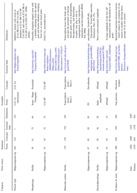
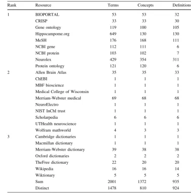
example, ‘‘action potential’’ and ‘‘spike’’ are synonyms for which multiple machine-readable definitions exist. This mapping yielded 810 distinct concepts from the 1478 unique terms, with a total of 924 unique definitions from 1378 distinct resource links. Table1 summarizes the neu-ron term counts, including number per category (i.e., morphological, molecular, and electrophysiological) and unique instances. Table2 organizes this information by resources providing the machine-readable external links to the term definitions.
3.3 Neuron type naming
The classification schema introduced by Hippocam-pome.org [11] defines neuron types based on their properties, starting from morphological patterns and with the added specification of molecular and electrophysio-logical features. For example, Hippocampome.org defines dentate gyrus granule cells as excitatory neurons with axons in the hilus, CA3 lucidum/pyramidale, and CA2 pyramidale, dendrites in the inner and outer molecular layer, and soma in the granular layer. These definitions are now available as an explicit list (hippocampome.org/ neuron-types) and linked from the term definition portal described above.
It is difficult to quantify how many unique neuron types have been defined to date in the hippocampal formation due to ambiguity and overlap of descriptors across research labs. We constrain the number of Hippocampome.org [11] neuron types (e.g., 122 in the initial release) by limiting the primary characterization properties to axonal/dendritic patterns and excitatory/inhibitory neurotransmitters.
Furthermore, Hippocampome.org neuron types are assigned both a formal name and a unique number identi-fier (e.g., DG (e) 2201p-CA3_00110 Granule; type 1000). The formal name contains several components (hip-pocampome.org/formal-name): (a) the abbreviation of the subregion where the soma is located, (b) a symbol speci-fying the putative major neurotransmitter (i.e., ‘‘e’’ for glutamatergic, excitatory neurons or ‘‘i’’ for GABAergic, inhibitory neurons), and (c) a numeric encoding for the presence or absence of neurites within the subregion of soma location. In neuron types whose axons extend outside of their home subregion, the numerical encoding continues with a ‘‘p’’ (for projecting) followed by codes analogous to (a) and (c) to specify the subregions receiving the projec-tion. Finally, the formal name ends with a unique, human-friendly label that attempts to maximize usability and understanding of neuron types within the research com-munity. Figure6 illustrates the selection process for determining this ‘‘common name.’’
In the most clear-cut cases, a single name dominates the literature as universally recognized and understood. In such
‘‘canonical’’ cases, we adopt these standard names, as in Granule, Mossy, CA3 basket, and CA1 pyramidal cells. In other situations, a neuron type may not be as broadly known, but is only cited in a single way. In these cases, we straightforwardly adopt the single cited name, such as in Semilunar Granule, CA3 Giant, and CA3 Granule cells. The remaining cases represent the confusing scenarios in which the literature describes the same neuron types with multiple names and different neuron types with the same name.
If one name or acronym is clearly dominant, with more frequent citations than all other names, we adopt it as the common name, as in the cases of HIPP, MOPP, HICAP, and MOLAX interneurons. Other neuron types, however, have multiple, approximately equally cited names, espe-cially in the less-studied entorhinal cortex. In these cases, to avoid playing favorites, we hybridize the cited names, as is LI-II Multipolar-Pyramidal, LI-II Pyramidal-Fan, and MEC LII-III Pyramidal-Multiform. Lastly, there are neuron types for which all cited names entail potential confusion with similar or identical names already assigned to other neuron types based upon the rules above. In these scenar-ios, we are forced to either modify a cited name in order to differentiate it (e.g., Mossy MOLDEN, DG Basket CCK?, and CA3c Pyramidal) or to create a new name altogether (e.g., AIPRIM, HIPROM, MOCAP, CA3 SO–SO). We try to use this final clause sparingly (only 4 names out of 122 in Hippocampome.org are entirely new), but minor modi-fications of pre-existing names are often unavoidable (46 out of 122).
4 Discussion
We have striven to find human-friendly names that are recognizable to, at the least, those who are familiar with hippocampal neurons. In many cases, however, these names have minimal informational content to those unfamiliar with the type. The part of the formal name that is most informative is the numeric encoding of the neurite pattern (detailed description: hippocampome.org/find-term). Knowledge of the pattern of dendrites and axons confers information about potential connectivity of the neuron type within the circuit. Therefore, incorporation of this pattern into the name allows instantaneous envisioning of the location of the neurites and by extension the connectivity of the type. In addition, this numeric encoding is unique for most neuron types with only subtypes discriminated by their primary neurotransmitter, post-synaptic target specificity, or molecular marker and/or electrophysiology profiles having the same pattern. In these cases, the human-friendly part of the name provides uniqueness (e.g., ‘‘CA1 2232 Basket’’ and ‘‘CA1 2232 Basket CCK?’’). This method of naming neurons results in extremely informative, concise names without necessitating the memorization of many
acronyms. Furthermore, it is applicable to any brain region that is divisible into parcels.
Going beyond Hippocampome.org, the same approach to defining neuron types can be extended outside the hip-pocampal formation. For example, CA1 neurons that pro-ject to other brain regions such as the lateral septum, medial septum, and/or hypothalamus can be characterized by extending the axonal/dendritic patterns to encompass those regions.
Nomenclature confusion could be mitigated with increased awareness of the neurons, molecules, and proper-ties and how they fit in the historical context. This is a lot to ask of researchers, but resources like Hippocampome.org provide significant assistance. Hippocampome.org demon-strates that the necessary and sufficient discriminating property of neurite patterning is a workable and advanta-geous foundation upon which to build a neuron type library. Enhancing such a library with a terms definition portal fur-ther reduces terminology confusion. Coupled, these resour-ces begin clarifying the muddied state of the literature and re-illuminating the path to neuroscience progress.
Table 2 Term resource
summary Rank Resource Terms Concepts Definitions
1 BIOPORTAL 53 53 32
CRISP 33 33 30
Gene ontology 119 100 105
Hippocampome.org 649 130 130
MeSH 176 168 111
NCBI gene 112 111 6
NCBI protein 103 102 7
Neurolex 429 354 311
Protein ontology 121 120 6
2 Allen Brain Atlas 35 35 33
ChEBI 1 1 1
MBF bioscience 1 1 1
Medical College of Wisconsin 1 1 1
Merriam-Webster medical 69 68 68
NeuroElectro 1 1 1
NIST InChI trust 1 1 1
Scholarpedia 6 6 6
UTHealth neuroscience 1 1 1
Wolfram mathworld 4 3 3
3 Cambridge dictionaries 1 1 1
Macmillan dictionary 1 1 1
Merriam-Webster dictionary 39 38 38
Oxford dictionaries 2 2 2
TheFree dictionary 22 20 20
Wikipedia 16 16 14
Wiktionary 5 5 5
Sum 2001 1372 935
Acknowledgments This work was supported by National Institutes of Health (NIH) National Institute of Neurological Disorders and Stroke (NINDS) Grant R01NS39600, National Science Foundation (NSF) Robust Intelligence Grant IIS-1302256, Air Force Office of Scientific Research (AFOSR) Grant FA9550-10-1-0385, Keck Nakfi, Office of Naval Research (ONR) Grant MURI N00014-10-1-0198R01.
Open Access This article is distributed under the terms of the
Creative Commons Attribution 4.0 International License (http://crea tivecommons.org/licenses/by/4.0/), which permits unrestricted use, distribution, and reproduction in any medium, provided you give appropriate credit to the original author(s) and the source, provide a link to the Creative Commons license, and indicate if changes were made.
References
1. Kushchayev SV, Moskalenko VF, Wiener PC, Tsymbaliuk VI, Cherkasov VG, Dzyavulska IV, Kovalchuk OI, Sonntag VK, Spetzler RF, Preul MC (2012) The discovery of the pyramidal neurons: Vladimir Betz and a new era of neuroscience. Brain 135(Pt 1):285–300. doi:10.1093/brain/awr276
2. Elston GN (2003) Cortex, cognition and the cell: new insights into the pyramidal neuron and prefrontal function. Cereb Cortex 13(11):1124–1138
3. Freund TF, Buzsa´ki G (1996) Interneurons of the hippocampus. Hippocampus 6(4):347–470
4. Ascoli GA, Alonso-Nanclares L, Anderson SA, Barrionuevo G, Benavides-Piccione R, Burkhalter A, Buzsa´ki G, Cauli B, Defe-lipe J, Faire´n A, Feldmeyer D, Fishell G, Fregnac Y, Freund TF, Gardner D, Gardner EP, Goldberg JH, Helmstaedter M, Hestrin S, Karube F, Kisva´rday ZF, Lambolez B, Lewis DA, Marin O, Markram H, Mun˜oz A, Packer A, Petersen CC, Rockland KS, Rossier J, Rudy B, Somogyi P, Staiger JF, Tamas G, Thomson AM, Toledo-Rodriguez M, Wang Y, West DC, Yuste R (2008) Petilla terminology: nomenclature of features of GABAergic interneurons of the cerebral cortex. Nat Rev Neurosci 9(7): 557–568. doi:10.1038/nrn2402
5. Bowden DM, Song E, Kosheleva J, Dubach MF (2012) Neu-roNames: an ontology for the BrainInfo portal to neuroscience on the web. Neuroinformatics 10(1):97–114. doi: 10.1007/s12021-011-9128-8
6. Bug WJ, Ascoli GA, Grethe JS, Gupta A, Fennema-Notestine C, Laird AR, Larson SD, Rubin D, Shepherd GM, Turner JA, Martone ME (2008) The NIFSTD and BIRNLex vocabularies: building comprehensive ontologies for neuroscience. Neuroin-formatics 6(3):175–194. doi:10.1007/s12021-008-9032-z
7. Hamilton DJ, Shepherd GM, Martone ME, Ascoli GA (2012) An ontological approach to describing neurons and their relation-ships. Front Neuroinform 27(6):15. doi:10.3389/fninf.2012.00015 8. Arman˜anzas R, Ascoli GA (2015) Towards the automatic clas-sification of neurons. Trends Neurosci 38(5):307–318. doi:10. 1016/j.tins.2015.02.004
9. Battaglia D, Karagiannis A, Gallopin T, Gutch HW, Cauli B (2013) Beyond the frontiers of neuronal types. Front Neural Circuits 7(7):13. doi:10.3389/fncir.2013.00013
10. DeFelipe J, Lo´pez-Cruz PL, Benavides-Piccione R, Bielza C, Larran˜aga P, Anderson S, Burkhalter A, Cauli B, Faire´n A, Feld-meyer D, Fishell G, Fitzpatrick D, Freund TF, Gonza´lez-Burgos G, Hestrin S, Hill S, Hof PR, Huang J, Jones EG, Kawaguchi Y, Kisva´rday Z, Kubota Y, Lewis DA, Marı´n O, Markram H, McBain CJ, Meyer HS, Monyer H, Nelson SB, Rockland K, Rossier J, Rubenstein JL, Rudy B, Scanziani M, Shepherd GM, Sherwood CC, Staiger JF, Tama´s G, Thomson A, Wang Y, Yuste R, Ascoli GA (2013) New insights into the classification and nomenclature of cortical GABAergic interneurons. Nat Rev Neurosci 14(3):202–216. doi:10.1038/nrn3444
11. Wheeler DW, White CM, Rees CL, Komendantov AO, Hamilton DJ, Ascoli GA (2015) Hippocampome.org: a knowledge base of neuron types in the rodent hippocampus. Elife. doi:10.7554/eLife. 09960
12. Kumar SS, Buckmaster PS (2006) Hyperexcitability, interneu-rons, and loss of GABAergic synapses in entorhinal cortex in a model of temporal lobe epilepsy. J Neurosci 26(17):4613–4623 13. Lingenho¨hl K, Finch DM (1991) Morphological characterization
of rat entorhinal neurons in vivo: soma-dendritic structure and axonal domains. Exp Brain Res 84(1):57–74
14. Canto CB, Witter MP (2012) Cellular properties of principal neurons in the rat entorhinal cortex. II. The medial entorhinal cortex. Hippocampus 22(6):1277–1299. doi:10.1002/hipo.20993 15. Buhl EH, Halasy K, Somogyi P (1994) Diverse sources of
hip-pocampal unitary inhibitory postsynaptic potentials and the number of synaptic release sites. Nature 368(6474):823–828 16. Daw MI, Tricoire L, Erdelyi F, Szabo G, McBain CJ (2009)
Asynchronous transmitter release from cholecystokinin-contain-ing inhibitory interneurons is widespread and target-cell inde-pendent. J Neurosci 29(36):11112–11122. doi:10.1523/ JNEUROSCI.5760-08.2009
17. Sik A, Penttonen M, Ylinen A, Buzsa´ki G (1995) Hippocampal CA1 interneurons: an in vivo intracellular labeling study. J Neu-rosci 15(10):6651–6665
18. Vida I, Halasy K, Szinyei C, Somogyi P, Buhl EH (1998) Unitary IPSPs evoked by interneurons at the stratum radiatum-stratum lacunosum-moleculare border in the CA1 area of the rat hip-pocampus in vitro. J Physiol 506(Pt 3):755–773
19. Klausberger T, Marton LF, O’Neill J, Huck JH, Dalezios Y, Fuentealba P, Suen WY, Papp E, Kaneko T, Watanabe M, Csicsvari J, Somogyi P (2005) Complementary roles of chole-cystokinin- and parvalbumin-expressing GABAergic neurons in hippocampal network oscillations. J Neurosci 25(42):9782–9793 20. Ha´jos N, Papp EC, Acsa´dy L, Levey AI, Freund TF (1998) Distinct interneuron types express m2 muscarinic receptor immunoreactivity on their dendrites or axon terminals in the hippocampus. Neuroscience 82(2):355–376
21. Zemankovics R, Ka´li S, Paulsen O, Freund TF, Ha´jos N (2010) Differences in subthreshold resonance of hippocampal pyramidal cells and interneurons: the role of h-current and passive mem-brane characteristics. J Physiol 588(Pt 12):2109–2132. doi:10. 1113/jphysiol.2009.185975
22. Svoboda KR, Adams CE, Lupica CR (1999) Opioid receptor subtype expression defines morphologically distinct classes of hippocampal interneurons. J Neurosci 19(1):85–95
23. Ali AB, Thomson AM (1998) Facilitating pyramid to horizontal oriens-alveus interneurone inputs: dual intracellular recordings in slices of rat hippocampus. J Physiol 507(Pt 1):185–199 24. Losonczy A, Zhang L, Shigemoto R, Somogyi P, Nusser Z (2002)
Cell type dependence and variability in the short-term plasticity of EPSCs in identified mouse hippocampal interneurones. J Physiol 542(Pt 1):193–210
25. Ha´jos N, Mody I (1997) Synaptic communication among hip-pocampal interneurons: properties of spontaneous IPSCs in morphologically identified cells. J Neurosci 17(21):8427–8442 26. Amaral D, Lavenex P (2007) Hippocampal neuroanatomy. In:
Andersen P, Morris R, Amaral D, Bliss T, OKeefe J (eds) The hippocampus book. Oxford University Press, Oxford, pp 35–112 27. Laszto´czi B, Tukker JJ, Somogyi P, Klausberger T (2011) Ter-minal field and firing selectivity of cholecystokinin-expressing interneurons in the hippocampal CA3 area. J Neurosci 31(49):18073–18093. doi:10.1523/JNEUROSCI.3573-11.2011 28. Gulya´s AI, Miles R, Ha´jos N, Freund TF (1993) Precision and
variability in postsynaptic target selection of inhibitory cells in the hippocampal CA3 region. Eur J Neurosci 5(12):1729–1751 29. Ascoli GA, Brown KM, Calixto E, Card JP, Galva´n EJ,
Perez-Rosello T, Barrionuevo G (2009) Quantitative morphometry of electrophysiologically identified CA3b interneurons reveals robust local geometry and distinct cell classes. J Comp Neurol 515(6):677–695. doi:10.1002/cne.22082
30. Mott DD, Turner DA, Okazaki MM, Lewis DV (1997) Interneurons of the dentate-hilus border of the rat dentate gyrus: morphological and electrophysiological heterogeneity. J Neurosci 17(11):3990–4005
31. Lu¨bke J, Frotscher M, Spruston N (1998) Specialized electro-physiological properties of anatomically identified neurons in the hilar region of the rat fascia dentata. J Neurophysiol 79(3):1518–1534
32. Szabadics J, Soltesz I (2009) Functional specificity of mossy fiber innervation of GABAergic cells in the hippocampus. J Neurosci 29(13):4239–4251. doi:10.1523/JNEUROSCI.5390-08.2009 33. Hamam BN, Amaral DG, Alonso AA (2002) Morphological and
electrophysiological characteristics of layer V neurons of the rat lateral entorhinal cortex. J Comp Neurol 451(1):45–61 34. Kiss J, Csaba Z, Csa´ki A, Hala´sz B (2013) Demonstration of
estrogen receptoraprotein in glutamatergic (vesicular glutamate transporter 2 immunoreactive) neurons of the female rat hypothalamus and amygdala using double-label immunocyto-chemistry. Exp Brain Res 226(4):595–602. doi: 10.1007/s00221-013-3474-8
35. Lei W, Deng Y, Liu B, Mu S, Guley NM, Wong T, Reiner A (2013) Confocal laser scanning microscopy and ultrastructural study of VGLUT2 thalamic input to striatal projection neurons in rats. J Comp Neurol 521(6):1354–1377. doi:10.1002/cne.23235 36. Yokoyama T, Nakamuta N, Kusakabe T, Yamamoto Y (2014)
Vesicular glutamate transporter 2-immunoreactive afferent nerve terminals in the carotid body of the rat. Cell Tissue Res 358(1):271–275. doi:10.1007/s00441-014-1921-x
37. Favier M, Carcenac C, Drui G, Boulet S, El Mestikawy S, Savasta M (2013) High-frequency stimulation of the subthalamic nucleus modifies the expression of vesicular glutamate trans-porters in basal ganglia in a rat model of Parkinson’s disease. BMC Neurosci 5(14):152. doi:10.1186/1471-2202-14-152 38. Dingledine R, Borges K, Bowie D, Traynelis SF (1999) The
glutamate receptor ion channels. Pharmacol Rev 51(1):7–61 39. Traynelis SF, Wollmuth LP, McBain CJ, Menniti FS, Vance KM,
40. Yepes AJJ, Plaza L, Carrillo-de-Albornoz J, Mork JG, Aronson AR (2015) Feature engineering for MEDLINE citation catego-rization with MeSH. BMC Bioinform. doi: 10.1186/s12859-015-0539-7
41. Larson SD, Martone ME (2013) NeuroLex.org: an online framework for neuroscience knowledge. Front Neuroinform 7:18. doi:10.3389/fninf.2013.00018
42. Whetzel PL, Noy NF, Shah NH, Alexander PR, Nyulas C, Tudorache T, Musen MA (2011) BioPortal: enhanced function-ality via new Web services from the National Center for Biomedical Ontology to access and use ontologies in software applications. Nucleic Acids Res 39:W541–W545. doi:10.1093/ nar/gkr469
43. Collins KA (1989) The CRISP system: an untapped resource for biomedical research project information. Bull Med Libr Assoc 77(1):8–14
44. Coˆte´ R, Reisinger F, Martens L, Barsnes H, Vizcaino JA, Her-mjakob H (2010) The Ontology Lookup Service: bigger and better. Nucleic Acids Res 38:W155–W160. doi:10.1093/nar/ gkq331
45. Gene Ontology Consortium (2015) Gene Ontology Consortium going forward. Nucleic Acids Res 43:D1049–D1056. doi:10. 1093/nar/gku1179
46. Somogyi P, Klausberger T (2005) Defined types of cortical interneurone structure space and spike timing in the hippocam-pus. J Physiol 562(Pt 1):9–26
47. Shen EH, Overly CC, Jones AR (2012) The Allen Human Brain Atlas: comprehensive gene expression mapping of the human
brain. Trends Neurosci 35(12):711–714. doi:10.1016/j.tins.2012. 09.005
D. J. Hamilton MS current research interests include
neuroinfor-matics, hippocampal formation, neuron classification.
D. W. Wheeler PhD current research interests include network
dynamics, hippocampal formation, neuron classification
C. M. White PhD current research interests include molecular
biomarkers, hippocampal formation, neuron classification
C. L. Rees MS current research interests include graph-theoretic
analysis, hippocampal formation, neuron classification
A. O. Komendantov PhDcurrent research interests include
electro-physiological models, hippocampal formation, neuron classification
M. Bergamino PhD current research interests include biomedical
engineering, hippocampal formation, neuron classification
G. A. Ascoli PhDcurrent research interests include computational
![Fig. 1 Relationships between cited names [3, 15–29] and neuron types. This bipartite graph highlights the naming confusion that is typicalwithin the neuroscience community today](https://thumb-us.123doks.com/thumbv2/123dok_us/815403.1578938/3.595.83.513.378.686/relationships-neuron-bipartite-highlights-confusion-typicalwithin-neuroscience-community.webp)
![Fig. 2 Examples of confusing nomenclature.rent injections.suprathreshold depolarizing ( a Morphological terms[18,47].bPhysiologicalproperties.Neuronalresponsestotop) and hyperpolarizing (bottom) cur- Green and red labels show different definitions ofelectrophysiological parameters (action potential amplitude and sag](https://thumb-us.123doks.com/thumbv2/123dok_us/815403.1578938/4.595.56.543.60.295/nomenclature-suprathreshold-morphological-bphysiologicalproperties-neuronalresponsestotop-hyperpolarizing-denitions-ofelectrophysiological.webp)