PERCUTANIOUS PINNING AND EXTERNAL FIXATION IN
THE TREATMENT OF UNSTABLE INTRAARTICULAR DISTAL
RADIUS FRACTURES
Abhilekh MishraA, Sandeep BhindeB, Sameer GuptaC,
A- Assistant Professor, Department of Orthopaedics, Jaya Arogya Hospital and G. R. Medical College, Gwalior, Madhya Pradesh.
B- Senior resident, Department of Orthopaedics, Jaya Arogya Hospital and G. R. Medical College, Gwalior, Madhya Pradesh
C- Professor & HOD, Department of Orthopaedics, Jaya Arogya Hospital and G. R. Medical College, Gwalior, Madhya Pradesh
Abstract:
Introduction: We retrospectively compared the outcomes of external fixation and percutaneous pinning in treating similar unstable distal radius fractures with a minimum 12 month follow-up.
Methods: Both groups included 40 patients with an average follow-up of 12 months. The external fixation group comprised 20 patients and percutanious pinning comprised 20 patients. Average age at presentation was 34 years in the external fixation group and 30 years in the percutanious group. The male/female ratios were 12:8 among the external fixation group and 11:9 in percutanious group. The two groups were compared for clinical and functional outcomes measured by the disabilities of the arm, shoulder, and hand (DASH) score. Pain scores were similar. Radiographic measurements were also evaluated between groups.
Results: Final ranges of motion and grip strengths were similar between the two groups. The mean DASH score of the percutanious pinning group was 9 compared to 20 for the external fixation group. Radiographically, volar tilt and radial length were significantly better in the patients treated with external fixation group. The percutanious pinning group required less therapy visits. Complications occurred in percutanious pinning group, two patients had pin tract infections and Significant radial shortening was observed in 2 cases, Loosening of one of the K-wires was observed in 3 cases at the time of removal of the pins, and 2 had prolonged finger stiffness in external fixation group.
Conclusion: external fixation compares favorably to and pinning for amenable fracture patterns. Whereas grip and range-of-motion data were similar, DASH scores, frequency of rehabilitation, and some radiographic parameters were superior in patients treated with external fixation. Although augmented external fixation represents a popular first line treatment for unstable fractures of the distal radius this study suggests that for fractures with minimal articular displacement similar clinical results can be obtained with percutaneous pinning and casting.
Keywords: Distal Radius fracture. Percutaneous pinning ,External fixation, intra-articular fractures
Orthopaedics
Corresponding Author
Dr. Abhilekh Mishra Department of Orthopaedics, Jaya Arogya Hospital and G.R.Medical College, Gwalior-474009, Madhya Pradesh
Article Submitted on: 10 November 2017
Introduction
Distal radius fractures are a serious medical problem. The incidence of these injuries is expected to increase with an aging population.1 Optimal management of fractures of the distal end of the radius continues to be debated among the orthopedic community. Popular surgical options for unstable distal radius fractures include closed reduction
and pin fixation with and without external fixation1, There are published reports comparing variations and types of
pinning/external fixation1 . Many other investigators have attempted to compare different types of ORIF ,various
types of internal fixation1,2. and open reduction and internal
fixation (ORIF) with dorsal-, volar-, and fragment-specific
approaches3,2
One of the major challenges in effectively comparing treatments for distal radius fractures lies in the wide variation of injury patterns. Closed reduction and cast immobilization6,7 often leads to collapse of the radius. Percutaneous K-wire fixation provides additional stability and is one of the earliest forms of internal fixation.4,5
Depalma described ulno-radial pinning drilled at 45° angle, 4 cm proximal to ulnar styloid. Kapandji,(5) described
double intrafocal pinning into the fracture surface and Rayhack described ulno-radial pinning with fixation of distal radioulnar joint.5 Bridging external fixators and
ligamentotaxis indirectly reduce the fracture.6 Ruch et al5,
and many others described open reduction and internal fixation of distal radius fracture. Doi et al. recommended itfor comminuted intraarticular fractures.
Most of the work done with percutaneous pinning emphasizes that there is significant residual stiffness of the hand and wrist. The acute palmar flexed position of the wrist during the postoperative immobilization period was blamed as the main reason for stiffness.
The advantages for Dorsal spanning plate fixation has been described as an effective treatment option for unstable comminuted distal radius fractures, bilateral wrist fractures with metaphyseal bone loss or diaphyseal extension, and complex injuries requiring extensive soft tissue and bony reconstruction.7 In addition, this method allows for
early weight bearing and transfer in polytrauma patients with pelvic and lower extremity fractures. The pin is temporarily fixed to the second or third metacarpal under the extensor tendons. After fracture healing is confirmed at
approximately 12 weeks, the plate is removed and therapy is initiated for wrist motion and strengthening. The goals of surgical treatment are anatomic reduction of the distal radius, placement of a stable construct to enable fracture healing, and restoration of normal wrist kinematics.8
Materials and Methods
This study was done prospectively in the Department of Orthopaedics and Trauma Centre in J. A. Group of Hospitals, Gwalior (M. P.) from july 2015 to 2016 for the period of 1 year. Total of 40 distal radius fractures, treated with External fixators and of percutanious pinning. Fractures were classified using Frykman classification9 and
Randomization was done to allocate the patient to one of the External fixators groups.
Pinning and External Fixation Technique
The fracture is reduced with traction and direct manipulation.
A series of k-wires are then used to maintain the reduction. Typically, at least three 0.62 k-wires are used to secure the radial styloid to the diaphysis. This is followed by two 0.45 subchondral k-wires from the radial styloid to the lunate facet fragments. On occasion, dorsal-to-volar
Kapandji-style pins10 are utilized to help maintain the reduction. After
k-wire stabilization, the external fixator is applied. Two pins are placed into the index finger metacarpal through
a dorsal–radial incision. The apparatus is measured out to length and an incision is placed over the radial–dorsal aspect of the radius. Two radius pins are placed between the extensor carpi radialis brevis and longus. The device
is then secured and the traction is removed. Final x-rays are used to confirm that the reduction is maintained. A
bulky dressing and splint is applied. Postoperatively, the
fixator remains in place for approximately 6 weeks. Finger
ROM is encouraged immediately. At 2 weeks after surgery, the sutures are removed and xrays obtained. A supportive removable splint is prescribed and pin care initiated. Wrist
ROM is started after external fixator removal. Strengthening
is initiated as ROM improves and symptoms normalize.
Results
to assess Irrespective of the direction and amount of initial displacement, a great majority of intra articular fractures of the distal radius can be managed with a Distractor and percutanious approach. Distraction fixation for distal radius fractures is safe with minimal complications.
Table 1 Demographic data between groups. Distractor group
Sex Number of patients %
Male 12 60%
Female 08 40%
Percutanious pinning group
Sex Number of patients %
Male 11 55%
Female 09 45%
Table 2 : Side Affected: Right Handed Versus Left Handed
Handedness Distractor Percutanious pinning
Right (Rt) 12 13
Left (Lt) 08 07
Total 20 20
Table:3 Clinical Outcome
Distractor fixator(n=20)
6 month 1 year P value
Flexion (°) 50° 56° 0.046
Extension(°) 55° 58° 0.070
Pronation (°) 85° 89° 0.230 Supination (°) 86° 89° 0.025 Radial
deviation (°) 22° 24° 0.004 Ulnar
deviation (°) 38° 38° 0.373
Percutanious pinning (n=20)
6 month 1 year P value
Flexion (°) 50° 54° 0.275
Extension(°) 49° 56° 0.080
Pronation (°) 88° 89° 0.828 Supination (°) 79° 85° 0.025 Radial
deviation (°) 20° 21° 0.603 Ulnar
deviation (°) 35° 37° 0.373
Table 3 Radiographic results at final follow-up to date Ex-fix Group
(N=20) Group (N=20)Pinning ValueP
Ulnar variance
(mm) −0.3 (−2–0) +1.3 (−0.3–3) 0.013 Articular step
off (mm) 0.2 (0–1) 0.7 (0–2) NS Volar tilt (°) 11 (3–20) 3 (−3–12) 0.041 Radial height
(mm) 11 (7–13) 10 (6–12) NS Radial
inclination (°) 23 (18–27) 21 (15–25) NS
Discussion
The use of external fixation and pinning has demonstrated
successful outcomes in multiple studies1. Cooney et al.
demonstrated 90% good and excellent results in their review of external fixation and pinning for unstable distal
radius fractures1. Superiority to closed reduction and casting has been demonstrated in several studies6. In the current study, the clinical outcomes and radiographic parameters
in patients treated with external fixation are comparable
with percutanious pinning. In our practice, this technique maintains an important role in the treatment of distal radius fractures.
Several prospective studies have included external
fixation and various methods of fixation. Hutchinson et al. prospectively evaluated external fixation and pins
with plaster technique1. Clinical outcomes were similar
between groups. The external fixation group was better at maintaining radial length long-term. McQueen et al1. prospectively evaluated four options in the treatment of distal radius fractures that had lost their reduction after
attempted closed treatment: (1) remanipulation and plaster, (2) open reduction and bone grafting, (3) closed reduction and application of external fixator (3) with mobilization at 3 weeks, and (4) without mobilization at 3 weeks. Despite
improved radiographic appearance in the open reduction and bone grafting group, clinical outcomes were similar
between groups at 1 year follow-up.4
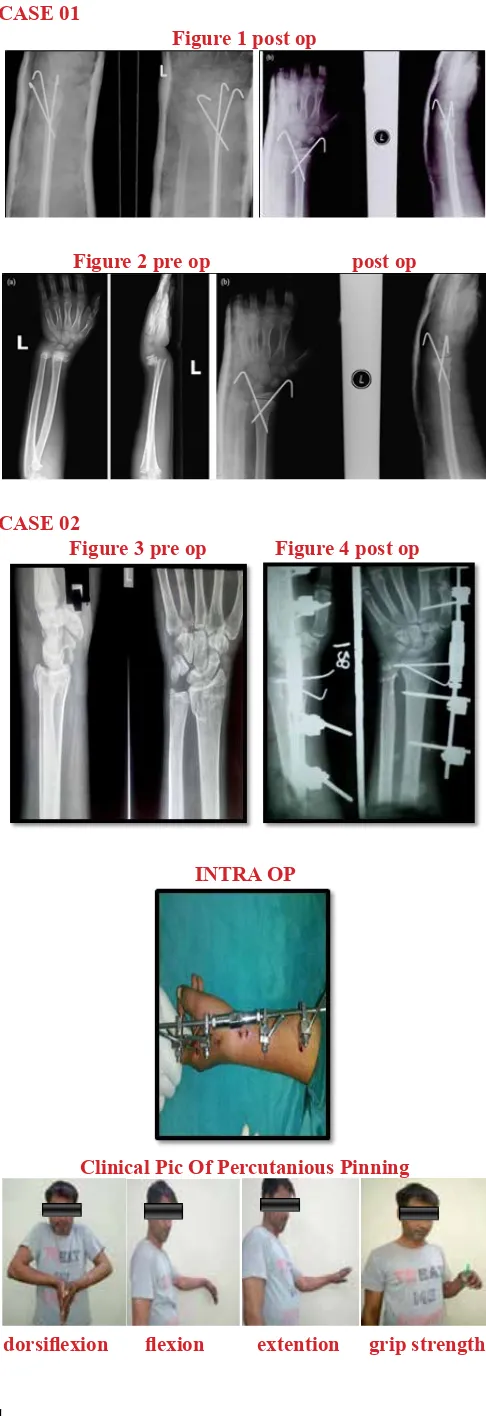
CASE 01
Figure 1 post op
Figure 2 pre op post op
CASE 02
Figure 3 pre op Figure 4 post op
INTRA OP
Clinical Pic Of Percutanious Pinning
dorsiflexion flexion extention grip strength
joint stiffness and painful wrist. Hence, this method is for low-demand elderly patients.5
Percutaneous pinning with K-wires was first recommended by Green as a simple and inexpensive procedure.11
Various techniques of percutaneous pinning are available. Most studies attribute poor results of this technique to radial shortening, wrist stiffness and reflex sympathetic dystrophy.5
In Percutaneous pinning with K-wires
wriststiffness and reflex sympathetic dystrophy occur because of the palmar-flexed position of the wrist in which postoperative immobilization of the fracture is done. Prolonged immobilization of the wrist for greater than 3 weeks increases the magnitude of the problem. Hence we developed our protocol for the treatment of extra-articular distal radius fractures. Fracture reduction was achieved by longitudinal traction and direct pressure over the displaced fragment followed by percutaneous pinning.12
Significant radial shortening was observed in 2 cases only.Radial shortening remains the main displacement in distal end radius fractures, especially intra-articular and comminuted fractures.5
Loosening of one of the K-wires was observed in 2 cases at the time of removal of the pins, but it did not jeopardize fracture alignment. Circumferential cast for additional immobilization was not necessary.12
Radiographic outcomes, the given values of which were compared with those for the contralateral extremity, may affect functional outcome. It is the surgeon’s goal to restore the distal radius to its normal anatomical alignment and congruity. How accurately this is achieved can be measured from radiographs by comparing volar tilt angle, radial inclination etc. with published normal values8. In our study, radial inclination and ulnar variance were superior in the EF group six months postoperatively.
Conclusion
Clinical Pic Of Distractor Fixator
Dorsiflexion Palmar Flexion
Radial Deviation Ulnar Deviation
References
1. Rizzo M, Katt BA, Carothers JT. Comparison of Locked Volar Plating Versus Pinning and External Fixation in the Treatment of Unstable Intraarticular Distal Radius Fractures. Hand N Y N. 2008 Jun;3(2):111–7.
2. Internal fixation versus other surgical methods for treating distal radius fractures in adults - Jariwala - 2014 - The Cochrane Library - Wiley Online Library [Internet]. [cited 2018 Feb 26]. Available from: http:// onlinelibrary.wiley.com/doi/10.1002/14651858. CD011212/full
3. Open reduction and internal fixation versus external fixation for unstable distal radial fractures: A meta-analysis - ScienceDirect [Internet]. [cited 2018 Feb 26]. Available from: https://www.sciencedirect.com/ science/article/pii/S187705681300025X
4. Franceschi F, Franceschetti E, Paciotti M, Cancilleri F, Maffulli N, Denaro V. Volar locking plates versus K-wire/pin fixation for the treatment of distal radial fractures: a systematic review and quantitative synthesis. Br Med Bull. 2015 Sep 1;115(1):91–110.
5. Das AK, Sundaram N, Prasad TG, Thanhavelu SK. Percutaneous pinning for non-comminuted extra-articular fractures of distal radius. Indian J Orthop. 2011;45(5):422–6.
6. Lee DJ, Elfar JC. External Fixation Versus Open Reduction With Locked Volar Plating for Geriatric Distal Radius Fractures. Geriatr Orthop Surg Rehabil. 2014 Sep;5(3):141–3.
7. Lee DJ, Elfar C. Dorsal Distraction Plating for Highly Comminuted Distal Radius Fractures. J Hand Surg. 2015 Feb;40(2):355–7.
8. Chung KC, Mathews AL. Management of Complications of Distal Radius Fractures. Hand Clin. 2015 May;31(2):205–15.
9. Gaillard F. Frykman classification of distal radial fractures | Radiology Reference Article | Radiopaedia.org [Internet]. Radiopaedia. [cited 2017 Sep 22]. Available from: https://radiopaedia. org/articles/frykman-classification-of-distal-radial-fractures
10. Bonnevialle N, Ibnoulkatib A, Mansat P, Bonnevialle P. Kapandji pinning and tuberosities fixation of three- and four-part fractures of the proximal humerus. Int Orthop. 2013 Oct;37(10):1965–71.
11. L1 kk, s2 mg, j3 mc, m4 r. Keywords radius fractures c26.088.268.556, Fracture fixation, internal e04.555.300.300, Arthritis c05.550.114. Study fract fixat distal end radius percutaneous pinning study fract fixat distal end radius percutaneous pinning study fract fixat distal end radius percutaneous pinning study fract fixat distal end radius percutaneous pinning. 2016 Jul 28;,