*Corresponding author:Sunila Hussain
PHARMACEUTICAL RESEARCH
ISSN: 2395-6429, Impact Factor: 4.656
Available Online at www.journalcmpr.com
Volume 4; Issue 7(A); July 2018; Page No. 3437-3443
DOI: http://dx.doi.org/10.24327/23956429.ijcmpr20180480
Research Article
IMMUNOHISTOCHEMICAL EXPRESSION OF VASCULAR ENDOTHELIAL GROWTH
FACTOR-C (VEGF-C) IN SALIVARY GLAND TUMOURS
Sunila Hussain
1., Rakia Sahaf
2., Muhammad Rashid Siraj
3., Fakeha Rehman
4., Sameer Anjum
5.,
Nida Ali
6., Ayesha Saeed
7., Ihtesham-Ud-Din Qureshi
8and Nadia Naseem
91
Department of Oral Pathology, Faryal Dental College, Lahore, Pakistan
2
Oral Pathology, University of Health Sciences, Lahore, Pakistan
3
Department of Surgery, Akhtar Saeed Medical & Dental College, Lahore, Pakistan
4
Department of Pathology, King Edward Medical University Lahore, Pakistan
5
Department of Morbid Anatomy and Histopathology / Oral Pathology,
University of Health Sciences Lahore, Pakistan
6,7,9
Department of Morbid Anatomy and Histopathology, University of Health Sciences Lahore, Pakistan
8
Department of Pathology, Akhtar Saeed Medical & Dental College, Lahore, Pakistan
ARTICLE INFO ABSTRACT
Context/Background:Vascular endothelial growth factor C expression has been focus of research for variety of neoplasms owing to its potential role played in lymphangiogenesis and metastasis through interactions with various molecules.
Objective:This study was designed to determine the immunohistochemical expression of VEGF-C in benign and malignant salivary glands tumours in local Pakistani population.
Materials & Methods:This descriptive study was conducted at the Department of Morbid Anatomy and Histopathology/ Oral Pathology, University of Health Sciences Lahore, Pakistan. Biopsies and detailed clinical data of 85 cases of salivary gland neoplasms (31 benign and 54 malignant) were obtained from different local tertiary care hospitals in Lahore from Jan. 2015 to Sep 2016. After confirming the histologic diagnosis,immunohistochemical expression of VEGF-C was determined in the salivary gland tumours. SPSS version 21.0 Chi-square and Fischer Exact tests were applied for statistical analysis and p<0.05 was considered to be statistically significant.
Results:Expression of VEGF-C and staining patternwas found to be significantly associated with benign and malignant salivary gland tumours (p=0.002 and p<0.0001).
Conclusion
Higher expression of VEGF-C in malignant salivary gland tumours may predict its potential advantage as a biologic marker and promising therapeutic target to restrict the metastatic spread of malignant salivary gland neoplasms to distant organs.
Copyright © 2018 Sunila Hussain et al. This is an open access article distributed under the Creative Commons Attribution License, which permits unrestricted
use, distribution, and reproduction in any medium, provided the original work is properly cited.
INTRODUCTION
Lymphangiogenesis is a crucial player for metastasis in various cancers and this role is verified by the expression of various lymphangiogenic markers. These lymphangiogenic markers enable the pathologists to predict behaviour and prognosis for various malignant neoplasms. Vascular endothelial growth factor-C (VEGF-C) is one such marker(1).
Vascular endothelial growth factor-C (VEGF-C), a 21-kD non-disulfide-linked homodimeric protein, is a member of vascular endothelial growth factor (VEGF) family and platelet-derived growth factor (PDGF) superfamily (2). It has high affinity for both vascular endothelial growth factor receptor 2 & 3
(VEGFR-2 & VEGFR-3). Through its interaction with
VEGFR3 and VEGFR2, VEGF-C promotes
lymphangiogenesis (both peritumoral and stromal) and angiogenesis (2). Molecular interaction of VEGF-C with chemokines facilitates entry of tumour cells into the lymphatic vessels (2), stimulate dilation of lymphatic vessels and hyperplasia of sentinel lymph nodes, thus ultimately leading to metastasis and tumour progression (3).
Salivary gland tumours exhibit tremendous morphological variability in their histologic profile including features like hybrid tumours, anaplasia, lack of proper grading systems and tendency for benign tumours to transform into malignant ones, Article History:
Received 5th April, 2018 Received in revised form 24th May, 2018
Accepted 20th June, 2018 Published online 28th July, 2018
Key words:
so necessitating the use of specialized techniques for proper diagnosis and prediction of their biological behavior(4).
It is a common notion that carcinoma spreads through lymphatics and sarcomas spread through blood vessels(5), the fact that lymphangiogenesis can be a mechanism during malignant transformation of benign salivary gland tumours(6) lead us to study the expression of VEGF-C in benign and malignant salivary gland tumours.
MATERIALS AND METHODS
This study was conducted at the Department of Morbid Anatomy and Histopathology/Oral Pathology, University of Health Sciences, Lahore. A total of 85 biopsies, 25 each of pleomorphic adenoma (PA), adenoid cystic carcinoma (AdCC)& mucoepidermoid carcinoma (MEC), 6 of Warthin tumour (WT) and 2 each of carcinoma ex pleomorphic adenoma (CEPA) and basal cell adenocarcinoma (BCA) of salivary glands reported at Histopathology Departments of University of Health Sciences, King Edward Medical College/Mayo hospital, Sheikh Zaid hospital and Fatima Jinnah Medical College /Ganga Ram Hospital, Lahore from January, 2015 to September, 2016 were included in the study. Detailed clinical data including clinical stage (wherever possible) was retrieved from the respective departmental records. After approval from the ethical committee of University of Health Sciences, Lahore, we proceeded towards the laboratory procedures.
Hematoxylin &eosin staining
Paraffin embedded tissue sections were made from biopsy specimens. Tissue sections of 4µm were cut using rotary microtome and were stained with hematoxylin and eosin stain. Diagnosis was confirmed by 2 oral pathologists/ Histopathologists. Subtype determination of PA was done according to the criteria provided by Seifert (7).
Histologic grading of AdCC was done according to the grading criteria provided by Spiro(8) where mostly tubular or cribriform pattern (no stipulations or minor solid components) was given grade I, 50% solid pattern was grade IIand mostly solid pattern was assigned grade III.
Grading of MECs was done on the basis of less than 20% cystic component (+2), presence of neural invasion (+2), necrosis (+3), ≥ 4 mitoses per 10 high power fields (+3) and anaplasia (+4). Sum of the point values was used to determine low (0-4), intermediate (5-6) or high (7-14) grade MEC(9).
Immunohistochemistry
About 4 µm thick tissue sections were cut with the help of rotary microtome and taken on Poly-L-lysine coated slides for immunohistochemical staining with anti-VEGF-C antibody. Two sections were taken from each block, dried at 600 C for 50 minutes followed by de-waxing in xylene and rehydration in alcohol. Next, the slides were placed in Coplin jars containing citrate buffer (pH 6.0) solution and then in hot water bath (950C) for 40 minutes in order to retrieve antigens (Heat Induced Epitope Retrieval). After removing the slides from hot water bath, they were allowed to cool at room temperature and hydrogen peroxide was added to block endogenous peroxidase activity followed by thorough washing with PBS (phosphate buffered saline).Sections were then incubated with 1-2 drops of protein blocker for 10 minutes to block endogenous enzymatic activity and then again washed
with PBS. This was followed by incubation with primary antibody, rabbit anti-human VEGFC polyclonal antibody (Abcam ab135506, Cambridge, UK) diluted to 1:25µg/ml (suggested dilution by the manufacturer) for 1 hour. Then, sections were incubated successively with Biotinylated Secondary Antibody for 10 minutes and Streptavidin Peroxidase Reagent for 10 minutes before application of DAB (di-amino-benzidine) (2 minutes) to avoid false positive staining. All incubation steps were separated by thorough washing with PBS. Counter staining with hematoxylin was done followed by dehydration and mounting of sections with coverslips using DPX. Positive (oral mucosa and skin) and negative (omission of primary antibody) controls were run with each batch of 20 histological sections of salivary gland tumours. VEGF-C staining was evaluated on the basis of extent and intensity immunolabeling of tumor cells (10).
The intensity (qualitative variable) of staining was scored: 0 (absent), 1 (weak), 2(moderate) and 3(strong)
The proportion (quantitative variable) of tumor cells staining was semi-quantitatively evaluated as:
0 (no positive tumor cells); 1 (1 - 10% positive tumor cells); 2 (11% to 49% positive tumor cells); 3 (>50% positive tumor cells)
Total/Final Score: The sum of the intensity and extent scores
was the final score (0-6). Negative: 0-1
Weak positive (1+): 2 Moderate positive (2+): 3-4 Strong positive (3+): 5-6
Statistical Analysis
The clinical, histological and immunohistochemical data was analyzed statistically using SPSS 21.0. Chi-square and Fischer Exact tests were applied and p-value <0.05 was considered to be statistically significant.
RESULTS
The clinical parameters of the salivary gland tumours studied are summarized in Table 1.
The mean age for benign tumours was found to be 32.52± 16.326 years with an age range of 12-70 years. Most patients were seen in 2nd& 3rd (25.8% each) decades of life. Almost equal gender predisposition was noted (F:M, 1.06:1). Parotid gland (61.3%) was the most frequent site affected followed by minor salivary glands (22.6%) with palate being the commonest site (71.4%).
Cell-rich or cellular subtype of PA (n=12; 48%) was the commonest subtype noted in PA, closely followed by classic (n=11; 44%). Only 2(8%) cases of stroma rich/hypo-cellular subtype were seen in the current study.
Regarding tumour morphology of malignant tumours, 17 (68%) cases of AdCC were of grade I and 8(32%) were grade III. Cribriform pattern (n=15; 60%) was the predominant pattern noted in AdCC followed by tubular (n=6; 24%) and solid (n=4; 16%). As for MEC, 9(36%) were grade I, 7(28%) were grade II and 9(36%) were grade III. Both cases of basal cell adenocarcinoma were of solid subtype characterized by solid nests of cells delineated by basement membrane like material.
Positive nodes were noted in 2(8%) of AdCC, 12(48%) cases of MEC and 2(100%) cases of carcinoma ex PA.
Perineural invasion (PNI) was noted in 9(36%) and 10(40%) cases of MEC and AdCC respectively. One (50%) case of basal cell adenocarcinoma showed PNI.
Vascular invasion (VI) was noted in 18(72%) cases in of AdCC and 1(50%) case of basal cell adenocarcinoma.
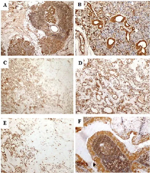
In the normal peritumoral salivary gland tissue, strong nuclear and cytoplasmic expression of VEGF-C was noted in ductal structure while strong nuclear reaction was noted in acinar cells (Figure 1A &B).
The total scores for VEGF-C in benign and malignant salivary gland tumours are summarized in Table 2.
The VEGF-C positive scores were significantly higher in malignant neoplasms (p=0.002). Significant association was noted among the total score of VEGF-C staining in the benign tumours (p=0.004), however, no significant association was noted within the malignant group (p=1.000).In contrast, the type of staining pattern was statistically significant (differed significantly) not only in benign and malignant tumours but also within the groups (p<0.0001) (Table 3).
Table 1 Frequencies, Percentages and p-value Regarding Clinical Data of the Salivary Gland Tumours (n=85)
Parameter PA WT AdCC MEC BCA CEPA Total p-value
F % F % F % F % F % F % F %
Age
Mean Age 30.36 ± 14.838 41.50 ± 20.550 41.32 ± 11.022 31.44 ±2.999 34.00 ± 19.799 19.00 ± 8.485 34.51 ± 14.949
0.028
Minimum (years) 12 18 22 9 20 13 09
Maximum (years) 70 70 70 70 48 25 70
Commonest decade 2nd& 3rd 6th 5th 3rd - - 5th
Gender
Female 14 56 2 33 9 36 13 52 1 50 1 50 40 47 0.724
Male 11 44 4 67 16 64 12 48 1 50 1 50 45 53
F:M 1.3 : 1 1 : 2 1 : 1.8 1.1 : 1 1 : 1 1 : 1 1:1.1
Gland
Parotid 15 60 04 66.7 4 16 17 68 01 50 1 50 42 49.4
<0.001
Submandibular 04 16 01 16.7 0 0 02 08 0 0 1 50 08 9.4
Sublingual 0 0 0 0 0 0 02 08 0 0 0 0 02 2.4
Minor 06 24 01 16.7 21 84 04 16 01 50 0 0 33 38.8
Laterality
Right 10 40 01 16.7 09 36 13 52 1 50 2 100 36 42
0.041
Left 10 20 05 83.3 09 36 1 4 1 50 0 0 33 39
Not mentioned 05 20 0 0 07 28 11 44 0 0 0 0 16 19
Clinical stage
Tumour size
T1-2 - - - - 16 64 17 68 - - - -
0.001
T3-4 - - - - 09 36 08 32 - - - -
Nodes
Positive - - - - 02 08 12 48 0 0 02 100 23 42.6
Negative - - - - 16 64 11 44 0 0 0 0 16 29.6
Not reported - - - - 07 28 02 8 02 100 0 0 15 27.8
Metastasis
M0 - - - - 25 100 24 96 - - - -
M1 - - - - 0 0 01 04 - - - -
Final stage
I/II - - - - 15 60 13 52 - - - -
III/IV - - - - 07 28 08 40 - - - -
Unknown - - - - 03 12 04 16 - - - -
Table 2 VEGF-C Total Score in Benign and Malignant Salivary Gland Tumours (n=85)
Tumour Total Score
VEGF-C Negative Weak positive
Moderate positive
Strong positive
p-value
F % F % F % F %
0.002
Pleomorphic adenoma 5.36±0.490 0 0 0 0 0 0 25 100
Warthin tumour 4.33±0.516 0 0 0 0 03 50 03 50
Adenoid cystic carcinoma 5.48±0.510 0 0 0 0 0 0 25 100
Mucoepidermoid
carcinoma 5.40±0.577 0 0 0 0 01 04 24 96
Basal cell
adenocarcinoma 5.50±0.707 0 0 0 0 0 0 02 100
Carcinoma ex
The VEGF-C positivity was strong in 28(90.3%) of benign tumours followed by moderate in 3(9.7%) (Table 2). In PA, both the epithelial component and mesenchymal/myoepithelial cells showed strong nuclear and cytoplasmic staining reaction (Table 3 &Figure 1C-1E). Predominant staining pattern was significantly associated with the type of tumour (p<0.0001). It was nuclear and cytoplasmic in all cases of PA while cytoplasmic alone in WT (Table 3 and Figure 1F). Nuclear reaction was noted in abluminal cells around the ductal structures. Stroma showed weak staining in most cases most cases of PA while WT showed strong reaction in stroma of all cases (Figure1C-F).
Figure 1 Photomicrograph (A) & (B) showing strong cytoplasmic and nuclear
staining in normal salivary gland (VEGF-C; 40X and 100X), (C) low power view of strong positive staining in PA (VEGF-C; 40X), (D) & (E) showing strong to moderate staining in epithelial and mesenchymal component of PA (VEGF-C; 100X) (F) showing moderate positive cytoplasmic staining in WT
(VEGF-C; 100X).
All the malignant tumours were strong positive for VEGF-C antibody, only 1 case of MEC was moderate positive.
The staining pattern was mostly nuclear & cytoplasmic (n=29;53.7%) followed by membranous and cytoplasmic 18(33.3%) cases. Only 1 (1.9%) case showed cytoplasmic localization alone. Most cases (n=21; 84%) of AdCC showed cytoplasmic &nuclear staining. Nuclei alone were positive in the abluminal cells lining the cribs and tubules in AdCC. On the other hand most MECs (n=18; 72%) showed membranous & cytoplasmic localization and only 7(28%) cases showed
nuclear & cytoplasmic reaction in addition to membranous. Basal cell adenocarcinomas showed membranous and cytoplasmic staining reaction in both cases.One case of carcinoma ex pleomorphic adenoma showed cytoplasmic reaction and the other showed nuclear and cytoplasmic staining.
Stroma in AdCC and MEC was moderately reactive in 13(52%) and 03(12%) cases respectively, strong in 7(28%) & 2(100%) cases of MEC & CEPA respectively. Weak stromal reaction was noted in 2(100%), 12(48%) and 15(60%) cases of BCA, AdCC & MEC respectively.
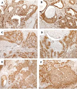
Vascular endothelial growth factor C staining pattern was significantly associated with the histological patterns in AdCC (p= 0.003) and histological grades in MEC (p<0.001). No significant association of VEFG-C antibody staining was noted with clinical stage, histological grade, lymph node involvement, perineural invasion or vascular invasion in both AdCC and MEC. Figures 2 &3 show anti-VEGF-C staining in grades of AdCC and MEC respectively while Figure 4 shows staining in CEPA (Figure 4A-4B) and BCA (Figure 4C-4D).
Figure 2 Photomicrograph (A) & (B) showing strong positive anti- VEGF-C
nuclear and cytoplasmic staining in tubular pattern of adenoid cystic carcinoma (VEGF-C;40X & 100X), (C)& (D) Strong positive staining in cribriform pattern of AdCC (VEGF-C;40X & 100X), (D) & (E) strong positive
cytoplasmic staining in solid pattern of AdCC (VEGF-C ;40X & 100X)
Table 3 VEGF-C Staining Pattern in Benign and Malignant Salivary Gland Tumours (n=85)
Tumour
Staining Pattern p-value
Cytoplasmic alone
Membranous & Cytoplasmic
Nuclear & Cytoplasmic
<0.0001
F % F % F %
Pleomorphic adenoma - - - - 25 100
Wartin tumour 6 100 - - - -
Adenoid cystic carcinoma 4 16 - - 21 84
Mucoepidermoid carcinoma - - 18 72 7 28
Basal cell adenocarcinoma - - 2 100 - -
Carcinoma ex
Figure 3 Photomicrograph (A)&(B) showing strong nuclear cytoplasmic positive staining reaction in grade I MEC (VEGF-C; 40X &100X), (C) & (D) Membranous staining in most cells of grade II MEC (VEGF-C; 40X &100X), (E) & (F) Strong positive staining in grade III MEC (VEGF-C; 40X & 100X)
Figure 4 Photomicrograph (A)& (B) showing strong positive cytoplasmic and
membranous staining reaction in BCA (VEGF-C ; 40X &100X), (C) & (D) Strong membranous and cytoplasmic staining in most cells of CEPA
(VEGF-C;40X & 100X). Note the strong stromal reactivity in CEPA.
DISCUSSION
Salivary gland tumours constitute 1-4% of all tumours occurring in human body (11). Among these, pleomorphic adenoma is the most commonly occurring benign salivary gland tumour in any location mostly involving the parotid gland (12). As regards the commonest malignancy of salivary glands, there is some debate. Some studies have reported adenoid cystic carcinoma to be the most commonly occurring malignant tumour of salivary glands(4,13) while others have named mucoepidermoid carcinoma as the commonest(14,15). VEGF-C is one of the lymphangiogenic marker being studied extensively to accurately determine its role in cancer metastasis. In the current study, we determined the expression
of VEGF-C in 85 cases (6 different types) of salivary gland tumours. Of these, 31 were benign (n=25 pleomorphic adenoma and n=06 Warthin tumour) and 54 were malignant (n=25 adenoid cystic carcinoma, n=25 mucoepidermoid carcinoma, n=02 basal cell adenocarcinoma and n=02 carcinoma ex pleomorphic adenoma).
The mean age of the patients for benign tumours (32.52± 16.326 years), malignant tumours (35.65±14.130 years) and total salivary gland neoplasms (34.51 ± 14.949 years) studied in the current study is quite lower than other studies conducted worldwide(13,16). Even when these tumours were considered individually (Table 1), they seem to affect younger age group in our population than reported in other studies(13,16-17). These differences may be attributed to geographical, racial or ethnic dissimilarities among the various populations or to the variations in sample size of the study.
In line with the findings of current study, Kɪzɪl(13)reported female predisposition for PA and MEC and male predilection for WT and AdCC. Other studies, however, have reported different findings(17).
Regarding site distribution of these salivary gland tumours, the present study is in accordance with other national and international studies with parotid being the commonest site for PA, WT, MEC, minor salivary glands (palate) for AdCC, both major and minor glands in basal cell adenocarcinoma and major glands for ca ex PA(13,16-17).
Byrd(18) reported lymph node involvement in 42.2% cases of MEC which is in accordance with the current study which reports nodal positivity in 48% cases.In contrast, Liu(19) reported only 13.8% positive nodes in MEC. Most cases of MEC were stage I & II which is line with the findings of Liu(19)who reported 68% cases of stage I/II. Bianchi(20) reported 9% positive nodes in AdCC which is almost same as found in the current study. In concordance with the findings of Ko21 almost 72% AdCC were of stage I/II.
McHugh(22) and Agarwal(23) reported peri-neural invasion in 28.7% and 32.4% cases of MEC and AdCC respectively which is close to the findings in the current study (MEC:36%, AdCC: 40%).
The staining reaction for anti-VEGF-C in the normal salivary gland tissue was strong nuclear and cytoplasmic in the ductal structures while in the acini the cytoplasmic intensity (when noted) was less profound than ducts however, nuclei of the acinar cells were strong positive. This localization of VEGF-C in normal salivary gland tissue is in accordance with other reported studies(5).
reported enhanced VEGF and HIF-1 in PA and concluded that hypoxia controls the VEGF expression in PA.
In contrast to the current study, Fujita(5) reported that VEGF-C was barely detectable in cases of AdCC. They also reported higher VEGF-C positive reaction in salivary glands than AdCC using real-time reverse transcriptase-polymerase chain reaction(5).
In line with the current study, Gleber-Netto(28) reported moderate to strong VEGF-C expression in all cases of MEC. The staining pattern was nuclear and cytoplasmic(28) which is slightly different from the current study as we also noted membranous expression in few cases. In addition, they also reported low lymphatic vessel density in MEC which cannot be explained by VEGF-C expression.
Mello(29) studied VEGF-C expression in different salivary gland carcinomas. They divided those carcinomas into high risk and low risk depending upon their metastasis to lymph nodes. They reported ≤ 25% positive cells in 29 cases and >25% in 16 cases. In line with the current study, they did not find any significant association between VEGF-C expression and high/low risk salivary gland cancers or peri-/ intra-tumoral lymph vessels. They suggested that either VEGF-C produced by salivary gland cancer cells is non-functional or there may be other markers involved in tumour lymphangiogenesis. They also concluded that low LVD in salivary gland cancers in contrast to head and neck squamous cell carcinomas may depict the different malignant behaviour of these cancers i.e. they may have different capacity to invade the lymphatic vessels in addition to the mere presence of these conduits.
CONCLUSION
As evident from the studies being conducted all over the world, VEGF-C is a potential lymphangiogenic marker in a large number of tumours. Its higher expression in malignant salivary gland tumours may dictate its potential advantage as a biologic marker in determining the lymphatic spread of these tumors. In addition, it may be utilized as a promising therapeutic target to restrict the metastatic spread of malignant salivary gland neoplasms to lymph nodes and distant organs1.
Acknowledgements
The authors acknowledge the encouragement extended by the Vice Chancellor of University of Health Sciences, Lahore Pakistan. Also, to Mr. Ghulam Rasool, the laboratory staff of Oral Pathology Department of University of Health Sciences, Lahore, Pakistan for their technical and logistic support. We also acknowledge the support of the Heads of Histopathology Departments of King Edward Medical University, Fatima Jinnah Medical College, Postgraduate Medical Institute and Sheikh Zaid Hospital, Lahore for their assistance in providing access to material required for this research project.
References
1. StackerSA, Williams SP, Karnezis T, Shayan R, Fox SB, Achen MG. Lymphangiogenesis and lymphatic vessel remodelling in cancer. Nature Reviews/Cancer 2014; 14: 159-172
2. Tammela T, Enholm B, Alitalo K, Paavonen K. The biology of vascular endothelial growth factors. Cardiovascular Research 2005; 65: 550- 563
3. Liersch R, Hirakawa S, Berdel WE, Mesters RM, Detmar M (2012) Induced lymphatic sinus hyperplasia
in sentinel lymph nodes by VEGF-C as the earliest premetastatic indicator. Int J Oncol 41(6): 2073-2078. 4. Vuhahula EAM. Salivary gland tumors in Uganda:
clinical pathological study. Afr. Health Sci 2004;4(1): 15-23.
5. Fujita G, Sato S, Kishino M, Iwai S, Nakazawa M, Toyosawa S, Yura Y, Ogawa Y. Lymphatic vessels and related factors in adenoid cystic carcinoma of the salivary gland. Modern Pathology 2011; (2011) 24: 885-891.
6. Soares AB, Ponchio L, Juliano PB, Arau´jo VC, Altemani A. Lymphatic vascular density and lymphangiogenesis during tumour progression of carcinoma ex pleomorphic adenoma. J ClinPathol 2007; 60: 995-1000.
7. Seifert G, Langrock I, Donath K. A pathological classification of pleomorphic adenoma of the salivary glands (author’s transl). HNO 1976; 24(12):41526. 8. Spiro RH, Huvos AG, Strong EW. Adenoid cystic
carcinoma of salivary origin. A clinicopathologic study of 242 cases. Am J Surg 1974; 128(4):512-20.
9. Auclair PL, Goode RK, Ellis GL. Mucoepidermoid carcinoma of intraoral salivary glands. Evaluation and application of grading criteria in 143 cases. Cancer 1992; 69(8):2021-2030.
10. Abdo MM, Gaballah ET, Fikry HE, Mosbah MM (2014) Expression of VEGF-C in oral precancerous lesions and oral squamous cell carcinoma. Mansoura Journal of Dentistry 1(3):105-108.
11. Alves FA, Pires FR, De Almeida OP, Lopes MA, Kowalski LP. PCNA, Ki67 and p53 expressions in submandibular salivary gland tumours. Int J Oral Maxillofac Surg 2004;33(6): 593-7.
12. Fu H, Wang J, Wang L, Zhang Z, He Y. Pleomorphic adenoma of salivary glands in children and adolescents. Journal of Pediatric Surgery 2012; 47:715-719. 13. Kızıl Y, Aydil U, Ekinci O, Dilci A, Köybaşюğlu A,
Düzlü M, Ĭnal E. Salivary Gland Tumors in Turkey: Demographic Features and Histopathological Distribution of 510 Patients. Indian J Otolaryngol Head Neck Surg 2013; 65(Suppl 1):112-20.
14. Long - Jiang L, Yi L, Yu - Ming W, Hua L, Hong - Wei Z. Clinical analysis of salivary gland tumour cases in West China in past 50 years. Oral oncol 2008; 44: 187192
15. Ettl T, Schwarz - Furlan S, Gosau M, Reichert TE. Salivary gland carcinomas. Oral Maxillofac Surg 2012; 16: 267-283.
16. Li LJ, Li Y, Wen YM, Liu H, Zhao HW. Clinical analysis of salivary gland tumor cases in West China in past 50 years. Oral Oncol 2008; 44(2): 187-92.
17. Zaman S, Majid S, Chugtai O, Hussain M, Nasir M. Salivary gland tumours: A review of 91 cases. J Ayub Med Coll Abbottabad 2014; 26(3):361-3.
18. Byrd AS, Spector ME, Carey TE, Bradford CR, McHugh JB. Predictors of recurrence and survival for head and neck mucoepidermoid carcinoma. Otolaryngol Head Neck Surg 2013; 149(3): 402-408.
20. Bianchi B, Copelli C, Cocchi R, Ferrari S, Pederneschi N, Sesenna E. Adenoid cystic carcinoma of intraoral minor salivary glands. Oral Oncol 2008; 44(11): 1026-1031.
21. Ko JJ, Siever JE, Hao D, Simpson R, Lau HY. Adenoid cystic carcinoma of head and neck: Clinical predictors of outcome from a Canadian center. CurrOncol 2016;23(1):26-33.
22. McHugh CH, Roberts DB, El-Naggar AK, Hanna EY, Garden AS, Kies MS, Weber RS, Kupferman ME. Prognostic factors in mucoepidermoid carcinoma of the salivary glands. Cancer 2011; 118(16):3928-36. 23. Agarwal JP, Jain S, Gupta T, Tiwari M, Laskar SG,
Dinshaw KA, Chaturvedi P, D’cruz AK, Shrivastava SK. Intraoral adenoid cystic carcinoma: Prognostic factors and outcome. Oral Oncol 2008; 44(10):986- 993.
24. Soares AB, Ponchio L, Juliano PB, Arau´jo VC, Altemani A. Lymphatic vascular density and lymphangiogenesis during tumour progression of carcinoma ex pleomorphic adenoma. J Clin Pathol 2007; 60: 995-1000.
25. Salzman R, Stárek I, Kučerová L, Skálová A, Hoza J.
Neither expression of VEGF-C/D nor lymph vessel density supports lymphatic invasion as the mechanism
responsible local spread of recurrent salivary
pleomorphic adenoma. Virchows Arch 2014: 464:29-34.
26. Teymoortash A, Schrader C, Shimoda H, Kato S,
Werner JA (2007) Evidence of lymphangiogenesis in
Warthin's tumor of the parotid gland. Oral Oncol
43:614-618.
27. Swelam W, Ida-Yonemochi H, Maruyama S, Ohshiro
K, Cheng J, Saku T (2005) Vascular endothelial growth factor in salivary pleomorphic adenomas: one of the
reasons for their poorly vascularized stroma. Virchows
Arch 446(6):653-662.
28. Gleber-Netto FO, Floreˆncio TNG, de Sousa SF, Guimara˜ es Abreu MHN, Mendonc¸a EF, Aguiar MCF.
Angiogenesis and lymphangiogenesis in
mucoepidermoid carcinoma of minor salivary glands. J Oral Pathol Med 2012; 41: 603-609.
29. Mello MF, Costa AF, Freitas LL, Soares AB, Araujo
VC, Tincani AJ, Martins AS, Altemani A (2011)
Lymphatic vessel density and expressions of
lymphangiogenic growth factors in salivary carcinomas.
Neoplasma 58(4):331-336.
How to cite this article:
Sunila Hussain et al (2018) 'Immunohistochemical Expression Of Vascular Endothelial Growth Factor-C (Vegf-C) In Salivary Gland Tumours', International Journal of Current Medical And Pharmaceutical Research, 04(7), pp. 3437-3443.