Retraction Notice.
Int J Med Res Prof.. www.ijmrp.com
Retraction notice to: Clinical Importance of Evaluation of Iron Profile
Parameters in Hypothyroid Patients
Neeraj Singla
1, Heena Singla
2*1MBBS, DNB Medicine, Assistant Professor,
Department of General Medicine, Government Medical College, Sector 32, Chandigarh, India. 2*MBBS, MD Biochemistry, Assistant Professor,
Department of Biochemistry, GGS Medical College, Faridkot, Punjab, India.
*Corresponding Author:
Dr Heena Singla, Opposite MLA Deep Malhotra house, Old Cantt. Road, Faridkot, Punjab, India. Email: [email protected] DOI of Original article: 10.21276/ijmrp.2018.4.1.017
This article (Accepted on 24 December 2017; Published 31 January 2018) in the Volume 4 Issue 1 January 2018 issue of the Journal by Dr. Neeraj Singla and Dr. Heena Singla, has been retracted with the agreement of the authors. It was brought to the attention of the Editor-in-Chief regarding misconduct by authors. Subsequently after thorough investigation and discussion by editorial team it was decided to retract the article because of Scientific Misconduct that involves Data fabrication and falsification.
Original Research Article.
88 |P a g e Int J Med Res Prof.2018 Jan; 4(1); 88-93. www.ijmrp.com
Clinical Importance of Evaluation of Iron Profile Parameters in
Hypothyroid Patients
Neeraj Singla
1, Heena Singla
2*1MBBS, DNB Medicine, Assistant Professor,
Department of General Medicine, Government Medical College, Sector 32, Chandigarh, India.
2*MBBS, MD Biochemistry, Assistant Professor,
Department of Biochemistry, GGS Medical College, Faridkot, Punjab, India.
ABSTRACT
Introduction: The prevalence of iron deficiency anaemia has been found to further increase along with increase in the incidence of thyroid disorders. Low thyroid function has been one of the most overlooked causes of iron deficiency anaemia. Also, normal thyroid status requires adequate levels of many trace elements, which include iodine and iron. This study was carried out for estimation of the levels of serum iron, TIBC and serum ferritin in hypothyroid patients.
Methods: This case-control study was carried out at a tertiary care center. 100 cases of hypothyroidism and 100 apparently healthy euthyroid controls were enrolled for the study. Apart from routine laboratory investigations, measurement of serum ferritin, iron and TIBC was done. Diagnosis of hypothyroidism was confirmed by measurement of levels of free T3, free T4 and TSH.
Results: Patients of hypothyroidism had significantly lower levels of iron and ferritin, while TIBC was significantly higher in these patients in comparison with euthyroid healthy controls. (p = 0.00, p =0.00, p = 0.00). Strong positive correlation was established between the levels of free T4 and serum ferritin (r = + 0.63, p< 0.05) in these patients.
Conclusion: Hypothyroidism may cause bone marrow
repression and/or decrease in production of erythropoietin. In our case-control study, a large number of hypothyroid patients had underlying iron deficiency anaemia as well. So screening for iron deficiency anaemia and its timely treatment should be regarded as an important part of treatment in hypothyroid patients to achieve best results.
Keywords: Primary Hypothyroidism, Iron Deficiency Anaemia, Erythropoiesis.
*Correspondence to:
Dr Heena Singla,
Opposite MLA Deep Malhotra house, Old Cantt. Road, Faridkot, Punjab, India.
Article History:
Received: 07-11-2017, Revised: 29-11-2017, Accepted: 24-12-2017
Access this article online
Website: www.ijmrp.com
Quick Response code
DOI:
10.21276/ijmrp.2018.4.1.017
INTRODUCTION
Thyroid dysfunction is a common endocrine disorder worldwide. In India too, there is a significant burden of thyroid diseases.
According to a recent study, about 11.5% of Indian population suffers from thyroid disease, while prevalence of undetected subclinical hypothyroidism is much higher.1Normal thyroid status
is dependent on adequate levels of many trace elements for both synthesis and metabolism of thyroid hormones. The most important ones being iodine, iron, selenium and zinc.2
An epidemiological study by Alvarez-Uria G et al found that iron deficiency is the major cause of anaemia in developing countries.3
A study by Pasricha et al illustrated that around 70 percent children suffered from anaemia in India.4 Another study by
Thankachan P et al has also shown high prevalence of iron deficiency anaemia in young women in India.5 Though
most readily available sign of iron deficiency anaemia is low concentration of haemoglobin, a significant fall in haemoglobin in circulation cannot be detected until the late stage of iron deficiency.6
So iron deficiency has been defined as occurring when body’s iron
stores become depleted, a restricted supply of iron to tissues become apparent.7
A study by Takamatsu J et al has established a very close inter-relation between low thyroid function and low iron or more specifically, low ferritin.8 Several studies have shown that
nutritional iron deficiency may cause significant reduction in the levels of T3 and T4 in circulation.9 Iron deficiency has been
reported to impair synthesis of thyroid hormones, which could increase the need for thyroid medication. One hypothesis is that iron is a component of thyroid peroxidase (TPO). This enzyme participates in the first two steps of thyroid hormone synthesis.10
Thyroid peroxidase is a membrane bound glycosylated haemoprotein which plays a key role in synthesis of thyroid hormones. This enzyme promotes oxidation of iodine to iodide radicals (organification), and further binding of iodide to tyrosyl residues of thyroglobulin. Hence mono-iodotyrosine (MIT) and di-iodotyrosine (DIT) are produced.
Neeraj Singla & Heena Singla. Iron Profile Parameters in Hypothyroid Patients
89 |P a g e Int J Med Res Prof.2018 Jan; 4(1); 88-93. www.ijmrp.com Thyroid hormone T3 is produced by coupling of mono-iodotyrosine
(MIT) and di-iodotyrosine (DIT), while thyroid hormone T4 is produced by coupling of two molecules of di-iodotyrosine (DIT). A separate coupling enzyme has not yet been identified. Since it is an oxidative process, it is assumed that the same thyroid
peroxidase (TPO) enzyme which promotes oxidation of iodine to iodide radicals, catalyses the coupling reactions also. This hypothesis is supported by the observation that the same drug which inhibits iodine oxidation also inhibits coupling step of thyroid hormone synthesis.11
Fig 1: Role of Thyroid Peroxidase enzyme in thyroid hormone synthesis.
Thyroid Peroxidase enzyme first generates I2 by oxidizing I- ions
present in the follicular lumen. Thyroid Peroxidase then "organifies" the generated I2 by covalently linking it with the
tyrosine residues present in Thyroglobulin. This generates either single or doubly-iodinated species of tyrosine termed "Monoiodotyrosine (MIT)" and "Diiodotyrosine (DIT)", respectively. Peroxidase then combines MIT and DIT residues to generate T4 or
T3 species within the thyroglobulin protein, a process termed
"Coupling". Importantly, peroxidase is much more efficient at combining of two DIT residues and thus generation of T4 occurs
much more readily, explaining why the thyroid gland primarily produces T4 rather than T3.
Another reason can be reduced peripheral conversion of T4 to T3 in iron deficiency. Only a small fraction of T4 gets converted into T3, while a larger proportion gets converted into rT3 which is a physiologically inactive metabolite. A study has shown increased in vitro hepatic rT3 deiodination in iron deficiency with decreased levels of T4-5’ deiodinase in circulation.12 Yet it is not clear how
iron deficiency exerts its effect on activity of T4-5’ deiodinase.
Hypothyroidism may itself lead to low iron levels due to poor gut absorption, which may be because of decreased levels of digestive acids, and/or presence of associated autoimmune diseases like celiac disease.13 Iron deficiency in hypothyroidism
may also be possibly due to heavy menstruation as seen in some female patients.14 Therefore, this study was planned to estimate
the levels of serum iron, ferritin and TIBC in hypothyroid patients.
MATERIALS AND METHODS
This case-control study was carried out in Department of Medicine, Govt. Medical College, Sector 32, Chandigarh. The study was conducted on 100 hypothyroid patients (age group
20-60 years) and 100 age and sex matched euthyroid healthy controls. An informed consent was taken from all the study subjects before enrolling them for the study. The diagnosis of hypothyroidism was made on the basis of clinical history, physical examination and measurement of thyroid profile parameters. A detailed clinical history of the subject was taken on a pre-designed proforma. Examination was done which included measurement of body weight, body mass index, pulse rate and palpation for enlarged thyroid gland. Then each patient was evaluated for hypothyroidism or hyperthyroidism by separate questionnaires.15,16
Summed scores of plus 20 or above in the hypothyroidism questionnaire indicated a high probability of thyroid dysfunction.
Zulewski’s Hypothyroidism clinical score. 15 SYMPTOMS
Diminished Sweating 12
Hoarseness 07
Paraesthesia 14
Dry skin 21
Constipation 14
Impairment of hearing 02
Weight increase 18
PHYSICAL SIGNS
Slow movements 13
Delayed ankle reflex 16
Coarse skin 14
Periorbital puffiness 15
Cold skin 14
Women <55 years 10
After an informed consent, all the subjects were advised to visit Medicine OPD after 12 hours overnight fasting. Around six ml of peripheral venous blood was taken under all aseptic precautions. The investigations included CBC, PBF, Hb, FBS, lipid profile, liver function tests and renal function tests. Apart from these basic routine laboratory parameters, measurement of thyroid profile (free T3, free T4 and TSH) was done on Access-2 chemiluminescence machine.
Estimation of serum iron and TIBC was done on Siemens Dimension fully autoanalyser. Serum ferritin was measured on Immulite 1000 chemiluminescence machine for all the study subjects.
Reference ranges for the thyroid hormones are17:
Free T3 (2.5-3.9 pg/ml) Free T4 (0.61-1.12 ng/dl) TSH (0.35- 5.5 mIU/L)
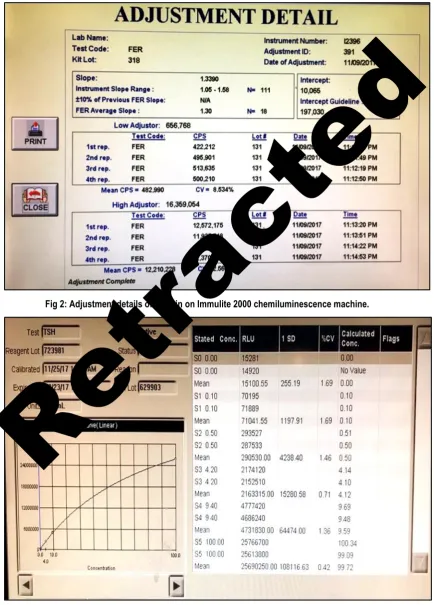
Fig 2: Adjustment details of Ferritin on Immulite 2000 chemiluminescence machine.
Fig 3: Calibration Curve of TSH on Access-2 chemiluminescence machine.
Neeraj Singla & Heena Singla. Iron Profile Parameters in Hypothyroid Patients
91 |P a g e Int J Med Res Prof.2018 Jan; 4(1); 88-93. www.ijmrp.com
Following subjects were excluded from the study:
1. Patients with hepatic disorders and renal diseases since thyroid hormones metabolism is affected in these conditions. 2. Those on drugs which may affect thyroid hormone
metabolism, including oral contraceptives since oestrogen has anti-thyroid action.
3. Pregnant and lactating females, and females with PCOD. 4. Those receiving iron supplements or taking treatment with
thyroxine.
Statistical Analysis
Statistical analysis of the results was done using SPSS version 19 software. The p value was calculated to know the level of significance between the two groups. A p value < 0.001 was
considered to be highly significant. Pearson’s correlation
coefficient (r value) was calculated to determine the level of correlation between the concerned parameters.
RESULTS
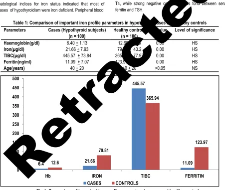
Haematological indices for iron status indicated that most of the cases of hypothyroidism were iron deficient. Peripheral blood
smear of these patients showed microcytic hypochromic anaemia. As shown in Table 1, we found significantly lower levels of iron and ferritin in most of the patients of hypothyroidism in our study. On other hand, TIBC was significantly higher in these patients. (p < 0.001, p < 0.001, p < 0.001) Iron and ferritin levels were significantly decreased in patients with deranged thyroid status. As depicted in table 2, mean value of FT4 was much lower in hypothyroid cases in comparison with healthy controls, while mean value of TSH was much higher in hypothyroid cases in comparison with healthy controls (p<0.01, p=0.00 respectively). There was a significant association of free T4 and TSH with only the serum ferritin (r = +0.63, p< 0.001, r = -0.34, p< 0.05 respectively). The statistical model showed that when serum ferritin was used as an independent variable, subjects with lower ferritin had a significantly lower free T4 to free T3 ratio (r = -0.51, p< 0.01).
As shown in Table 3 and the above scatter plot, there was found strong positive correlation between serum ferritin and free T4, while strong negative correlation was fond between serum ferritin and TSH.
Table 1: Comparison of important iron profile parameters in hypothyroid cases and healthy controls
Fig 4: Comparison of important iron profile parameters in cases and healthy controls Table 2: Mean value of thyroid profile parameters in cases and healthy controls Parameters
with range
Cases(Hypothyroid subjects)
(n = 100)
Healthy controls (n = 100)
p value Level of
significance FT3 (pg/ml)
(Range)
2.73 +0.82 (1.90 – 3.55)
2.85 + 0.40 (2.12-3.8)
>0.05 NS
FT4 (ng/dl) (Range)
0.21 -1.06 (0.64 +0.42)
0.90 + 0.15 (0.63-1.2)
<0.01 HS
TSH (uIU/ml) (Range)
22.10 +19.87 (12.12 – 51.87)
2.61 + 1.12 (0.66-4.7)
0.00 HS
6.4 21.66 445.57 11.09 12.6 79.81 365.94 123.97 0 50 100 150 200 250 300 350 400 450 500
Hb IRON TIBC FERRITIN
CASES CONTROLS
Parameters Cases(Hypothyroid subjects)
(n = 100)
Healthy controls (n = 100)
p value Level of significance
Haemoglobin(g/dl) 6.40 + 1.13 12.60 + 1.08 0.00 HS
Iron(µg/dl) 21.66 + 7.93 79.81 + 43.2 0.00 HS
TIBC(µg/dl) 445.57 + 73.84 365.94 + 77.97 0.00 HS
Ferritin(ng/ml) 11.09 + 7.07 123.97 + 54.76 0.00 HS
Age(years) 40 + 20 40 + 20 >0.05 NS
Fig 5: Comparison of thyroid profile parameters in cases and healthy controls
Table 3: Correlation between the levels of serum ferritin and thyroid profile parameters in hypothyroid subjects
HS = Highly significant
Fig 6: Scatter diagram showing correlation between Ferritin and TSH (r= - 0.34, p<0.001).
DISCUSSION
In our study, the cases of hypothyroidism had much lower serum ferritin, lower serum iron and higher TIBC. A larger fraction of hypothyroid subjects were deficient in iron. These results are in accordance with other studies which reported that iron deficiency anaemia is frequently associated with low levels of thyroid hormones.9,12,14
In our study, a significant negative correlation was observed between TSH and ferritin. Bremner AP et al found that serum iron levels were significantly lower in patients with subclinical hypothyroidism in comparison with euthyroid healthy controls. Their study also illustrated significant inverse relationship of TSH with serum iron and transferrin saturation.18 Banday et al reported
iron deficiency in a significant percentage of patients with primary hypothyroidism.19At same time, a study by Akhter S et al reported
that deranged thyroid hormone status in iron deficient people could be a reflection of disturbed activities of iron dependent enzyme thyroperoxidase. This enzyme has an important role in synthesis of thyroid hormones.20 A similar study by Hess Y et al
mentioned the role of iron in transportation of thyroid hormone into the circulation. According to their study, lack of iron causes pooling of thyroid hormones leading to metabolically hypothyroid condition due to thyroid resistance.21 Our study
illustrated negative correlation of TSH with iron, while positive correlation was observed between TSH and TIBC, but these were not statistically significant.
2.73
0.64
22.1
2.85
0.9
2.61
0 5 10 15 20 25
FT3 FT4 TSH
CASES CONTROLS
Parameter r value p value Level of significance
Ferritin and free T4 0.63 <0.05 HS
Ferritin and TSH -0.34 <0.001 HS
Ferritin and free T4 to free T3 ratio -0.51 <0.01 HS
Neeraj Singla & Heena Singla. Iron Profile Parameters in Hypothyroid Patients
93 |P a g e Int J Med Res Prof.2018 Jan; 4(1); 88-93. www.ijmrp.com Thyroid hormones have a well-known role in regulation of gene
expression for transferrin. The expression of gene for ferritin has also been reported to be induced by T3 hormone.22 These
hormones also play an important role in erythropoiesis. They stimulate erythroid colony development. Hypothyroidism may cause bone marrow repression and/or decrease in production of erythropoietin due to decreased oxygen requirement.23
Thyroxine administration in these patients has been reported to increase erythropoietin levels and improve erythropoiesis. This leads to increased iron requirement and may culminate in manifestations of iron deficiency.24 Already existing iron deficiency
in these hypothyroid patients may make the clinical picture worse. On other hand, the key enzyme in thyroid hormone synthesis, thyroperoxidase is iron dependent. Hence iron deficiency may be the underlying cause in development of hypothyroidism. This fact is of great importance while treating these patients. The symptoms of sympathetic overstimulation due to anaemia may worsen on administration of thyroxine. Thus the condition becomes a vicious cycle since iron deficiency may both be a cause and/or effect of hypothyroidism. An important limitation of our study was that anti-TPO Antibodies (Anti-thyroperoxidase Antibodies) could not be assayed in the study subjects. We tried to exclude the patients on any kind of anti-oxidants supplements. But many patients are in habit of taking different herbs as a routine in our country, which may be missed even by a careful drug history.
CONCLUSIONS
Our study established a significant cause and effect relationship between iron deficiency and thyroid dysfunction. Hence it can be concluded that screening for iron deficiency anaemia is very important in patients with hypothyroidism since it may guide the treatment protocol in these patients.
ACKNOWLEDGEMENTS
Authors are thankful to all the volunteers who participated in the study. Authors did not recieve any kind of technical assistance or writing assistance
REFERENCES
1. Tiwari N. Prevalence of hypothyroidism in adults: An epidemiological study in eight cities of India. Indian Journal of Endocrinology and Metabolism. 2013; 17(4): 647-652.
2. Metwalley KA, Farghaly HS, Hassan AF. Thyroid status in Egyptian primary school children with iron deficiency anemia: Relationship to intellectual function. Thyroid Res Pract.. 2013; 10: 91-95.
3. Gerardo Alvarez-Uria, Naik PK, Midde M, Yalla PS, Pakam R. Prevalence and severity of anaemia stratified by age and gender in rural India. Anaemia. Volume 2014, ID 176182. doi: org/10.1155/2014/176182.
4. Pasricha SR, Black J, Muthayya S. Determinants of anaemia among young children in rural India. Paediatrics.2010;126(1):e140-9. 5. Thankachan P, Muthayya S, Walczyk T, Kurpad AV, Hurrell RF. An analysis of the etiology of anaemia and iron deficiency in young women of low socioeconomic status in Bangalore, India. Food and Nutrition Bulletin. 2007; 28(3): 328-336.
6. Craig JIO, Mc Clelland DBL, Ludlam CA. Blood disorders. Davidson’s Principles and Practice of Medicine. 20th edition (2006); 999-1064.
7. Bothwell TH, Charlton RW, Cook JB, Finch CA. Iron metabolism in man. Oxford: Blackwell Scientific. 1979: 20-39.
8. Takamatsu J, Majima M, Miki K, Kuma K, Mozai T. Serum ferritin as a marker of thyroid hormone action on peripheral tissues. Journal of Clinical Endocrinology and Metabolism. 1985; 61(4): 672–76. 9. Beard JL, Borel MJ, Deer J. Impaired thermoregulation and thyroid function in iron deficiency anemia. American Journal of Clinical Nutrition. 1990; 52: 813-819.
10. Sonja YH, Michael BZ, Myrtha A, Wolfgang L, Richard FH. Iron deficiency anemia reduces thyroid peroxidase activity in rats. J Nutrition. 2002; 132: 1951-55.
11. Murray RK, Granner DK, Mayes PA, Rodwell VW. The diversity of the endocrine System. Harper's Illustrated Biochemistry. 26th ed. (2003): 434-455.
12. Smith SM, Johnson PE, Lukaski HC. In vitro hepatic thyroid hormone deiodination in iron-deficient rats: effect of dietary fat. Life Sciences. 1993; 53: 603-609.
13. Jason WH, Stephen FH, Rajasehkar R, Govind B, Peter HRG. Anemia in celiac disease is multifactorial in etiology. American Journal of Hematology. 2007; 82: 996–1000.
14. Das C, Sahana PK, Sengupta N, et al. Etiology of anemia in primary hypothyroid subjects in a tertiary care center in Eastern India. Indian Journal of Endocrinology and Metabolism. 2012; 16(2):361-63. 15. Crook SJ, Murray IPC, Waynes EJ. Staistical methods applied to the clinical diagnosis of thyrotoxicosis. The Quarterly Journal of Medicine. 1959; 28:211-34.
16. Billewicz WZ, Chapman RS, Crooks J. Statistical methods applied to the clinical diagnosis of hypothyroidism. The Quarterly Journal of Medicine.1969; 38: 255-66
17. Clinical Laboratory Technical Procedure Manuals: Approved Guideline; fourth edition 2006, Vol. 22#5(22).
18. Bremner AP, Feddema P, Joske DJ, et al. Significant association between thyroid hormones and erythrocyte indices in euthyroid subjects. Clinical Endocrinology (Oxf). 2012; 76(2): 304–311. 19. Banday TH, Bhat SB, Shah N, Bashir S. To study prevalence of incipient iron deficiency in primary hypothyroidism. International Journal of Research and Medical Sciences. 2014; 2(2): 472–475. 20. Akhter S, Nahar ZU, Parvin S, et al.Thyroid status in patients with low serum ferritin level. Bangladesh Journal of Medical Biochemistry. 2012; 5: 5-11.
21. Hess SY, Zimmermann MB, Arnold M, Langhans W, Hurrel RF. Iron deficiency anaemia reduces thyroid peroxidase activity in rats. J. Nutr.2002; 132: 1951-1955.
22. Torti FM, Torti SV. Regulation of ferritin genes and protein. Blood. 2002; 99: 3505-3516.
23. Refaat B. Prevalence and characteristics of anemia associated with thyroid disorders in non-pregnant Saudi women during the childbearing age: a cross-sectional study. Biomedical Journal. 2015; 38(4): 307–316.
24. Christ-Crain M, Meier C, Huber P. Effect of restoration of euthyroidism on peripheral blood cells and erythropoietin in women with subclinical hypothyroidism. Hormones (Athens). 2003; 2:237-242.
Source of Support: Nil. Conflict of Interest: None Declared.
Copyright: © the author(s) and publisher. IJMRP is an official publication ofIbn Sina Academy of Medieval Medicine & Sciences, registered in 2001 under Indian Trusts Act, 1882. This is an open access article distributed under the terms of the Creative Commons Attribution Non-commercial License, which permits unrestricted non-commercial use, distribution, and reproduction in any medium, provided the original work is properly cited.
Cite this article as: Neeraj Singla, Heena Singla. Clinical Importance of Evaluation of Iron Profile Parameters in Hypothyroid Patients. Int J Med Res Prof. 2018 Jan; 4(1):88-93.
DOI:10.21276/ijmrp.2018.4.1.017