HEPCIDIN AND SERUM IRON: A CORRELATIONAL STUDY IN OBSTETRIC PATIENTS
WITH IRON DEFICIENCY ANAEMIA IN WINDHOEK, NAMIBIA, IN 2017
Department of Biomedical Sciences, Namibia University of Science and Technology, Namibia A R T I C L E I N F O
INTRODUCTION
Anaemia, according to WHO (2016), is a condition in which the number of red blood cells or their oxygen
capacity is insufficient to meet physiologic needs, which vary by age, sex, altitude, smoking, and pregnancy status. Iron deficiency as stated by Shils et al., (2006), is the most common cause of anaemia. Although anaemia is multifactorial, it is primarily characterized by a reduction in haemoglobin (Hb) levels, regardless of an accompanying reduction in red cells content or hematocrit.
International Journal of Current Advanced Research
ISSN: O: 2319-6475, ISSN: P: 2319-6505,
Available Online at www.journalijcar.org
Volume 7; Issue 4(A); April 2018; Page No.
DOI: http://dx.doi.org/10.24327/ijcar.2018
Copyright©2018 Anna Tjivambi and Martin Gonzo
which permits unrestricted use, distribution, and reproduction in any medium, provid
*Corresponding author: Anna Tjivambi
Department of Biomedical Sciences, Namibia University of Science and Technology, Namibia
Article History:
Received 24th January, 2018 Received in revised form 13th
February, 2018 Accepted 8th March, 2018 Published online 28th April, 2018
Key words:
Haemoglobin Serum iron
Iron deficiency anaemia Hepcidin
Ferroportin Diagnostic tool Haemodilution Supplements
HEPCIDIN AND SERUM IRON: A CORRELATIONAL STUDY IN OBSTETRIC PATIENTS
WITH IRON DEFICIENCY ANAEMIA IN WINDHOEK, NAMIBIA, IN 2017
Anna Tjivambi and Martin Gonzo
Department of Biomedical Sciences, Namibia University of Science and Technology, Namibia A B S T R A C T
Background: Anaemia is a worldwide major health concern. It is especially important in pregnant women due to the increased requirements for both the mother and the baby. Horowitz et al., (2013) states that Iron deficiency anaemia accounts for 75% of all anaemias in pregnancy.
Aim and Objectives: The interest of this study was to hepcidin, the iron regulatory peptide. The study objectives were t
behavior of hepcidin in obstetric patients with IDA and to observe the relationship between haemoglobin, serum iron and hepcidin levels in pregnant women.
Methodology: Two hundred and eighty-three samples of pregnant women from the ANC ward were collected from NIP between June and September 2017. These samples were required to have an Hb of less than 12g/dL. The samples were tested
those with iron deficiency anaemia. Finally, samples with iron deficiency were tested for hepcidin levels. Additional samples were included to complete the hepcidin assay.
Results: 107 samples had an iron concentration of less than 7.
classified as iron deficient. Hepcidin analysis was then carried out on 176 samples (including the 108 with iron deficiency and some normal and high (higher than 26.85µmol/L) to observe if a direct proportionality exists in high iron conc
results indicated a poor, but significant correlation between haemoglobin and serum iron (r=0.163, p=0.032). According to the r value, there is a weak positive linear relationship between haemoglobin and serum iron. Furthermore, haemoglobin
weak uphill (positive) linear correlation (r=0.123, P=0.106).
Conclusion: The study found that hepcidin had a weaker correlation to haemoglobin than serum iron to haemoglobin. Hb is a poor indicator of IDA because of haemodynamic changes and the slow rate of iron being incorporated and raising Hb. Additionally, the weak relationship between haemoglobin and hepcidin is a result of erythropoeitc drive and hepcidin sensitivity, because while increased serum iron
drive lowers it. Inflammation, which is common in pregnancy also raises hepcidin, altering the normal homeostatic mechanism of iron. These factors have thereby influenced the degree of their direct proportionality. Therefore, serum iron is the be
IDA.
Anaemia, according to WHO (2016), is a condition in which the number of red blood cells or their oxygen-carrying capacity is insufficient to meet physiologic needs, which vary by age, sex, altitude, smoking, and pregnancy status. Iron (2006), is the most common cause of anaemia. Although anaemia is multifactorial, it is primarily characterized by a reduction in haemoglobin (Hb) levels, regardless of an accompanying reduction in red cells
This could be a result of either:
Blood loss
Decreased erythropoiesis (red cell production) or haemoglobin synthesis or
Increased red blood cell destruction
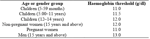
Table 1 Haemoglobin thresholds used to define anaemia according to the World Health Organization
Age or gender group
Children (5-59 months) Children (5.00–11 years)
Children (12–14 years) Non-pregnant women (15 years and above)
Pregnant women Men (15 years and above)
International Journal of Current Advanced Research
6505, Impact Factor: 6.614
www.journalijcar.org
; Page No. 11306-11315
//dx.doi.org/10.24327/ijcar.2018.11315.1954
Anna Tjivambi and Martin Gonzo. This is an open access article distributed under the Creative Commons Attribution License, which permits unrestricted use, distribution, and reproduction in any medium, provided the original work is properly cited.
Department of Biomedical Sciences, Namibia University of
HEPCIDIN AND SERUM IRON: A CORRELATIONAL STUDY IN OBSTETRIC PATIENTS
WITH IRON DEFICIENCY ANAEMIA IN WINDHOEK, NAMIBIA, IN 2017
Department of Biomedical Sciences, Namibia University of Science and Technology, Namibia
Anaemia is a worldwide major health concern. It is especially important in pregnant women due to the increased requirements for both the mother and the baby. Iron deficiency anaemia accounts for 75% of all
The interest of this study was to correlate serum iron levels to he iron regulatory peptide. The study objectives were to evaluate the quantitative behavior of hepcidin in obstetric patients with IDA and to observe the relationship between
vels in pregnant women.
three samples of pregnant women from the ANC ward were collected from NIP between June and September 2017. These samples were required to have an Hb of less than 12g/dL. The samples were tested for iron to determine those with iron deficiency anaemia. Finally, samples with iron deficiency were tested for hepcidin levels. Additional samples were included to complete the hepcidin assay.
107 samples had an iron concentration of less than 7.16µmol/L and were classified as iron deficient. Hepcidin analysis was then carried out on 176 samples (including the 108 with iron deficiency and some normal and high (higher than 26.85µmol/L) to observe if a direct proportionality exists in high iron concentrations. The results indicated a poor, but significant correlation between haemoglobin and serum iron (r=0.163, p=0.032). According to the r value, there is a weak positive linear relationship Furthermore, haemoglobin and hepcidin showed a weak uphill (positive) linear correlation (r=0.123, P=0.106).
The study found that hepcidin had a weaker correlation to haemoglobin than serum iron to haemoglobin. Hb is a poor indicator of IDA because of haemodynamic nges and the slow rate of iron being incorporated and raising Hb. Additionally, the weak relationship between haemoglobin and hepcidin is a result of erythropoeitc drive and hepcidin sensitivity, because while increased serum iron increases hepcidin, erythropoietic drive lowers it. Inflammation, which is common in pregnancy also raises hepcidin, altering the normal homeostatic mechanism of iron. These factors have thereby influenced the degree of their direct proportionality. Therefore, serum iron is the better diagnostic tool for
could be a result of either:
Decreased erythropoiesis (red cell production) or haemoglobin synthesis or
Increased red blood cell destruction
Haemoglobin thresholds used to define anaemia according to the World Health Organization
Haemoglobin threshold (g/dl)
11.0 11.5 12.0 pregnant women (15 years and above) 12.0 11.0 13.0
Research Article
International Journal of Current Advanced Research Vol 7, Issue 4(A), pp 11306-11315, April 2018
Hb levels vary with age, sex and physiological status. The table above indicates the thresholds for the Hb levels of different groups as stated by WHO (2011). Inversely, anaemia can also be caused by vitamin A, vitamin B12 and folate deficiency, bacterial and parasitic infection, or autoimmune disease.
It can, therefore, be considered a marker for poor health and nutrition. Some of the symptoms of anaemia are:
Fatigue
Malaise
Dyspnea
Increased cardiac output or tachycardia
Pallor
Jaundice
Pica (Hoffbrand and Moss, 2008)
What’s more, anaemia affects 2.2 billion people globally (Kassebaum et al., 2014). WHO (2001) statistics also designates 17.2 million (57.1%) Africans are affected by anaemia. WHO further outlines that 50% of the anaemia cases were due to dietary insufficiency and 40% of the anaemia cases in children are due to iron dietary insufficiency. Additionally, 41.8% pregnant women worldwide were affected (1993-2005) and 80% of the affected countries suffer from moderate to severe anaemia, proving that anaemia is indeed a major public health concern.
According to the National Heart, Lung, and Blood Institute (2014), anaemia is the most common blood disorder, affecting more than 3 million Americans, of which “iron deficiency is the most common type of anaemia worldwide” (WHO, 2016). Although, anaemia may result from a number of causes, the most significant contributor is iron deficiency.
LITERATURE REVIEW
As previously mentioned, iron is an essential element for the health and survival of all living things. Scholl (2005) articulates that, “< 50% of women do not have adequate iron stores for pregnancy.” Because pregnant women require substantial iron store of (3–4 mg/dL), risk of iron deficiency and iron deficiency anaemia should increase with gestation. It is, therefore, essential that iron levels should be adequate to supply both the mother and her unborn baby, as there is increased demand.
Frazer & Anderson (2005) outline that iron levels are carefully regulated by the iron regulatory peptide, known as hepcidin. The relationship is such that: when serum iron is elevated, hepcidin is up regulated in order for hepcidin to bind to ferroportin and reduce further absorption of iron in the gut. On the other hand, if hepcidin is low, it indicates that serum iron levels are low and ferroportin remains unbound in order to allow uptake of iron from serum (Ganz & Nemeth, 2012).
Moreover, hepcidin directly controls iron absorption and bioavailability in circulation, as specified by Girelli et al., (2016). However, in IDA, transcription of hepcidin is suppressed. The role and function of hepcidin and its interaction with ferroportin have not been entirely understood, as this is a newly discovered field of study. As such, it leaves a wide open field for further research in understanding the crucial role of hepcidin in regulation of iron and, possibly, the treatment of IDA.
Iron
Iron is an essential element in the synthesis of the haem component of the oxygen transporter haemoglobin (Hoffbrand & Moss 2011). They further stated that 65 % of iron is found in haemoglobin. It is considered as one of the most reliable indicators of anaemia, because it gives an indication of the severity of anaemia. Defects in the haemoglobin chains may result in hypochromic anaemias, i.e. sickle cell anaemia, thalassemia, anaemia of chronic disease and iron deficiency anaemia. Therefore, insufficient or impaired haemoglobin synthesis is the primary cause of hypochromic anaemias (Hoffbrand & Moss 2011).
Primarily, most of the iron in the body is in the haemoglobin of red cells, which contain about 1 mg of iron per millilitre of erythrocytes, or about 2-3 g of iron total. In contrast, Ganz & Nemeth (2012) outlined that blood plasma contains only 2– 3 mg of iron, bound to transferrin, the plasma iron carrier that is the exclusive source of iron for erythropoiesis. As erythrocytes become senescent (after 120 days), macrophages engulf the cells, releasing their contents to plasma ferritin. This generates about 25 mg of iron daily. Hepatocyte and macrophage iron is stored in cytoplasmic ferritin and is readily mobilized during period of high iron demand.
However, in individuals with too little or too much iron, cell damage is likely to occur. Individuals with insufficient iron (hypoferremia) may experience weakness, as iron is required for energy. Subsequently, Ganz & Nemeth (2012) further described the role of hepcidin in iron regulation, asserting that, “hepcidin inhibits iron efflux by directly binding to ferroportin presumably inducing a conformational change, and triggering the endocytosis of both molecules, with consequent lysosomal degradation.” Too much iron (hyperferremia) can cause cell damage due to toxicity.
Abbaspour et al., (2014) states that the mechanism of iron absorption greatly depends on its physical state. At physiological pH, ferrous iron (Fe+2) is rapidly oxidized to the insoluble ferric (Fe+3) form. Gastric acid lowers the pH in the proximal duodenum reducing Fe+3 in the intestinal lumen by ferric reductases, thus allowing the subsequent transport of Fe+2 across the apical membrane of enterocytes. This enhances the solubility and uptake of ferric iron. When gastric acid production is impaired, iron absorption is reduced substantially. Also, “intestinal iron uptake from the gut lumen through divalent metal transporter 1 (DMT1) is increased by the activation of hypoxia-inducible factor 2α.” (Camaschella, 2015).
Iron Deficiency Anaemia
Iron deficiency anaemia (IDA) is a result of iron insufficiency. This is mainly caused by dietary insufficiency and increased demand, both of which causes reduced erythropoiesis. The American Society of Haematology (2016) states that during pregnancy, the amount of blood in your body increases by about 20-30 percent, which increases the supply of iron and vitamins that the body needs to make haemoglobin. It also states that mild anaemia is normal during pregnancy due to the more rapid increase in blood plasma than in red cell mass. However, severe anaemia may result in preterm birth or low birth weight babies (Allen, 2000).
lack of iron results in impairment of processes requiring iron, as previously mentioned. Severe IDA is linked to increased risk of preterm labour, low birth weight, as well as child and maternal mortalityand infection. However, IDA manifests as a hypochromic anaemia, due to the decreased Hb, and microcytic, due to a reduction in cell size. Severe IDA is characterized by pencil cells, due to a reduced haemoglobin content. The symptoms result from impaired tissue oxygen delivery, because iron plays an essential part in the synthesis of the haem component of haemoglobin (Kassebaum et al., 2014).
Hepcidin
Hepcidin is an irregularly shaped polypeptide that is held together by four disulphide bonds, as determined from earlier work by Krause et al., (2000). Hepcidin exists as three main isomer forms, namely: Hepcidin 25, Hepcidin 22 and Hepcidin 20, which are made of 25, 22 and 20 amino acids respectively (Kenma et al., 2008). The mature forms of hepcidin originate from an 84 amino acid long precursor known as pre-prohepcidin. The conversion of prohepcidin to hepcidin is mediated by the prohormone convertase furin. This conversion may be regulated by alpha-1 antitrypsin.
Furthermore, Frazer & Anderson (2005) describe hepcidin as the iron regulatory peptide, which normally exists as a 25 amino acid peptide. It is primarily produced in the hepatocytes, but macrophages and adipocytes express hepcidin mRNA, but at a much lower level. According to Ganz & Nemeth (2012), “hepcidin regulates intestinal iron absorption, plasma iron concentration and tissue iron distribution by inducing degradation of the cellular iron exporter, ferroportin.” This membrane protein is the only known iron exporter.
Hepcidin was first discovered in Krause et al., (2000) in human blood ultra-filtrate and urine samples. It was originally called liver-expressed antimicrobial peptide (LEAP-1) because of its antimicrobial activity against some gram-positive Bacillus and Staphylococcus species as well as Neisseria sp., Neisseria cinerea which is a gram negative bacteria (Krause et al., 2000). The anti-microbial activity of Hepcidin goes beyond the bacterium kingdom; it also shows anti-fungal activity against Saccharomyces cerevisae, commonly known as brewer’s yeast.
Moreover, since its discovery, a lot of work has gone into characterizing this molecule. The name ‘Hepcidin’ originated from the anatomical site of its synthesis as well as its antimicrobial activity (Rossi, 2005). The prefix Hep-; is derived from the word hepatocytes which are liver cells and the suffix -cidin which is usually designated to antibiotics, emanates the antimicrobial activity of the molecule.
Additionally, Brancatisano et al., (2014) also demonstrates the anti-fungal effect of Hepcidin 20, against Candida species and this goes on to show its broad antimicrobial properties over and above its role in iron metabolism. Since its discovery and the unearthing of its antimicrobial activity, it took a further year to confirm the existence of the link between Hepcidin and iron metabolism (Pigieon et al., 2001). The linkage forms the basis of this research, which will look at the behavior of hepcidin with reference to IDA in obstetric patients.
Relationship between iron and hepcidin
Hepcidin is part of the feedback mechanism for iron. It also functions as an acute-phase reactant that adjusts fluctuations in plasma iron levels caused by absorptive enterocytes and macrophages in the spleen. This is done by binding to and inducing the degradation of ferroportin, which exports iron from cells as described by Nemeth et al., (2004). Ferroportin- the iron transporter present on cells of the intestinal duodenum, macrophages, and cells of the placenta- regulates iron uptake through a complex mechanism.In the duodenum, ferroportin is located on the basolateral membrane of enterocytes but dietary iron absorption is dependent on iron uptake on their apical surfaces.
“To coordinate apical absorption of iron with the basolateral transfer of iron to plasma, the effect of hepcidin on basolateral ferroportin must be communicated to the apical iron absorption mechanisms to decrease apical uptake.” Ganz & Nemeth (2012). Spottiswoode et al., (2014) articulates that hepcidin serves to block iron absorption from the diet and to route iron in the body into macrophages and away from the serum. In people with iron deficiency, the body stimulates an increase in iron absorption from the gastrointestinal tract. Whereas, in people suffering from iron overload, hepcidin binds to ferroportin, resulting in decreased iron uptake. The iron concentration in biological fluids should be tightly regulated to only make iron available as needed and to avoid toxicity, because too much iron can generate free radicals, causing damage to cells.
Furthermore, the relationship between hepcidin, ferroportin and iron is such that when iron is high, the hepcidin level in the liver significantly increases, so as to down-regulate serum-iron. While the ferroportin level in the duodenum substantially decreases, to inhibit absorption of iron from the diet. However, “body iron status, erythropoietic drive, and inflammation are the main regulators of hepcidin production and operate via a complex array of molecules and signalling pathways.” (Frazer & Anderson. 2016). Additionally, the hepcidin levels in plasma are also regulated by different stimuli, including cytokines, hypoxia and anaemia. This research therefore aims to evaluate if normal iron homeostasis takes place in pregnant women, seeing as there is an increased demand in nutrients for different functions i.e. iron, for DNA synthesis, erythropoiesis, in order to supply the necessary nutrients for the growth and development of the unborn baby.
Problem statement
Iron deficiency anaemia accounts for 75% of all anaemias in pregnancy (Horowitz et al., 2013). It is preliminarily diagnosed using haemoglobin and confirmed by doing iron studies to determine of the patient has sufficient iron, ferritin and transferrin in serum. A Full blood count (FBC) and blood smear should also be requested in order to observe the haemoglobin content and red blood cell morphology. Serum iron is then tested, followed by an assessment of bone marrow iron stores, in order to diagnose IDA. However, this is a long process.
International Journal of Current Advanced Research Vol 7, Issue 4(A), pp 11306-11315, April 2018
anemia. The findings of this study will contribute to the establishment of a new biomarker that is in line with international trends of introducing diagnostic tests that are sensitive, cost effective, of good prognostic value and not influenced by haemodilution.
However, because of haemodilution and the prolonged date of iron incorporation into haemoglobin, we believe that haemoglobin is not a very reliable diagnostic tool for IDA. Therefore, this researched aimed to observe the usefulness of hepcidin in diagnosis of IDA in obstetric patients.
Aim
This research aimed to evaluate and correlate the hepcidin and serum iron levels in obstetric patients with IDA in Namibia, in 2017. The findings of this study will aid in the implementation of hepcidin as the new biomarker for IDA in pregnant women, in order to effectively manage them and avoid unnecessary use of iron supplements, which may have severe complications for both the mother and the unborn baby. This development will therefore ensure that patients are diagnosed and treated in a timely and effective manner.
Research questions
1. How does hepcidin behave in obstetric patients with IDA?
2. Is there a significant association between the haemoglobin, hepcidin and serum iron levels in obstetric patients who have IDA?
Specific Objectives
1. To evaluate the quantitative behavior of hepcidin in obstetric patients with IDA.
2. To observe the relationship between haemoglobin, serum iron and hepcidin levels in pregnant women.
MATERIALS AND METHODS
Study design
The study was an experimental study which involved record review and analysis. This study was used to support, refute or validate the hypothesis that hepcidin and iron have a directly proportional relationship, in healthy patients, where hepcidin increases when iron is high and decreases when iron is low. The study included controls for both the iron and hepcidin analysis, each time an assay was run. This was done to limit confounding factors and variables, in order to increase reliability and validity of results.
Sample strategy and population
The sampling method used was convenience sampling and the entire sample size was included in the study. Specific samples from obstetric patients with a low Hb were selected. The study population were obstetric patients whose samples were sent to NIP (Katutura State and Central hospital) and records, i.e. FBC/ Hb levels were available. The tests for this research were conducted on the leftover samples of obstetric patients. Basically, the study population was pregnant women (this was confirmed by careful observation of the ward, i.e. antenatal care (ANC) and sex on Meditech, who were anaemic (i.e. Hb should be less than 12 g/dL) and whose samples were sent to NIP for testing.
Patients were selected based on the following inclusion criteria:
Only samples of pregnant women (from ANC) were collected.
Only specimens with a low Hb (<12g/dL) were collected.
No specimens older than 24 hours were collected.
No haemolysed specimens were collected, because red blood cells contain iron and therefore may falsely elevate serum iron.
Sample size
The convenient sample size was determined using a confidence level of 95% and 5% margin of error using a statistical formula proposed by Judd (2009):
n=z²pq/d2, where:
n= desired minimum sample z= 1.96 (at 95% confidence interval)
p= population of study population with a particular character being studied, in this case it is the 75% of pregnant women with iron deficiency anaemia, amongst the 30.6% of pregnant women with anaemia (WHO, 2008). That is 75% x 30.6%= 23.3%
q= 1-p
d= 0.05 (5% margin of error) Therefore, the sample size is:
= (1.96)2x 0.23 x 0.77/ (0.05)2 =272
However, 283 samples were collected.
Specimen collection
Specimens, whose Hb was less that 12g/dL, were centrifuged, then an alliqoute was collected and immediately frozen at -80°C after analysis at NIP. This is done in order to maintain sample integrity, because hepcidin is very unstable. Samples were aliquoted within 24 hours of collection, because fresh specimens were ideal for this study. Specimens were acquired, separated and the plasma frozen as soon as all testing at NIP was completed.
Specimen transportation
Specimens were then transported to NUST in a hamper, which was clearly labelled biohazard, according to NUST SOP for specimen transportation. The specimens were transported on ice, in order to retain specimen integrity, and testing commenced.
Data collection
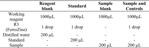
Table 2 Preparation procedure of standards, samples and controls for FerroZine assay
Reagent
blank Standard
Sample blank
Sample and Controls
Working
reagent 1000µL 1000µL 1000µL 1000µL
R3
(FerroZine) 1 drop 1 drop - 1 drop
Distilled water 200 µL - - -
Standard - 200 µL - -
Sample - - 200 µL 200 µL
The table above outlines how samples, standards and controls were prepared for this assay. They are then mixed and incubated for 5 minutes at 37°C or 10 minutes at 25°C. The ferrous ions form a colored complex in FerroZine whose absorbance is read at a wavelength of 562nm. The concentration was then calculated using the following equation:
( ) − ( ) − ( )
( ) − ( )
=µ /
Conversion factor= µg/dL*0.179µmol/L
The second experiment, which was conducted, is an enzyme-linked Immunosorbent assay, better known as ELISA. As described by Bishop et al., (2010), the ELISA method is a type of labelled immunoassay that employs enzymes as the “label”. Enzymes are biological catalysts that increase the rate of substrate to product conversion, without taking part or being consumed by the reaction. The CUSABIO hepcidin assay is quantitative immunoassay, which employs the sandwich technique. A microplate is coated with antibodies specific for Hepc25. One hundred microliters of standards and samples were added into the wells and incubated at 37°C for two hours. The liquid contents of the wells are decanted. One hundred microliters of a biotin-conjugated antibody specific for Hepc25 is then added to the wells and the plate is incubated for one hour at 37°C.
Any unbound substances are removed by washing. After washing, One hundred microliters of avidin conjugated Horseradish Peroxidase (HRP) is added to the wells. After excess unbound avidin-enzyme reagent is washed off by aspirating five times, 90µL of tetramethylabenzide (TMB) substrate solution is added to the wells and colordevelops in proportion to the amount of Hepc25 bound in the initial step. The color development is stopped by the addition of 50µL and the intensity of the color is measured at 450nm. The final concentrations were calculated from a standard curve of the logarithmic values of the concentrations and optical densities.
Specimen disposal
Once testing was complete, the remaining specimens were discarded according to the SOP for discarding biohazardous materials. The researcher was fully clothed in personal protective equipment (PPE), i.e. laboratory coats, gloves and goggles. The used specimens were disposed of in the red waste bag for biological material. The bags were properly tied or sealed with sellotape and transported on a trolley to the waste room, where the laboratory technicians disposed of the waste according to the NUST waste disposal SOP.
Measures to ensure Validity
Primarily, the SPINREACT kit for iron has a detectable range of 0.850 µg/dL to a linearity limit of 1000 µg/dL. The sensitivity of the kit is one µg/dL. This indicates that this test is highly sensitive and has a low detection limit; it made detection of very low amounts of iron possible. Hence why it was possible to distinguish the different levels of iron in the test samples.
Additionally, the CUSABIO kit for hepcidin has a wide detectable range of 4.69 ng/ml-300 ng/ml. However, the minimum detectable dose of human Hepc25 is typically less than 1.17ng/ml. The systematic procedure was followed as stipulated by the package inserts for both assays, under supervision. Trial or pilot tests for both tests were carried out, prior to the actual testing of specimens. The pilot tests were performed under the careful supervision of Mr. Gonzo, in order to assess the researcher’s level of understanding and competency to carry out the assays. Each run included known controls and the researcher ensured that reagents used were not expired. To prevent using specimens that are not within the stated criteria, records were carefully reviewed.
Additionally, quality controls and standards were included in each run, to ensure that reagents were prepared correctly and samples were handled with vigilance. Repeated freeze thawing was avoided as this influences the stability of the analytes and reduces the quality of the specimens. Testing was conducted in a neat space with sufficient reagents and consumables at hand to avoid contamination and over-incubation.
Data Management
All data collected for this study was kept in a secured folder on an encrypted device/ memory stick. Collected data was managed using the Statistical Package for Social Sciences (SPSS) software version 24. The findings were then entered in Microsoft Excel spreadsheets that include haemoglobin, serum iron and hepcidin results amongst other parameters. The findings of the study were kept confidential and only disclosed and reported upon completion of the study.
Furthermore, in order to ensure that the aims and objectives of this research were achieved, the study involved descriptive statistics and correlation analysis between haemoglobin and serum iron and serum iron and hepcidin respectively, so as to confirm and assess the extent to which iron and hepcidin have a linear relationship, if it exists.
RESULTS AND DISCUSSION
Results
Table 3 Number of samples against haemoglobin concentrations
Hb (g/dL) Number of
samples
5-7.9 5
8-9.9 35
10-10.9 67
11-11.9 164
12+ 9
International Journal of Current Advanced Research Vol 7, Issue 4(A), pp 11306-11315, April 2018
The following histogram displays the haemoglobin concentrations in the samples collected during the study.
Figure 3 A histogram of haemoglobin concentration
As indicated by the histogram, most (164) samples had a concentration of 11-11.9g/dL, whereas only five samples had a concentration less than 8g/dL.
The following figure is a graphical presentation of serum iron concentrations, as obtained from the FerroZine assay:
Figure 4 Distribution of serum iron results
The figure above displays the serum iron results, with most samples having a normal serum iron concentration. This is further discussed in the discussion section.
The underlying figure is a graphical presentation of hepcidin concentrations, as obtained from the CUSABIO assay:
Figure 5 Distribution of Hepcidin concentrations
Figure 5 above illustrates the distribution of hepcidin concentrations across the study population. These results are further discussed in the results section.
Table 4 Descriptive statistics
Mean Standard
Deviation N
Haemoglo
bin 10.77 0.98 173
Serum iron 7.95 6.23 176
Hepcidin 119.08 256.38 176
Table 4 above displays the descriptive analysis of haemoglobin, serum iron and hepcidin, respectively. It indicates that the standard deviation of hepcidin is very high, followed by serum iron and haemoglobin being the lowest.
Table 5 Correlation results
Haemoglobin Serum iron Hepcidin
Haemoglobin
Pearson
Correlation 1 0.163
* 0.123
Sig. (2-tailed) 0.032 0.106
N 173 173 173
Serum iron
Pearson
Correlation 0.163
* 1 0.175*
Sig. (2-tailed) 0.032 0.020
N 173 176 176
Hepcidin
Pearson
Correlation 0.123 0.175
* 1
Sig. (2-tailed) 0.106 0.020
N 173 176 176
*. Correlation is significant at the 0.05 level (2-tailed).
Table 5 above displays the correlation between haemoglobin, serum iron and hepcidin, in obstetric patients, as per tests carried out for this research, using SPSS.
DISCUSSION
Results obtained from study
Table 2 displays the series of cumulative results of haemoglobin concentrations. According to table 2 and figure 3, one hundred and sixty-four samples had a concentration between 11-11.9 g/dL, followed by 67 with a concentration of between 10 and 10.9g/dL. Five samples had a concentration of less than eight (i.e. 5-7.9 g/dL). However, according to WHO classification, the 164 would not be classified as anaemic, because their Hb is higher than 11g/dL, 67 would have mild anaemia, 35 have moderate anaemia and five have severe to moderate anaemia (WHO, 2011).
Figure 4 displays the distribution of serum iron results as determined using the SPINREACT FerroZine assay. Out of 283 samples, 108 samples had a serum iron concentration of less than 7.16 µmol/L, which is the lower limit for females, for this particular assay. This is followed by 167 samples with a normal serum iron concentration, of between 7.16 and 26.85 µmol/L. Out of the remaining eight samples, four had a concentration above 26.85 µmol/L and four were insufficient, so they could not be tested for iron concentration. These results indicate that, most of the serum iron concentrations of this particular population are normal.
The difference in the actual number of samples collected and those with iron deficiency, according to the assay, supports the statement made by Chaudhari et al., (2013) that during pregnancy, there is an expansion in blood volume and red cell content, in order to compensate for the increased oxygen demand. This expansion results in haemodilution, which lowers haemoglobin concentration. Since haemoglobin was the parameter used in sample selection for this particular study, it 0
20 40 60 80 100 120 140 160 180
N
u
m
b
e
r
o
f
sa
m
p
le
s
Haemoglobin concentration g/dL Histogram of haemoglobin concentrations
5-7.9 8-9.9 10-10.9 11-11.9 12+
0 50 100 150 200
<7.16 7.16-26.85 >26.85
N
u
m
b
e
r
o
f
sa
m
p
le
s
Serum iron concentration (µmol/L)
Distribution of serum iron results
0 10 20 30 40 50 60 70 80
<17.186 17.186-91.237 >91.237
N
u
m
b
e
r
o
f
S
a
m
p
le
s
Hepcidin concentrations (ng/mL)
influenced the number samples collected. That is why, out of 283 samples, only 108 have iron deficiency. That is 38.7% of 279 samples. In addition, haemoglobin is not solely affected by iron content, therefore the low Hb may be a result of haemodilution.
Moreover, this may be attributed to the oral iron supplements that are administered to the pregnant women in Namibia. Because the iron supplements are administered orally, it takes longer for them to become effective. They also have to be taken long enough to correct anaemia and replenish iron stores, depending on the severity of anaemia. From these findings, we can deduce that anaemia is a serious problem because 108 samples out of 279 that were assayed, which is nearly 50%, have iron concentrations below the normal lower limit for this particular assay.
Table 4 displays the descriptive analysis of haemoglobin, serum iron and hepcidin, respectively. Haemoglobin has the smallest standard deviation (0.98), followed by serum iron (6.23). This indicates that the deviation scores of the distribution is low. This means that the degree of variation in the results from the mean is low. However, concerning hepcidin, the standard deviation is 256.38, which indicates that the degree of deviation of the scores from the mean is high. This indicates that the degree of result distribution is high. It is understandable, because the detection range of hepcidin is 4.69-300 ng/mL, with a minimum detection limit of less than 1.17 ng/mL.
The results in table 5 indicate that there is a poor, but significant correlation between haemoglobin and serum iron (r=0.163, P=0.032). According to the r-value, there is a weak uphill (positive) linear relationship between haemoglobin and serum iron. Normally, 65% of iron in the body is found in haemoglobin, because iron is an essential element in haemoglobin synthesis (Hoffbrand & Moss, 2011). The weak correlation may be attributed to the fact that as part of the management of pregnant women in Namibia, pregnant women are given ferrous fumarate supplements for iron, regardless of her iron status, as the ANC test panel does not require serum iron testing.
This is credited to the mechanism of iron absorption and the required duration of oral supplementation. Primarily, Hoffbrand & Moss (2011) states that inorganic iron is reduced to the Fe2+ form and transported to the cell by DMT-1. It is first in oxidized to Fe 3+ and transported out of the cell by ferroportin. However, haem iron is absorbed via a haem receptor at the cell surface, followed by digestion of haem by haem oxygenase and other enzymes in the enterocyte. Secondly, oral iron supplementation takes longer to have an effect on Hb levels. The iron supplements contain iron that is in its ferrous state and is thus in its “absorbable” state. However, it takes long to absorb therefore Hb slowly increases by 2g/dL after 3 weeks when taking iron supplements. It should, therefore, be administered long enough to both correct the anaemia and replenish iron stores. This normally takes about 6 months (Hoffbrand & Moss, 2011).
However, the effectiveness of the supplementation is influenced by the severity of anaemia, because it will take longer to correct anaemia (i.e. return Hb to normal) and replenish stores in conditions of severe anaemia. It is unknown how long it takes iron to be incorporated into haemoglobin, but haemoglobin levels rise at an approximately 2g/dL every 3
weeks (Hoffbrand & Moss, 2011). This is therefore an indication that although iron levels may return to normal in the patients, their haemoglobin levels may only slightly rise after approximately 3 weeks. Hence why, for example it will take an extended period for an Hb of 5g/dL to be corrected.
Additionally, although a woman is anaemic, she may not immediately present with iron deficiency anaemia, until the body iron stores have been depleted. Ganz and Nemeth (2006) outline that when serum iron levels are low, it causes hypoxia. Hepcidin is down-regulated to increase both the uptake from the gut and release of iron from hepatocytes and macrophages, respectively. This iron is utilized in erythropoiesis to compensate for the hypoxia. However, in a study by Chaudhari et al., (2013), serum iron level correlated well with Hb concentration. Whereas, a Finnish study showed that iron supplementation during pregnancy improved maternal serum ferritin, but not haemoglobin, concentrations for ≥6 month postpartum (Allen, 2000).
Moreover, another study by Milman et al., (1995) indicated decreasing correlations between haemoglobin, transferrin saturation and serum ferritin values at inclusion displayed steadily declining correlation coefficients with values obtained later in pregnancy. There was no data indicating the stage of pregnancy, however out of 283 samples, 107 had iron levels less than 7.16 µmol/L. Therefore, according to the reference range, those samples were assumed to be of patients with iron deficiency anaemia.
Based on a study by Gonzo et al., (2017) the reference ranges for hepcidin concentrations in the healthy blood donor population was 17.186-91.237ng/mL. According to figure 5, 59 out of 176 samples (33.5%) had a hepcidin concentration of less than 17.186ng/mL. This, in comparison to the 38.7% of samples with a low iron, supports the relationship between serum iron and hepcidin, as outlined by Ganz & Nemeth (2012); that when iron stores are low, hepcidin expression is suppressed in order to increase iron absorption from the duodenum. Whereas, when iron stores are adequate, hepcidin synthesis is increased. It then binds ferroportin, causing its internalization and subsequent degradation, to prevent excess iron uptake and release from macrophages and hepatocytes.
Additionally, a study by Kulik-Rechberger et al., (2016) assumed that a relatively low hepcidin concentration in women in late pregnancy facilitates their iron accumulation. According to van Santen et al., (2013) this was likely determined by the occurrence of iron deficiency. Although, Koenig et al., (2014) argues that pregnancy-specific regulators of hepcidin production may exist. There are currently no other reference ranges established in Namibia for this particular population. As such, the above-mentioned reference ranges established by Gonzo et al., (2017) were used for this study.
Furthermore, serum iron and hepcidin showed a weak uphill (positive) linear correlation (r=0.175, P=0.020). The low correlation may be attributed to:
International Journal of Current Advanced Research Vol 7, Issue 4(A), pp 11306-11315, April 2018
of the reduced oxygen carrying capacity, i.e. reduced haemoglobin. It is believed that in hypoxic conditions, hypoxia inducible factor/von Hippel-Lindau (HIF/vHL) pathway can inhibit hepcidin expression in hepatocytes (Kwapisz et al., 2009). This leads back to the essentiality of iron in proper haemoglobin synthesis.
Iron supplementation and the directly proportional relationship that exists between serum iron and hepcidin. When serum iron is low, hepcidin is down-regulated to increase iron uptake by enterocytes in the gastrointestinal tract. As iron increases, hepcidin synthesis is increased, as part of the homeostatic mechanism, because as a result of the iron supplementation, serum iron levels are increased which results in increased hepcidin levels. Although, elevated hepcidin limits the availability of iron for erythropoiesis by reducing iron absorption in the gut and limiting iron release from splenic macrophages and the liver (Schulze et al., 2008).
The study also found that there was a (r=0.123, P=0.106) This is attributable to the sensitivity of hepcidin and the above mentioned relationships, where erythropoetic drive and cytokines produced in inflammation lower hepcidin, while an increase in serum iron increases hepcidin. The above mentioned reasons contribute to the weak relationship observed between haemoglobin, serum iron and hepcidin. The normal relationship is influenced by an external variable, which is the iron supplementation that is administered to all pregnant women in Namibia.
Additionally, some samples with a normal and high iron concentration were also included in the hepcidin analysis. This was done to compare if the relationship between iron and hepcidin remains directly proportional. The findings were inconsistent because some were inversely proportional as opposed to the normal relationship between serum iron and hepcidin. This may be attributed to the fact that the hepcidin levels in plasma are regulated by different stimuli, including cytokines, hypoxia (inadequate oxygenation of tissue) and anaemia.
Expected outcome
As previously mentioned, the typical relationship between iron and hepcidin is directly proportional. Therefore, in patients with iron deficiency anaemia or low iron, a low hepcidin value is expected. This study aided in the investigation of diagnostic and treatment regimens of IDA in that hepcidin use for diagnosis is faster than doing iron studies, bone marrow aspirates and FBC. The findings of this study, however, do not discourage the use of hepcidin as a diagnostic tool for iron deficiency anaemia, because the findings were influenced by the external variable, which is the iron supplementation.
CONCLUSION
This study found that there was a weak, but significant relationship between haemoglobin and serum iron and attributed it to the fact that iron is not readily incorporated into haemoglobin. When one takes iron supplementation, it takes about 3 weeks to raise haemoglobin by 2g/dL. Hence why the Iron supplements should be taken long enough to compensate for the anaemia and replete iron stores.
Additionally, 38.7% of samples had iron deficiency and 33.5% had low serum hepcidin concentrations, based on the reference ranges established by Gonzo et al., (2017). The closeness of these values indicates a weak, but significant correlation between serum iron and hepcidin in pregnant women with IDA. However, hepcidin had a weaker correlation to haemoglobin. This is attributed to erythropoietic drive and iron supplements, because erythropoietin causes a decrease in hepcidin, whereas iron supplements increase hepcidin concentrations. These factors have thereby influenced the degree of their direct proportionality.
Therefore, the iron supplements that participants received as part of the management of pregnant women in Namibia had a significant influence in the findings of the study. However, the research does not solely attribute the findings of the study to the iron supplements. Nevertheless, the findings of this study do not refute the use hepcidin as diagnostic tool for iron deficiency anaemia. However, for this particular study and population, taking into account the iron supplements, we concluded that serum iron was the more suitable diagnostic tool.
Acknowledgements
Firstly, I would like to thank my Heavenly Father and Lord for the opportunity to study Biomedical sciences. The past 4 years have not been easy, but by His grace and strength, I have made it this far. Secondly, I would like to thank Mr. Martin Gonzo, my supervisor, for providing the hepcidin kits, which I used for sample analysis and for his patience and ongoing motivation and assistance throughout the research process.
I would also like to thank the technical staff at NUST for the assistance and provision of consumables and instruments needed for sample testing. I am also very grateful for the encouragement during the process of sample analysis. I would also like to thank the NIP staff members for guidance and assistance during the sample collection process. I would like to thank my family for the ongoing support and motivation during the toughest times and when I felt demotivated. Thank you for the constant encouragement and prayers. Finally, to my classmates and friends, this road has not been easy or the most fun, but we have made it this far, because of our commitment, hard work and teamwork.
References
Abbaspour, N., Hurrell, R., & Kelishadi, R. (2014). Review on iron and its importance for human health. Journal of Research in Medical Sciences: The Official Journal of Isfahan University of Medical Sciences.
Allen, L. H. (2000) Anaemia and iron deficiency: effects on
pregnancy outcomes. Retrieved from:
http://ajcn.nutrition.org/content/71/5/1280s.full Accessed on 01/10/2017
Alton, I., (2005), Iron Deficiency Anaemia. Guidelines for Adolescent Nutrition Services Page 101-108.
American Pregnancy Association (2005) Anaemia during
pregnancy. Retrieved from:
http://americanpregnancy.org/pregnancy-concerns/anemia-during-pregnancy/ Accessed on 10/10/2017
American Society of Haematology (2016). Anemia and
http://www.hematology.org/Patients/Anemia/Pregnancy .aspx# Accessed on 05/10/2016
Bishop, L., Fody, E. P. & Schoeff, L. E. (2010). Clinical Chemistry: Principles, Procedures, Correlations. (6th Ed). Philadelphia: Lippincott.
Brancatisano, L. F., Maisetta, G., Di Luca, M., Semih, E., Bottai, D., Bizzarri, R., … Batoni, G. (2014). Inhibitory effect of the human liver-derived antimicrobial peptide hepcidin 20 on biofilms of polysaccharide intercellular adhesin (PIA)-positive and PIA-negative strains of Staphylococcus epidermidis. Biofouling. 30. 10.1080/08927014.2014.888062.
Camaschella, C., Longo, D. L., (2015). Iron deficiency Anaemia. The New England Journal of Medicine Chaudhari, H., Dixit, R. & Jadeja, J. M. (2013). Serum
Level of Iron and Transferrin in Normal and Anaemic Pregnant Women. International Journal of Basic and Applied Physiology. Vol. 2 Issue 1. Page 123.
Duhiq, K., Chappell, L. C. & Shennan, A. H. (2016). Oxidative stress in pregnancy and reproduction.
Retrieved from:
http://www.ncbi.nlm.nih.gov/pubmed/27630746 Accessed on 29/08/2017
Frazer DM, Anderson GJ (2005) Iron imports. I. Intestinal iron absorption and its regulation. Retrieved from: https://www.ncbi.nlm.nih.gov/pubmed/16160078 Accessed on 08/08/2017
Frazer DM, Anderson GJ (2016), Hepcidin and the Hormonal Control of Iron Homeostasis. Retrieved from:
http://www.sciencedirect.com/science/article/pii/B9780 128021682000154 Accessed on 29/08/2017
Ganz, T. & Nemeth, E. (2006). Iron imports. IV. Hepcidin and regulation of body iron metabolism. American Journal of Physiology - Gastrointestinal and Liver Physiology Vol. 290 no. 2
Ganz, T., & Nemeth, E. (2012). Hepcidin and iron homeostasis. Biochimica et Biophysica Acta, 1823(9), 1434–1443.
http://doi.org/10.1016/j.bbamcr.2012.01.014 Accessed on 29/08/2017
Girelli, D., Nemeth, E., & Swinkels, D. W. (2016). Hepcidin in the diagnosis of iron disorders. Blood, 127(23), 2809–2813. http://doi.org/10.1182/blood-2015-12-639112
Gonzo, M., Maramba, A. & Taylor, G., (2017). Hepcidin testing; establishing reference ranges values for the Namibian blood donor population. International Journal of Blood transfusion and Immunohematology. Volume 7
Hoffbrand, A. V. & Moss, P.A.H. (2011) Essential Haematology 6th ed. NJ, Hoboken
Hoffbrand, A. V., Pettit, J. E. & Vyas, P. (2011) Color Atlas of Clinical Haematology 4th ed. PA, Philadelphia. Horowitz K. M., Ingardia, C.J. & Borgida AF. (2013),
Anaemia in pregnancy. Retrieved from: https://www.ncbi.nlm.nih.gov/pubmed/23702118Access ed on 05/10/2016
Judd, D., R. (2009). Survey Dilemmas: Determining Sample Size Improving Response Rate, Measuring Satisfaction.
Retrieved from:
http://www.nacep.org/confdownloads/SurveyMethods.p df
Kassebaum, N. J., Jasrasaria, R., Naghavi, M., Wulf, S. K., Johns, N., … Murray, C. J. L. (2014). A systematic analysis of global anemia burden from 1990 to
2010. Blood, 123(5), 615–624.
http://doi.org/10.1182/blood-2013-06-508325 Accessed on 29/08/2017
Kenma, E. H., Tjalsma, H., Willems, H. L. & Swinkels, D. W., (2008). Hepcidin: from discovery to differential
diagnosis. Retrieved from:
https://www.ncbi.nlm.nih.gov/pubmed/18166790 Accessed on 29/08/2017
Koenig, M. D., Tussing-Humphreys, L., Day, J., Cadwell, B., & Nemeth, E. (2014). Hepcidin and Iron Homeostasis during Pregnancy. Nutrients, 6(8), 3062– 3083. http://doi.org/10.3390/nu6083062 Accessed on 10/10/2017
Krause, A., Neitz, S., Mägert, H.J., Schulz, A., Forssmann, W.G., Schulz-Knappe, P. & Adermann, K. (2000). LEAP-1, a novel highly disulfide-bonded human peptide, exhibits antimicrobial activity. FEBS Lett. 2000; 480: 147-150.
Kulik-Rechberger, B., Kosceisza, A., Szponar, E. & Domosud, J. (2016). Hepcidin and iron status in pregnant women and full-term new-borns in the first
days of life. Retrieved from:
https://www.ncbi.nlm.nih.gov/pubmed/27321101 Accessed on 29/08/2017
Kwapisz, J., Slomka, A. & Zekanowska, E., (2009). Hepcidin and its role in iron homeostasis. Retrieved from:
https://www.ncbi.nlm.nih.gov/pmc/articles/PMC497527 9/#ref10 Accessed on 01/10/2017
Levy, A., Fraser, D., Katz, M., Mazor, M. & Sheiner, E., (2005). Maternal anaemia during pregnancy is an independent risk factor for low birth weight and preterm delivery. European Journal of Obstetrics & Gynecology and Reproductive Biology. Volume 122, Issue 2, 1 October 2005, Pages 182-186.
Millman, N., Graudal. N. & Agger, A. O. (1995) Iron status markers during pregnancy No relationship between levels at the beginning of the second trimester, prior to delivery and postpartum.Journal of internal medicine. Volume 237, Issue 3, Pages 261-267.
Ministry of Health and Social Services (2011). Namibia Standard Treatment Guidelines. Page 711-712.
National Heart, Lung, and Blood Institute (2014).What Is Iron-Deficiency Anemia? Retrieved from: http://www.nhlbi.nih.gov/health-topics/topics/ida Accessed on 29/08/2017
Nemeth, E., Tuttle, M.S. & Powelson, J. (2004) Hepcidin regulates cellular iron efflux by binding to ferroportin and inducing its internalization. Science.; 306(5704):2090-3. DOI:10.1126/science.1104742 NHS (2016), Iron Deficiency Anaemia. Retrieved from
http://www.nhs.uk/conditions/Anaemia-iron-deficiency-/Pages/Introduction.aspxAccessed on 29/08/2017 Pigeon, C., Ilyin, G., Courselaud, B., Leroyer, P., Turlin, B.,
Brissot, P. & Loreal, O, (2001). A new mouse liver-specific gene, encoding a protein homologous to human antimicrobial peptide hepcidin, is overexpressed during iron overload. J Biol Chem; 276: 7811-7819.
International Journal of Current Advanced Research Vol 7, Issue 4(A), pp 11306-11315, April 2018
Scholl, T. O. (2005). Iron status during pregnancy: setting the stage for mother and infant. The American Journal of Clinical Nutrition. Volume 81 no. 5.
Schulze, K. J., Christian, P., Ruczinski, I., Ray, A. L., Nath, A., Wu, L. S.-F., & Semba, R. D. (2008). Hepcidin and iron status among pregnant women in Bangladesh. Asia Pacific Journal of Clinical Nutrition, 17(3), 451-456. Shils, M. E., Shike, M., Ross, A.C., Caballero, B. &
Cousins, R. J. (2006) Modern Nutrition in Health and Disease
Spottiswoode, N., Duffy, P. E., & Drakesmith, H. (2014). Iron, anemia and hepcidin in malaria. Frontiers in
Pharmacology, 5, 125.
http://doi.org/10.3389/fphar.2014.00125 Accessed on 09/09/2017
Van Santen, S., Kroot, J. J., Zijveld, G., Wiegerink, E. T., Spaanderman, M. E. & Swinkels, D. W. (2013) Iron regulatory peptide decreased in pregnancy: A prospective longitudinal study. Retrieved from: https://www.ncbi.nlm.nih.gov/pubmed/23241678 Accessed on 01/10/2017
WHO (2008). The global prevalence of anaemia 1993-2005. © World Health Organization (2008). Retrieved from: http://apps.who.int/iris/bitstream/10665/43894/1/97892 41596657_eng.pdf Accessed on 09/09/2017
World Health Organization (2011). Haemoglobin concentrations for the diagnosis of anaemia and assessment of severity. Geneva, Switzerland.
World Health Organization (2016). Anaemia. Retrieved
from:
http://www.emro.who.int/health-topics/anaemia/index.htmlAccessed on 29/09/2017