University of Pennsylvania
ScholarlyCommons
Publicly Accessible Penn Dissertations
1-1-2012
Hippocampal Circuitry Dysfunction in the
5-HT1A Knockout Mouse
Kayla Lynn Metzger
University of Pennsylvania, kayla.metzger@gmail.com
Follow this and additional works at:http://repository.upenn.edu/edissertations Part of theNeuroscience and Neurobiology Commons
This paper is posted at ScholarlyCommons.http://repository.upenn.edu/edissertations/549
For more information, please contactlibraryrepository@pobox.upenn.edu.
Recommended Citation
Metzger, Kayla Lynn, "Hippocampal Circuitry Dysfunction in the 5-HT1A Knockout Mouse" (2012).Publicly Accessible Penn Dissertations. 549.
Hippocampal Circuitry Dysfunction in the 5-HT1A Knockout Mouse
Abstract
Anxiety disorders are the most prevalent class of mental illness, yet currently available treatments are often ineffective or inadequate, leaving many patients with lingering symptoms. The serotonin 1A receptor (1AR) has been implicated in the etiology of these disorders, which often show comorbidity with cognitive dysfunction. Mice with the 1AR genetically deleted or "knocked out" (1AKO) during a critical period in development (postnatal days 13-21) exhibit anxiety-like behavior and learning and memory deficits, and may therefore represent a useful genetic model in studying the neurobiological effects of this receptor. The hippocampus has been shown to highly express the 1AR and to be a key mediator in memory and the regulation of emotion. The experiments in this thesis focus on the structural and functional hippocampal changes in the 1AKO mouse compared to wild-type mice in order to elucidate the cellular mechanisms behind the alterations in behavior. Electrophysiology was used in the CA1 region of the hippocampus to show that pyramidal neurons in the 1AKO mouse receive less glutamatergic input than control mice during the critical period, resulting in decreased AMPA-mediated excitation and LTP in the adult. Interestingly,
morphological analyses demonstrated a significant enhancement in proximal dendritic branching in both the juvenile and adult 1AKO mouse that may be the result of the developmental effects of increased serotonergic efflux. Additional experiments focused on the role of corticotropin-releasing factor (CRF) in the 1AKO mouse, based on the fact that peak hippocampal levels of this neuropeptide coincide in time with the critical period of development when 1AR deletion has pronounced effects. We found that adult 1AKO mice showed increased numbers of CRF-containing interneurons and that CRF1 receptor antagonism restored CA1 LTP to control levels. Taken together, these results reveal a complex interplay of decreased synaptogenesis and number of AMPA receptors, and excessive activation of CRF1 receptors that may underlie the cognitive deficits and anxiety-like behavior of the 1AKO mouse. The experiments support the continued research into the neurobiological mechanisms of human anxiety disorders.
Degree Type
Dissertation
Degree Name
Doctor of Philosophy (PhD)
Graduate Group
Neuroscience
First Advisor
Sheryl G. Beck
Keywords
5-HT1A receptor, anxiety, electrophysiology, hippocampus, mouse
Subject Categories
Neuroscience and Neurobiology
HIPPOCAMPAL CIRCUITRY DYSFUNCTION IN THE
5-HT1AKNOCKOUT MOUSE
Kayla Lynn Metzger
A DISSERTATION
In
Neuroscience
Presented to the Faculties of the University of Pennsylvania
In Partial Fulfillment of the Requirements for the
Degree of Doctor of Philosophy
2012
________________________________________________ Sheryl G. Beck, PhD
Research Associate Professor of Anesthesiology Supervisor of Dissertation
________________________________________________ Rita Balice-Gordon, PhD
Professor of Neuroscience
Neuroscience Graduate Group Chairperson
Dissertation Committee
Irwin Lucki, PhD, Professor of Psychology in Psychiatry
Gregory C. Carlson, PhD, Assistant Professor of Neuroscience in Psychiatry Isabel C. Muzzio, PhD, Assistant Professor of Psychology
ii
ABSTRACT
HIPPOCAMPAL CIRCUITRY DYSFUNCTION IN THE
5-HT1AKNOCKOUT MOUSE
Kayla Lynn Metzger
Sheryl G. Beck, Ph.D.
Anxiety disorders are the most prevalent class of mental illness, yet currently available
treatments are often ineffective or inadequate, leaving many patients with lingering
symptoms. The serotonin 1A receptor (1AR) has been implicated in the etiology of these
disorders, which often show comorbidity with cognitive dysfunction. Mice with the 1AR
genetically deleted or “knocked out” (1AKO) during a critical period in development
(postnatal days 13-21) exhibit anxiety-like behavior and learning and memory deficits,
and may therefore represent a useful genetic model in studying the neurobiological
effects of this receptor. The hippocampus has been shown to highly express the 1AR and
to be a key mediator in memory and the regulation of emotion. The experiments in this
thesis focus on the structural and functional hippocampal changes in the 1AKO mouse
compared to wild-type mice in order to elucidate the cellular mechanisms behind the
alterations in behavior. Electrophysiology was used in the CA1 region of the
hippocampus to show that pyramidal neurons in the 1AKO mouse receive less
glutamatergic input than control mice during the critical period, resulting in decreased
iii
demonstrated a significant enhancement in proximal dendritic branching in both the
juvenile and adult 1AKO mouse that may be the result of the developmental effects of
increased serotonergic efflux. Additional experiments focused on the role of
corticotropin-releasing factor (CRF) in the 1AKO mouse, based on the fact that peak
hippocampal levels of this neuropeptide coincide in time with the critical period of
development when 1AR deletion has pronounced effects. We found that adult 1AKO
mice showed increased numbers of CRF-containing interneurons and that CRF1 receptor
antagonism restored CA1 LTP to control levels. Taken together, these results reveal a
complex interplay of decreased synaptogenesis and number of AMPA receptors, and
excessive activation of CRF1 receptors that may underlie the cognitive deficits and
anxiety-like behavior of the 1AKO mouse. The experiments support the continued
iv
TABLE OF CONTENTS
Abstract………..………..……….……….……ii
Table of Contents…..…………...……….……….…...iv
List of Tables……….……….……….….……….vi
List of Figures………..………..………..…vii
Chapter 1: General Introduction..……….………..1
Dissertation goals and hypotheses………...……….………..14
Chapter 2: Altered synaptogenesis during development may underlie hippocampal circuitry changes and impaired memory in the 5-HT1A knockout mouse……...….16
Abstract………..17
Introduction………18
Materials and Methods………...………....20
Results………24
Discussion………..30
Figure Legends………...…37
Table Legends………45
v
Appendix: 5-HT1A deletion alters hippocampal CRF expression and synaptic
plasticity………..………..……56
Abstract………..…57
Introduction………58
Materials and Methods………...61
Results………..…..66
Discussion………..…69
Figure Legends……….………..74
vi
LIST OF TABLES
Chapter 2
Table 1. Cellular characteristics of CA1 pyramidal cell neurons recorded from adult and
juvenile WT and 1AKO mice………..………..45
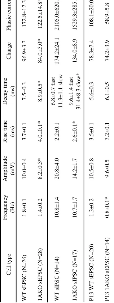
Table 2. Spontaneous post-synaptic currents from pyramidal cell neurons recorded from
vii
LIST OF FIGURES
Chapter 2
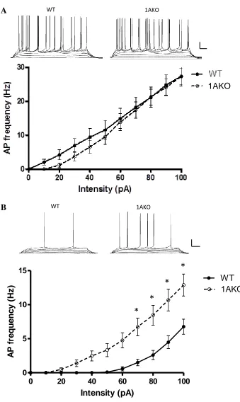
Figure 1. Frequency-intensity plots of the frequency of AP firing rate in response to
increasing depolarizing current pulses in neurons recorded from WT and 1AKO
mice………..……..37
Figure 2. Morphological analyses of CA1 pyramidal neurons in WT and 1AKO……...39
Figure 3. LTP in adult WT and 1AKO mouse hippocampal slices………..41
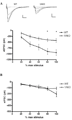
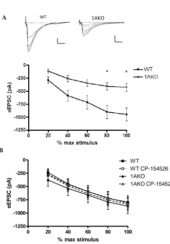
Figure 4. Evoked post-synaptic currents in WT and 1AKO CA1 pyramidal cells from
adult mice………...43
Appendix
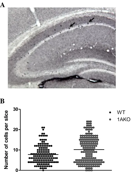
Figure 1. CRF-immunoreactive neurons in the 1AKO and WT hippocampal pyramidal
cell layer……….74
Figure 2. Differential effects of CRF on 1AKO and WT pyramidal cells………76
Figure 3. The CRF1 antagonist CP-154526 rescues the 1AKO deficit in LTP………….78
1
CHAPTER 1:
2
ANXIETY DISORDERS
Anxiety is a normal, adaptive reaction to stressful situations. However, when
anxiety becomes excessive or irrational it can become a chronic, disabling problem. The
five major types of anxiety disorders are Generalized Anxiety Disorder (GAD),
Post-traumatic Stress Disorder (PTSD), Obsessive-Compulsive Disorder (OCD), Panic
Disorder, and Social Phobia. Collectively, these disorders are the most common class of
mental illness in the general population (Kessler et al, 2009). Anxiety disorders show a
lifetime prevalence of 28.8% of the US adult population, with a mean onset at age 11
years (Kessler et al, 2005a). The estimated societal cost of anxiety disorders in the US
was approximately $42.3 billion in the early 1990s, representing an enormous burden at
nearly one-third of the total $148 billion mental health bill (Greenberg et al, 1999).
These disorders are also strongly associated with suicidal ideation and suicide attempts.
One large-scale study showed that among those who had attempted suicide, 64.1% had at
least one anxiety disorder (Sareen et al 2005).
Anxiety disorders have a high incidence of comorbid psychiatric and physical
illnesses. Approximately half of patients with GAD also have Major Depressive
Disorder (MDD) and vice versa (Grant et al, 2005; Kessler et al, 2005b). Furthermore,
both anxiety and depression are associated with cognitive deficits, particularly learning
and memory dysfunctions (Bremner et al, 1993; Deckersbach et al, 2011; Deckersbach et
al, 2000; Hickie et al, 2005; Johnsen and Asbjornsen, 2008; Mantella et al, 2007;
3
In spite of a number of efficacious treatments available to those who suffer from
these disorders, it is estimated that as many as 40% of patients find no relief from current
treatments, and many more have lingering symptoms (Bandelow et al, 2004; Cowley et
al, 1997; Pallanti et al, 2002). Of the two classes of drugs most commonly prescribed for
anxiety, benzodiazepines can induce sedation, memory loss, tolerance, and dependence,
while selective serotonin reuptake inhibitors (SSRIs) are characterized by high levels of
relapse and adverse side effects, and a delayed therapeutic onset of several weeks (Cryan
and Sweeney, 2011; Ravindran and Stein, 2010; Sartori et al, 2011). This highlights the
need for researchers to better understand the etiology of anxiety in order to identify new
targets for therapeutic intervention.
Animal models of anxiety
Given the ethical and legal issues involved in human brain research, most
preclinical work on psychiatric disease focuses on animal studies. Psychiatric disorders
are likely caused by a complicated interplay of genetic and environmental factors. In
view of the complexity and our poor understanding of the biological basis of these
disorders, it would be impossible to model the full human psychiatric condition in
animals. Scientists take advantage of genes and structures that are conserved across
species to study simplified conditions in a controlled environment in order to further our
knowledge of the causes and treatment possibilities for psychiatric disorders. Animal
4
responsiveness of human disease, although any single model is unlikely to replicate all
aspects of the disease process.
The laboratory mouse is a powerful system for biomedical research, and is the
most widely used mammalian model. In spite of outward appearances, mice are
remarkably similar to humans in physiology, anatomy, and genetics. Approximately 99%
of mouse genes have a detectible human homolog, and vice versa, making mice ideal for
use in genetic studies (Sands, 2003). A short lifespan and rapid reproductive rates
facilitate the ability for researchers to study a large number of individuals across many
lifetimes, and generated gene mutations can be maintained through selective breeding
using large numbers of mice at relatively little expense. Furthermore, inbred strains
minimize subject variation and provide controls for finely-tuned genetic manipulations.
Tests of behavioral anxiety in animals
Human anxiety cannot be fully duplicated in the mouse because we simply do not
know the true emotions of non-human animals, or whether they feel anxious or
depressed. However, aside from the human cerebral cortex, vertebrate brains have
structural and organizational similarities which allow comparisons to be made across
species (Cryan and Holmes, 2005; Jones, 2002; Tecott, 2003). Particularly among
mammals, neural structures and the circuitry that connects them have been evolutionarily
conserved. Therefore, in addition to genetic similarities with humans, mice have
5
A number of tasks have been developed for mouse research, which provoke these
behavioral or physiological responses in order to study the neural circuits and genetic
factors underlying human disease states. Many current rodent tests of anxiety-related
behavior fall into two major categories. Approach-avoidance tasks are based on a
balance between the innate rodent desire to explore, and their fear of open, elevated, or
illuminated areas. These tasks include the open field test, the elevated plus maze, and the
light/dark box. Mice in these tasks that are considered anxious tend to stay away from
aversive areas to a greater degree than the appropriate control. These tests have face
validity in that human anxiety disorders are often characterized by avoidance of feared or
threatening stimuli or situations. The power behind these experiments comes from their
predictive validity, in that avoidance behavior is reduced by treatment with clinically
effective anxiolytics, and intensified by anxiogenic drugs that cause anxious feelings in
humans (Belzung and Griebel, 2001; Lister, 1990).
Another commonly used type of behavioral task involves Pavlovian conditioned
fear responses, which are used to model the cognitive aspects of depression and anxiety
disorders (Fendt and Fanselow, 1999; Maren, 2001). These tasks involve repeated
pairings of an innocuous stimulus or environment with a threatening or painful stimulus
so that the rodent eventually displays fearful behavior when presented with the former.
Although mainly a learning and memory task, conditioned fear is known to activate the
same neural circuitry involved in human anxiety and depression, thereby providing
construct validity to this line of testing (LeDoux, 2000). Therefore these tasks may be
6
fear responses, particularly the extinction of the fear response, are sensitive to drugs that
also work clinically to extinguish panic or phobias. (Davis, 1990; Ledgerwood et al,
2005; Ressler et al, 2004).
Anxiety is a highly adaptive and conserved response to a potential threat across
species, and anxiety disorders can be considered to be the pathological end of the
spectrum of this normal, adaptive behavior. A person with an anxiety disorder exhibits
fear or panic in situations where most people would not feel anxious or threatened. On
the other hand, depression is a clinically defined human disease state with psychological
symptoms such as feelings of hopelessness, despair, or anger, and often accompanied by
low energy and other neuroendocrine and somatic symptoms. Therefore what is assayed
in the mouse are specific, measurable behaviors known as endophenotypes that appear
relevant to human depression (Holmes, 2003). The most commonly used behavioral
tasks for depression are the forced swim test and tail suspension test, both of which use
immobility as the dependent variable. These tests have both face validity and some
amount of predictive validity: they are sensitive to clinically efficacious antidepressants,
however these drugs reduce immobility after acute exposure, whereas only chronic
exposure is effective clinically (Duman, 2010; El Yacoubi et al, 2003; Naudon et al,
2002). The novelty suppressed feeding paradigm measures a rodent’s aversion to eating
in a novel environment. Although lacking in face validity, this test has high predictive
validity in that it is sensitive to acute anxiolytics and chronic antidepressants, but not
7
Although many of these tests have been standardized and validated to some
extent, there are limitations to every model. Several studies have shown that results of
behavioral tasks can be sensitive to even subtle variations in testing procedure, housing
conditions, and laboratory environment (Crabbe et al, 1999; Cryan and Mombereau,
2004; Wahlsten et al, 2003). Furthermore, as newer classes of drugs are made available,
researchers may need to utilize novel behavioral tasks to test them.
Animal models of anxiety: environmental and genetic
An animal model can be defined as an (non-human) organism or particular state
of an organism that reproduces aspects of the human pathology, providing some degree
of predictive validity. On the other hand, a test, such as those described above, provides
only an end-point behavioral or physiological measure designed to assess the effect of a
genetic, pharmacological, or environmental manipulation (Urani et al, 2005). Several
rodent models have been designed to study the contribution of environmental factors
towards the development of anxiety disorders. Most types of anxiety disorders are
thought to involve exposure to prolonged or recurring stressors rather that to a single
acute event. By repeatedly activating the stress pathways, these models reproduce
conditions under which pathological anxiety might develop. Many models use stressors
designed to provoke fear behaviors. For example, chronic stress paradigms such as
exposure to predator odors or an aggressive conspecific elicits defensive activities or
postures (Blanchard et al, 2005; Blanchard et al, 2003; Slattery et al, 2012; Staples,
8
behavioral changes in tests of anxiety (Lee et al, 2008). Early-life stress such as maternal
deprivation is also known to produce anxiety-like behavior that manifests in the juvenile
or adult animal (Rice et al, 2008; Sherrin et al, 2009; Troakes and Ingram, 2009). This
model suggests developmental experiences may result in life-long behavioral changes.
Since the median age of onset in human anxiety disorders is 11 years, there is great
interest in understanding the developmental aspects of the disorder and focusing on early
interventions (Kessler et al, 2005a).
The genetics of anxiety disorders are recognized to be complex, polygenic, and
epistatic, which have made them challenging to model in a straightforward way (Lesch,
2001). One traditional method has been to selectively breed animals for high versus low
trait anxiety behavior, allowing for the identification of candidate genes correlated with
the human disorder (Landgraf and Wigger, 2002; Muigg et al, 2009; Wegener et al,
2012). Environmental stressors may further differentiate these extremes, suggesting that
animals bred for high anxiety behavior may be more vulnerable to the effects of stress
(Savignac et al, 2011; Stedenfeld et al, 2011).
The targeted deletion or “knockout” of a single gene is currently used as one of
the primary approaches to elucidate the genetic basis of anxiety disorders. The classic
constitutive knockout has the gene mutation present in all cells and throughout the
lifetime of the animal, including during development. Some gene knockouts are
developmentally lethal, with deletion of an essential gene resulting in death of the animal
9
there may be multiple effects from the mutation, which can complicate the interpretation
of neurobiological research. Compensatory mechanisms in other neural systems may
obscure the function of the gene. However, the developmental effects following gene
deletion can themselves provide important insights into genetic function as well as into
the plasticity of neural systems. Since human anxiety disorders often present with an
early age of onset (average age 11 years), this disease class is particularly suited for this
strategy (Kessler et al, 2005a). The constitutive knockout mouse is a powerful method of
modeling the genetic contribution to a complex neurological disorder.
The 5-HT1A receptor
Brain serotonin (5-HT) plays a major role in a number of physiological processes
including the regulation of mood, impulse control, sleep, eating, libido, and cognitive
functions such as learning and memory. 5-HT is synthesized from the amino acid
L-tryptophan in serotonergic neurons in the raphe nuclei and released widely throughout the
cortex and limbic systems. There are 7 distinct families and at least 14 known 5-HT
receptor subtypes, many of which can be further divided into subpopulations (Berger et
al, 2009; Nichols and Nichols, 2008).
The 5-HT1A receptor (1AR) is the most extensively distributed of the 5-HT
receptors, showing high density in raphe nuclei, hippocampus, amygdala, and septum (el
Mestikawy et al, 1991). Low to moderate levels of the 1AR are also seen in cerebral
cortex, basal ganglia, hypothalamus, and thalamus (Kusserow et al, 2004). All except
10
with Gi/Go. Activation of the 1AR causes the opening of G-protein-gated
inwardly-rectifying potassium (GIRK) ion channels, hyperpolarizing the cell and making it less
likely to fire an action potential. Additionally, activation of this G-protein inhibits
adenylate cyclase, which decreases levels of the second messenger cAMP and results in
decreased transcription.
The 1AR functions as both a presynaptic autoreceptor and a postsynaptic
heteroreceptor (Blier et al, 1987). Presynaptic autoreceptors are located on the cell body
and dendrites of serotonergic neurons in the raphe nuclei where they serve to negatively
inhibit their own activity and suppress the release of serotonin (Aghajanian and Lakoski,
1984). Postsynaptic heteroreceptors are found mainly on glutamatergic and GABAergic
pyramidal cells in limbic and cortical regions innervated by serotonergic raphe cells,
where they inhibit the firing of target neurons and regulate the function of other
neurotransmitter systems (Azmitia et al, 1996; Hall et al, 1997; Palchaudhuri and Flugge,
2005).
The 5-HT1A receptor in anxiety
The first evidence for the role of the 1AR in anxiety came in 1979 with a clinical
study showing the anxiolytic properties of buspirone, a 1AR partial agonist (Goldberg
and Finnerty, 1979). Since then, SSRIs have been shown to be effective in many cases in
the treatment of anxiety disorders (Ballenger, 1999; Liebowitz, 1999). This class of
drugs blocks the 5-HT transporter and is thought to work by increasing the synaptic level
11
partial agonists can improve the effectiveness of SSRIs by halting the 1A autoreceptor
inhibition of cell firing that SSRIs induce early in treatment (Artigas et al, 2006; Stahl,
1997).
In addition to treatment studies, clinical findings from genetic analyses and
imaging data have shown the 1AR to be involved in anxiety disorders. For example,
patients with panic disorder show decreased 1AR binding in PET imaging studies
(Neumeister et al, 2004). A single nucleotide polymorphism (SNP) that occurs in the
transcriptional control region of the human 1AR gene is associated with anxiety-related
personality traits, severe depression, and suicide (Lemonde et al, 2003; Strobel et al,
2003). This allele abolishes repression of 1AR expression to reduce serotonergic
neurotransmission.
Recent evidence also shows a role for the 1AR in cognitive deficits and mood
associated with dementia. PET imaging has shown significantly decreased 1AR density
in the hippocampi of patients with mild cognitive impairment (24% average loss) or
Alzheimer’s disease (49% average loss) (Kepe et al, 2006).
5-HT1A receptor knockout mouse
In 1998, three lines of 1AR knockout mice were independently created on
different genetic backgrounds and tested under similar conditions in a variety of
anxiety-like behavioral tasks (Heisler et al, 1998; Parks et al, 1998; Ramboz et al, 1998). Each of
12
with decreased exploratory behavior and antidepressant-like effects. Subsequent findings
also demonstrated impairments in hippocampal-dependent learning and memory tasks
such as the Morris water maze and contextual fear conditioning (Klemenhagen et al,
2006c; Sarnyai et al, 2000). However, in learning tasks mediated by the prefrontal
cortex, such as the serial reversal learning paradigm, 1AKO mice performed similarly to
wild-type (WT) mice (Pattij et al, 2003; Wolff et al, 2004).
In many behavioral anxiety tests, the 1AKO mouse responds to treatment with
therapeutics used to treat anxiety and depression in humans, showing that this model has
predictive validity for human affective disorders. One background strain of the 1AKO
mouse was discovered to be insensitive to the anxiolytic effects of benzodiazepines,
which was attributed to a pathway from 1AR deficit to GABAA receptor subunit
abnormalities (Sibille et al, 2000a). This strain may be useful for modeling
benzodiazepine treatment-resistant anxiety. However, the anxiolytic effects of
8-OH-DPAT and SSRIs are blocked in the 1AKO in the novelty-suppressed feeding task, which
is sensitive to acute anxiolytics and chronic but not acute antidepressants. This indicates
that SSRIs exert their anxiolytic effects by activating 1ARs (Santarelli et al, 2003).
Prior studies using the 1AKO mouse have shown there to be a critical period for
the development of anxiety-like behavior. Genetic manipulations of 1AR expression
demonstrated that deletion of this receptor in the forebrain of the adult mouse did not
produce an anxiety phenotype (Gross et al, 2002). This is consistent with the inability of
13
and Rodgers, 1997; Fletcher et al, 1996). However, when mice had 1AR expression
disabled in forebrain regions during the embryonic and postnatal period (E0-P21), they
were behaviorally indistinguishable from 1AKO mice as adults (Gross et al, 2002).
Furthermore, pharmacological blockade from P13-P34 resulted in anxiety-like behavior
in the adult mouse, thereby narrowing the critical time for the development of
anxiety-like behavior in the 1AKO to P13-P21 (Lo Iacono and Gross, 2008).
A mouse line was recently developed for conditional deletion of the 1AR both
spatially (autoreceptor versus heteroreceptor) and temporally (whole-life versus adult).
Deletion of the autoreceptor throughout life produced a mouse that had an anxious
phenotype. In contrast, lifetime elimination of the heteroreceptor led to a non-anxious
mouse that exhibited a depression-like phenotype (Richardson-Jones et al, 2011). When
the autoreceptor was knocked out only during adulthood, mice exhibited depression-like
behavior, but adult suppression of the heteroreceptor resulted in no behavioral
abnormality (Richardson-Jones et al, 2010; Richardson-Jones et al, 2011). The
autoreceptor is therefore important during development for the predisposition to anxiety
whereas the heteroreceptor is important for the expression of behavioral depression.
However, changes that occur in forebrain regions following deletion of the autoreceptor
may contribute towards the anxiety-like behavior and cognitive deficits seen in the
1AKO.
In mice with 1AR constitutive genetic deletions, compensatory effects in
14
circuitry and induce the behavioral abnormalities seen in these mice. For example,
previous studies have shown that 5-HT1B receptors in the striatum appear to increase
their responsiveness in response to 1AR deletion (Knobelman et al, 2001a). In fact, some
of the most interesting or important findings that may be gained from constitutive
deletions are the compensatory or related consequences of life-long changes in the target
gene.
Goals of thesis research
The primary goal of this thesis was to characterize hippocampal dysfunction on a
cellular level that might contribute to the behavioral anxiety and cognitive deficits in the
1AKO mouse. These studies were undertaken because of the pivotal yet poorly
understood role the 1AR plays in the development and treatment of anxiety disorders.
The 1AKO mouse shows anxiety-like behavior along with cognitive deficits similar to
those that are often comorbid in patients with anxiety disorders. Prior studies in the
1AKO mouse have demonstrated a period in development that is crucial to formation of
the behavioral phenotype, indicating utility as a model for genetic predisposition to
anxiety. I used cellular and electrophysiological techniques to investigate alterations in
hippocampal cellular characteristics and activity that result from genetic deletion of the
5-HT1A receptor.
Clinical and animal studies have shown the hippocampus to be a key region in the
regulation of mood. This area of the brain is also known to be crucial to various aspects
15
1AKO mouse, I hoped to determine the cellular mechanism by which deletion of the 1AR
affects behavior and cognitive abilities. Chapter 2 describes my evaluation of cellular
properties and synaptic activity of CA1 pyramidal cells using whole-cell patch-clamp and
extracellular electrophysiology, and single-cell morphological analyses. Previous studies
show increased hippocampal theta activity in anxiety-provoking tasks in the 1AKO
mouse, and that 1AR agonists and anxiolytics decrease hippocampal activity (Adhikari et
al, 2011; Gordon et al, 2005b; Hirose et al, 1990; Tada et al, 1999; Zhu and
McNaughton, 1994). However, the learning and memory deficits of the 1AKO indicate
that hippocampal activity may be reduced. Based on reports that 1AKO hippocampal
activity is state-dependent, I examined both the baseline and stimulated hippocampal
systems to resolve this discrepancy.
The experiments described in the Appendix examine the effects of CRF or a CRF1
receptor antagonist on the same cellular characteristics and activity, as well as the
expression of CRF in the 1AKO mouse hippocampus. The CRF neuropeptide and CRF1
receptor have been shown to peak in expression in the rodent hippocampus during the
same critical time period when the 1AR is necessary for normal behavior in the adult
mouse. Therefore, I hypothesized that CRF plays an important role in the development
and maintenance of the behavioral phenotype of the 1AKO mouse.
These studies were designed to advance the knowledge of the development of
anxiety disorders. With continued research, these findings could help uncover more
16
CHAPTER 2:
ALTERED SYNAPTOGENESIS DURING DEVELOPMENT
MAY UNDERLIE HIPPOCAMPAL CIRCUITRY CHANGES
AND IMPAIRED MEMORY IN THE 5-HT1A
KNOCKOUT MOUSE
Kayla L. Metzger, B.A.1,2, Kelly A. Siderio, B.A.3, Sheryl G. Beck, Ph.D.1,2
1
Department of Neuroscience, University of Pennsylvania, Philadelphia, PA
2Department of Anesthesiology, Children’s Hospital of Philadelphia, Philadelphia, PA
3
17
ABSTRACT
The serotonin 1A receptor is linked to the etiology of mood disorders and
cognitive deficits. Mice in which the 5-HT1A receptor has been genetically deleted
(1AKO) during a limited developmental period demonstrate a robust anxiety phenotype
and deficits in learning and memory. The CA1 region of the hippocampus shows the
greatest expression of 5-HT1A receptors, and has been shown to be crucial to
development of the behavioral phenotype. This study used electrophysiology and
morphological analyses to examine differences in cell properties and synaptic activity in
the juvenile and adult 1AKO mouse and their wild-type (WT) controls. We showed that
both juvenile and adult 1AKO mice displayed increased dendritic branching in CA1
pyramidal cells compared to WT mice. Adult 1AKO mice showed cellular
characteristics that reflect this change in morphology, as well as deficits in LTP and
reduced AMPA receptor-mediated responses compared to the WT mice. In contrast,
juvenile mice exhibited no differences in cell membrane properties but showed decreased
glutamatergic input. Together these data indicated that morphological differences in the
1AKO already in place at the beginning of the critical period drive alterations in synaptic
development that led to deficits in the adult. These changes may underlie the behavioral
18
INTRODUCTION
The serotonin 1A receptor (1AR) has been linked to the etiology and treatment of
mood disorders and cognitive deficits. Clinical studies have demonstrated a 1AR
deficiency in people with anxiety or depression (Drevets et al, 1999; Lesch et al, 1992;
Meltzer and Maes, 1995; Sargent et al, 2000; Savitz et al, 2009). Patients with panic
disorder, a form of anxiety, and those with Alzheimer’s disease, show decreased 1AR
binding in PET imaging studies (Kepe et al, 2006; Neumeister et al, 2004; Truchot et al,
2008). An allelic variation in the 1AR gene that abolished repression of a transcriptional
regulator is associated with anxiety-related personality traits (Strobel et al, 2003).
Similarly, animals studies have shown that constitutive genetic deletion, or knockout, of
the 1AR in mice (1AKO) results in an anxiety-like phenotype, and learning and memory
deficits in the adult animal (Heisler et al, 1998; Klemenhagen et al, 2006b; Parks et al,
1998; Ramboz et al, 1998; Sarnyai et al, 2000).
1ARs are inhibitory G-protein coupled receptors found as autoreceptors on
serotonergic cells in the raphe nuclei, and as heteroreceptors on non-serotonergic neurons
in raphe and forebrain regions. The 1AR exhibits highest expression in the CA1 region
of the hippocampus (Chalmers and Watson, 1991; Gross et al, 2002). Gross et al (2002)
demonstrated that forebrain-specific ectopic expression of 1AR is sufficient to rescue the
anxiety-like phenotype of 1AKO mice. In these forebrain 1AKO “rescue” mice, the
pattern of the rescued receptor was similar to wild-type (WT) mice only in the
hippocampus. Recently, a mouse line was developed for selective elimination of the 1AR
19
roles. The knockout of the autoreceptor throughout life produced a mouse that had an
anxious phenotype but not reduced behavioral despair. In contrast, lifetime elimination
of the heteroreceptor led to a non-anxious mouse that exhibited a depression-like
phenotype (Richardson-Jones et al, 2011). When the autoreceptor was knocked out only
during adulthood, mice exhibited depression-like behavior, but adult suppression of the
heteroreceptor resulted in no behavioral abnormality (Richardson-Jones et al, 2010;
Richardson-Jones et al, 2011). The autoreceptor is therefore important during
development for the predisposition to anxiety whereas the heteroreceptor is important for
the expression of behavioral depression.
Changes driven by the heteroreceptor that occur in the hippocampus during
development are important for behavioral despair. Other changes may be important for
predisposition to anxiety or depression, possibly due to altered 5-HT input in the
autoreceptor knockout. For example, serotonin efflux is increased in some brain regions,
hippocampal pyramidal neurons demonstrate increased proximal dendritic branching,
theta power is augmented during an anxiety test, and GABA receptor subunits are altered
(Ferreira et al, 2010b; Gordon et al, 2005a; Parsons et al, 2001; Richardson-Jones et al,
2011; Sibille et al, 2000b). In addition, 1AKO mice have deficits in
hippocampal-mediated learning and memory tasks, but not in memory tests that are hippocampal-mediated by other
brain regions (Klemenhagen et al, 2006a; Sarnyai et al, 2000). These experiments
demonstrate that the hippocampus is crucial to the formation of the phenotype in the adult
1AKO animal. However, the cellular mechanisms underlying those changes are
20
The critical period for development of anxiety-like behavior in the 1AKO occurs
within a defined temporal window. Genetic manipulations showed that turning off 1AR
expression in the forebrain of the adult mouse did not produce an anxiety-like phenotype
(Gross et al, 2002). This is consistent with the inability of 1AR antagonist WAY-100635
to produce anxiety-like behavior in the adult mouse (Cao et al, 1997; Fletcher et al,
1996). However, mice with 1AR expression disabled in forebrain regions during the
embryonic and postnatal period (E0-P21) are behaviorally indistinguishable from 1AKO
mice as adults (Gross et al, 2002). A report using pharmacological blockade with
WAY-100635 showed that 1AR activity is necessary from P13-P34 for normal adult behavior,
thereby narrowing the critical time for the development of anxiety-like behavior in the
1AKO to P13-P21 (Lo Iacono et al, 2008).
The present study used whole-cell patch-clamp and extracellular
electrophysiology from hippocampal slices as well as morphological analyses to evaluate
the differences in cellular characteristics, excitatory, and inhibitory activity between
1AKO and WT mice at the beginning of the critical period and in adulthood. Our
findings suggest that changes taking place in the hippocampus of the 1AKO mouse
during this critical period contribute to cognitive impairments in the adult.
MATERIALS AND METHODS
21
Founders from an established colony in the laboratory of Dr. Mark Geyer,
University of California, San Diego, CA were obtained to generate a colony at Children’s
Hospital of Philadelphia. Male offspring from heterozygous pairings were used in this
study. Litters were weaned, separated by sex, and genotyped at 3 weeks of age. For all
experiments, adult (2-5 months) and juvenile (12-14 postnatal days) homozygous
wild-type (WT) and knockout (1AKO) male mice on a 129/Sv background (Ramboz et al,
1998) were used in accordance with the National Institutes of Health Guide for the Care
and Use of Laboratory Animals and approved by the institutional IACUC committee.
Electrophysiology
Brain slices were prepared as previously described (Richardson-Jones et al, 2011;
Tsetsenis et al, 2007). Briefly, mice were decapitated and their brains dissected out in
cold artificial cerebrospinal fluid (aCSF) in which NaCl was replaced with sucrose
(248mM). The forebrain was blocked and cut on a Leica VT1000s vibratome (Leica
Microsystems, Bannockburn, IL). Coronal slices (200 μm thick for whole-cell
patch-clamp, 400 μm for field recordings) containing hippocampus were placed in aCSF (in
mM, 124 NaCl, 3.2 KCl, 1.25 NaH2PO4, 1.3 MgSO4, 2.5 CaCl2, 10 dextrose and 26
NaHCO3) at 37 °C bubbled with 95% O2/5% CO2. After one hour, slices were kept at
room temperature. Individual slices were placed in a recording chamber and perfused
with aCSF at 2 ml/min at 32 °C maintained by an in-line solution heater (TC-324,
22
and signals were collected and stored using a Multiclamp 700B, Digidata 1322A
analog-to-digital converter, and pClamp 9.0 software (Molecular Devices, Sunnyvale, CA). All
drugs were made in stock solutions, diluted on the day of the experiment, and added
directly to the aCSF.
Whole-cell recordings were obtained using electrodes filled with an intracellular
solution of (in mM) 130 K-gluconate, 5 NaCl, 10 Na phosphocreatine, 1 MgCl2,
0.2 EGTA, 10 HEPES, 2 MgATP, 0.5 Na2GTP, biocytin 0.1%, pH 7.3 for measuring
EPSCs, and 70 K-gluconate, 70 KCl, 2 NaCl, 10 Na phosphocreatine, 4 EGTA, 10
HEPES, 4 MgATP, 0.3 Na2GTP, biocytin 0.1%, pH 7.3 for measuring IPSCs. For
evoked PSCs, a cut was made between CA3 and CA1 to prevent recurrent excitation.
CA1 pyramidal neurons were patched and cell characteristics recorded using current
clamp techniques as previously described (Beck et al, 2004). Voltage clamp recordings
were conducted holding the membrane potential at -60 mV (Lemos et al, 2011). To
isolate AMPA-mediated eEPSCs, bicuculline methiodide (BMI, 20 µM) and (2R
)-amino-5-phosphonopentanoate (AP5, 30µM) were added to the superfusing aCSF.
GABAA-mediated IPSCs were isolated similarly with the addition of AP5 and
6,7-dinitroquinoxaline-2,3-dione (DNQX, 20µM).
For extracellular evoked potential recording, glass electrodes were filled with 4M
NaCl and placed in stratum radiatum of area CA1. The Schaffer collateral afferent
pathway was stimulated with a concentric bipolar platinum/iridium electrode (FHC,
Bowdoin, ME) and field excitatory postsynaptic potentials (fEPSPs) were recorded.
23
half maximal response was used. Baseline potentials were monitored for 30 min before
tetanic stimulation. Long-term potentiation (LTP) was induced by four trains of
conditioning stimulation (1 s at 100 Hz) at 30-s intervals (high-frequency stimulation
(HFS)). Responses were subsequently followed for 60 min at 20-s intervals.
Data analysis
MiniAnalysis software (Synaptosoft, Inc., Decatur, GA) was used to analyze spontaneous
and miniature EPSC events for frequency, amplitude, rise time, decay time, baseline
holding current, and area. The slope and amplitude of fEPSP and ePSCs were measured
using pClamp 9 software. fEPSP was expressed as percent of pre-tetanus baseline
values, and ePSCs expressed as percent of maximal stimulus before an action potential
was elicited. LTP was quantified as the mean of the last 10 min of 60 min post-tetanus.
Statistica software was used for all statistical tests. Passive and active membrane
characteristics were analyzed using Clampfit software (Molecular Devices) and then
compared between WT and 1AKO using unpaired Student’s t-test. Repeated measures
ANOVA was used to examine frequency-intensity (F-I) plot and all evoked responses,
followed by Tukey post-hoc analysis.
24
After electrophysiological recording, slices were placed in 4% paraformaldehyde
and stored at 4°C until staining. To visualize the biocytin-filled neuron, slices were
incubated in streptavidin-conjugated Alexa Fluor 647 (1:200; Invitrogen) in PBS with
0.25% Triton X-100 and 0.5% bovine serum albumin for 90 min at room temperature.
After several washes, sections were mounted on Superfrost slides and coverslipped with
Fluoromount-G mounting media (Southern Biotech, Birmingham, AL). Labeled cells
were visualized using a Leica confocal DMIRE2 microscope (Leica, Allendale, NJ).
A 20X scan consisting of several serial, optical sections (0.6 μm) was acquired at the
level of the cell body of the biocytin-labeled neuron. For morphological analysis, the
total number of optical sections taken during the scan depended on the length of
the dendrites and the depth that the dendrites traveled through the section, as more optical
sections were needed to scan longer, deeper extending dendrites. The entire extent of the
dendritic tree of each neuron was obtained for analysis. Images were captured using a
digital camera and Leica Confocal software (Version 2.5, Leica). The xyz confocal stacks
were collected and analyzed using the Neurolucida software program (MicroBrightfield,
Williston, VT).
RESULTS
Cellular properties in the adult mouse
Cellular characteristics were measured in 26 WT and 28 1AKO pyramidal cells
25
1 contains the summary data (means ± SEM). Input resistance (p<0.001) and tau
(p<0.05) were significantly higher in the 1AKO as compared to the WT, showing
possible differences in morphological characteristics. There was no difference between
the genotypes in resting membrane potential (RMP) or action potential (AP) threshold.
However, the activation gap (i.e. the difference between the resting membrane potential
and the action potential threshold) was significantly smaller in the 1AKO, indicating a
higher intrinsic excitability in these cells. AP duration was longer in the 1AKO compared
to WT cells (p<0.005). A frequency-intensity plot was constructed from the mean firing
frequency in response to square current pulses (400 ms in duration) of increasing
amplitude (20 pA steps starting at 0 pA up to 100 pA). 1AKO neurons demonstrated
increased excitability compared with those of WT cells, in that they reached higher firing
rates given the same magnitude of input (Figure 1B). This difference was significant at
current steps ranging from 70 to 100 pA (p<0.05).
Spontaneous excitatory post-synaptic currents (sEPSCs) were measured in voltage
clamp at -60mV. The results are summarized in Table 2 (means ± SEM). The frequency
of excitatory events did not differ between 1AKO and WT pyramidal cells, however
sEPSC amplitude was greater in the WT cells as compared to the 1AKO cells (p<0.001).
Both rise and decay times of sEPSCs were longer in the 1AKO neurons than in the WT
cells (p<0.005 and p<0.05 respectively). In spite of these differences in kinetics, the
average charge per event, as defined by the area under the curve, was significantly
smaller in the 1AKO cells (p=0.005). When this value was multiplied by the sEPSC
26
neurons as compared to the WT cells (p<0.05), indicating that 1AKO cells receive less
total glutamatergic input.
In these recordings of sEPSC activity, larger amplitudes were correlated with
faster rise times only in the WT pyramidal neurons (R2=0.47, p<0.001). This indicated
that these cells exhibited electrotonic filtering (Ling & Benardo, 1999, Crawford et al
2011). To determine if differences in sEPSC characteristics between WT and 1AKO
cells were due to the activity of local neurons within the slice, recordings were done in a
small subset of cells in the presence of the AP blocker tetrodotoxin (1µM), and represent
the spontaneous release of neurotransmitter at the synapse. Miniature EPSCs (mEPSCs)
in the WT cells (N=6) displayed significantly lower amplitude (p<0.01) and phasic
current (p<0.05) as compared to sEPSCs, as well as increased mEPSC decay time
(p<0.05). The decreased amplitude indicated that AP-independent component makes up a
small portion of the total glutamatergic input to these cells. Conversely, there were no
differences between mEPSCs and sEPSCs in the 1AKO (N=5) cells, showing that
independent input occurs in greater proportion in 1AKO neurons. However, the
AP-independent glutamatergic input displayed electrotonic filtering in both the WT (R2=0.70,
p<0.05) and 1AKO (R2=0.96, p<0.005) cells.
In a separate set of experiments, spontaneous inhibitory post-synaptic currents
(sIPSCs) were recorded in the presence of DNQX and AP5 in order to isolate
GABAergic input (Table 2). The rise time (p<0.05) and slow decay time (p<0.05) were
significantly longer in 1AKO cells as compared to WT cells. This difference in kinetics
27
sIPSC frequency, amplitude, fast decay time, average charge per event, and phasic
current did not differ significantly between 1AKO and WT cells. Overall, 1AKO
pyramidal cells exhibited greater intrinsic excitability but decreased glutamatergic input
as compared to WT neurons, and evidence for changes in AMPA and GABAA receptor
subunits.
Cellular properties in the juvenile mouse
Whole-cell electrophysiology was performed on 20 WT and 14 1AKO cells from
5 WT and 5 1AKO P13 mice. Unlike the adult pyramidal cells, there were no significant
differences in passive or active membrane properties between the P13 WT and 1AKO
cells (Table 1). Additionally, there were no differences in excitability of pyramidal cells
between the genotypes as evidenced by the F-I plot (Figure 1A). This indicates that ion
channels in the P13 mouse are still developing. Further evidence for this comes from a
significantly higher resistance in the P13 pyramidal cells as compared to that in adult
(p<0.0001 for both genotypes). Analysis of sEPSCs showed that 1AKO cells have a
lower frequency of excitatory events than WT neurons (p<0.05), signifying that 1AKO
neurons receive less glutamatergic input at this time (Table 2).
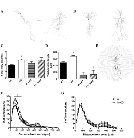
28
Morphological analysis was conducted on 19 WT and 24 1AKO pyramidal cells
that were filled with biocytin during whole-cell electrophysiology recordings (Figure 2).
1AKO neurons had a greater number of dendritic branches than WT cells (p<0.0001,
Figure 2C), as well as a higher number of nodes (p<0.0001) and ends (p<0.0001). The
total dendritic length when all processes were summed also differed between genotypes,
with 1AKO cells having significantly longer total dendrites than WT cells (p<0.0001,
Figure 2D).
A Sholl analysis was performed to determine the number of times the dendrites
crossed each radial segment (20µ) as well as the length of each dendritic segment within
each radii (Figure 2E). The dendritic trees of 1AKO neurons were significantly more
complex than those of WT cells. The number of intersections within each radii was
higher in the 1AKO cells (p<0.0001, Figure 2F), as was dendritic length within each
segment (p<0.0001). Post-hoc analyses showed that this significant increase in
complexity occurred from 60 to 180µ from the soma. This indicates that 1AKO cells
exhibit greater branching and complexity in the proximal dendrites as compared to the
WT neurons.
Morphology in the juvenile mouse
Pyramidal cell dendrites in the juvenile mice were shorter in length than dendrites
from the adult hippocampus. Total dendritic length was greater in P13 1AKO cells as
29
mice. There were also a greater number of dendritic ends in P13 1AKO mice compared
to P13 WT (p<0.05). The quantity of dendritic branches (Figure 2C) and total number of
nodes did not differ between genotypes. A Sholl analysis showed that there was a main
effect of genotype with 1AKO neurons having significantly more intersections (p<0.05)
and greater dendritic length (p<0.05) than WT cells (Figure 2G). However, there was no
interaction with radius.
Evoked responses
Extracellular field excitatory postsynaptic potentials (fEPSPs) were recorded from
the stratum radiatum of CA1 before and after high-frequency tetanic stimulation of the
Schaffer collaterals. The percent change of fEPSP slope from baseline, as averaged over
the last 10min of the 60min post-tetanus interval, was 54.8±0.6% in hippocampal slices
from adult WT mice (N=10) and 28.2±0.9% in slices from adult 1AKO mice (N=10,
Figure 3). This demonstrated a significant deficit (p<0.0001) in CA1 LTP in the 1AKO
mouse.
Evoked glutamatergic (eEPSC) and GABAergic (eIPSC) responses were obtained
using whole-cell electrophysiology in order to further understand the mechanism
underlying this LTP deficit in the 1AKO slices. The Schaffer collaterals were stimulated
using increasing levels of current in order to obtain input-output curves for each cell. WT
pyramidal cells (N=12) exhibited a significantly higher eEPSC response than 1AKO cells
30
receptors in the 1AKO hippocampus that may contribute to the deficit in LTP.
Conversely, there were no differences in eIPSCs between 1AKO (N=16) and WT (N=14)
cells, showing that GABA is not a contributing factor in the difference in LTP between
genotypes (Figure 4B).
DISCUSSION
The current study aimed to characterize cellular properties in the hippocampus of
the 1AKO mouse that may contribute to the behavioral phenotype of anxiety and
cognitive deficits. Several main conclusions can be drawn from these experiments. The
first is that genetic deletion of the 1AR resulted in increased CA1 pyramidal cell
proximal dendritic branching. Since there were already significant morphological
differences between the 1AKO and WT at P13, this change began to occur prior to the
start of the critical period for the 1AKO behavioral phenotype. Secondly, pyramidal
neurons of 1AKO mice received less glutamatergic input than those of WT mice. In the
juvenile mouse, this was shown by decreased spontaneous frequency of excitatory events,
indicating a pre-synaptic mechanism (Del Castillo and Katz, 1954; Redman, 1990;
Stevens, 1993). In the adult mouse, the change in amplitude and kinetics of spontaneous
EPSCs points towards alterations in postsynaptic receptor number and subunits. Finally,
activation of the hippocampal circuitry through stimulation of the Schaffer collaterals
revealed a deficit in LTP in the 1AKO mouse. Analysis of the evoked components
31
diminished compared to WT cells. This indicates that a decrease in the number of
AMPA receptors may be the underlying mechanism behind the reduced LTP and smaller
sEPSCs in the 1AKO.
Morphology in adult and juvenile mice
Pharmacological blockade of the 1AR during the third to fifth postnatal weeks has
been shown to phenotypically copy the behavior of the 1AKO mouse (Lo Iacono et al,
2008; Tsetsenis et al, 2007). A recent study by Ferreira et al (2010) demonstrated that
the same pharmacological treatment also resulted in increased dendritic arborization in
the stratum radiatum (SR) of 1AKO mice in hippocampal pyramidal cells at P35. Using
hippocampal cell cultures, they demonstrated that normal dendritic growth dynamics
were a result of 5-HT acting through the 1AR to reduce actin polymerization and restrict
growth cones. Our experiments add important data showing that genetic deletion of the
1AR produced increased proximal dendritic branching in the 1AKO that was already
present at the beginning of the third postnatal week.
A mechanism occurring prior to the critical period for the 1AKO phenotype that
leads to the differences in morphology seen at P13 therefore must be present. One
possible explanation is increased serotonin efflux in the hippocampus. Several studies
have looked at extracellular serotonin levels in the adult 1AKO mouse and have found no
differences in ventral hippocampus (Knobelman et al, 2001b; Richardson-Jones et al,
32
hippocampus that peak on day 14 (Mitchell et al, 1990). This may be due to a lack of
functional coupling of the 1AR in animals before that time in spite of high expression
levels of the receptor, a mechanism previously proposed based on studies in rat cortex
(Beique et al, 2004). By the time the animal reaches adulthood, serotonin levels are
greatly reduced, such that there is little tonic activation of 1ARs (Haddjeri et al, 2004).
This may account for the time course of the critical period for the 1AKO (Ferreira et al,
2010a; Gross et al, 2002; Lo Iacono et al, 2008).
Cellular properties in adult and juvenile mice
In the first two weeks of postnatal development, cell signaling in the rodent
hippocampus lacks an AMPA receptor-mediated component (Durand et al, 1996; Liao
and Malinow, 1996; Wu et al, 1996). During this time, GABAergic transmission is
excitatory and works in concert with NMDA receptor activation, allowing the
depolarizing activity necessary for the formation of mature synapses (Ganguly et al,
2001; Leinekugel, 2003). Excitation in these immature neurons alters synaptic efficacy
and strengthens connections in a Hebbian manner, much like in the adult (Ben-Ari,
2002). At age P13, 1AKO mice showed decreased frequency of spontaneous EPSCs,
indicating that pyramidal cells received less excitatory input at this time. This data
indicates that in the 1AKO mouse there was less synaptogenesis, which would lead to
decreased synaptic activity in the adult 1AKO. In support of this conclusion, we showed
AMPA-33
mediated EPSCs compared to WT mice, demonstrating that the number of AMPA
receptors was diminished in the 1AKO. Additionally, the kinetics of AMPA receptors
were altered, as evidenced by increased rise and decay times in 1AKO pyramidal cells.
These changes may be due to modifications in AMPA receptor subunit composition that
occurred early in the development of the 1AKO mouse hippocampus.
Within SR, the number or density of AMPA receptors is the major contributing
factor to synaptic strength (Nicholson et al, 2006). While the electrophysiology data
indicated that AMPA receptor-mediated input in this region was reduced in the 1AKO
mouse, the morphology of pyramidal cells showed that in the SR there was increased
dendritic branching and complexity compared to WT mice. Unlike WT cells,
patch-clamped pyramidal cells in the 1AKO did not demonstrate electrotonic filtering.
Filtering occurs when events generated distally but recorded somatically are delayed and
diminished due to the cable properties of the dendrites (Rall, 1969). There was no
change in capacitance between 1AKO and WT neurons (results not shown), indicating
that the larger membrane resistance of 1AKO cells is due to a decreased number of ion
channels. Therefore 1AKO neurons would have less loss of current and thus decreased
filtering. Additionally, this could indicate that synaptic events in the 1AKO may be
generated closer to the cell body within the complex dendritic arborization.
Decreased synaptogenesis in the 1AKO would also lead to an overall reduction in
the number of ion channels. CA1 pyramidal cells in the adult 1AKO mouse showed
greater resistance and tau, both of which contribute to the increased excitability seen in
34
be reflective of an increase in the surface area of the cell, stemming from the larger
dendritic branching, and/or from the reduction in the number of ion channels due to
decreased synaptogenesis. Additionally, the smaller activation gap in 1AKO cells would
contribute to the greater intrinsic excitability, which is independent of synaptic input.
These differences in membrane properties were not present in the juvenile 1AKO mouse,
indicating that changes in morphology occur concurrently with and may drive the
reduction in the number of channels and modifications in cellular properties seen in the
adult.
Long-term potentiation
LTP is a phenomenon by which synaptic strength between pre- and post-synaptic
neurons is increased. The most commonly studied type of LTP at the CA3-CA1 synapse
is NMDA receptor-dependent. However, there is evidence that postsynaptic modification
in CA1 may occur based on AMPA receptor recruitment (Malinow and Malenka, 2002).
Here we found that 1AKO mice exhibited a deficit in CA1 LTP. Both spontaneous and
evoked EPSCs in the 1AKO reflected a decrease in amplitude of glutamatergic signaling
likely due to a decreased number or density of postsynaptic AMPA receptors.
Furthermore, the kinetics of sEPSCs indicate possible changes in AMPA receptor
subunits. AMPA subunits are known to affect the trafficking properties and functionality
of the receptor, and phosphorylation of the GluR1 subunit is required for synaptic
35
Therefore, decreased number and activity of AMPA receptors may underlie the deficit in
LTP in the 1AKO mouse hippocampus. As memories are thought to be encoded by
synaptic strength, this also presents a mechanism that explains the
hippocampal-dependent deficits in spatial learning and memory that have previously been shown in the
1AKO mouse (Bliss and Collingridge, 1993; Sarnyai et al, 2000).
Conclusion
There have been few electrophysiological studies examining the hippocampus of
the 1AKO mouse in adulthood and during the critical period for the development of the
behavioral phenotype. Previous work from our laboratory shows correlations between
high glutamatergic activity and anxiety-like behavior in mice. However, in the current
study, electrophysiological and morphological comparisons were made between the two
genotypically verified groups. Juvenile 1AKO mice have decreased glutamatergic input
and increased dendritic branching, suggesting that synaptogenesis and cell architecture
drive changes during the critical period, possibly due to increased serotonin efflux from
the raphe. Additionally, these experiments show decreased AMPA-mediated responses
in the adult 1AKO that likely underlie the smaller LTP response and learning and
memory deficits seen in these mice. These findings provide further evidence for the role
of the 1AR in the development of mood and cognitive disorders.
36
We thank Mark Geyer for providing the founders for the colony of mice used in these
experiments, Akiva Cohen for critical advice on LTP experiments, and Zachary Spangler
37
FIGURE LEGENDS
Figure 1. Frequency-intensity plots of the frequency of AP firing rate in response to
increasing depolarizing current pulses in neurons recorded from WT and 1AKO
mice. (A) Representative data traces in current-clamp (top) used to generate
frequency-intensity plots (bottom), scale bar: 20 mV, 50 ms. P13 WT cells (N=20) did not differ in
excitability from 1AKO (N=14). (B) Adult WT cells (N=26) show significantly more
38
1AKO WT
WT 1AKO
Figure 1
A
B
*
*
*
*
0 20 40 60 80 100
0 5 10 15
WT 1AKO
Intensity (pA)
A
P
f
re
q
u
e
n
c
y
(
H
z
)
39
Figure 2:Morphological analyses of CA1 pyramidal neurons in WT and 1AKO. (A)
Representative traces of neurons from adult WT (left, N=19) and 1AKO (right, N=24)
cells, and (B) from P13 WT (left, N=11) and 1AKO (right, N=9) cells. Scale bars
represent 100µ. (C) Number of dendritic branches differs between WT and 1AKO in the
adult but not the juvenile neurons. (D) Total dendrite length represents the average sum
of all dendritic processes. 1AKO neurons had longer dendrites in total than WT cells in
both the adult and P13 animals. (E) Schematic of representative neuron used for Sholl
analysis. (F) Dendrites in the adult 1AKO showed significantly more complexity as
compared to WT dendrites. This increase in branching took place in proximal dendrites
from 60-180µ from the soma. (G) Dendrites of P13 1AKO cells had a greater overall
number of intersections as compared to WT, although the interaction with radius was not