Cardiac Muscle Differentiation in
Xenopus laevis
Embryos
A thesis submitted to the University of London
for the degree of Doctor of Philosophy
April 1995
Malcolm Patrick Oliver Logan
Department of Developmental Biochemistry
National Institute for Medical Research
Mill Hill
ProQuest Number: 10055345
All rights reserved
INFORMATION TO ALL USERS
The quality of this reproduction is dependent upon the quality of the copy submitted.
In the unlikely event that the author did not send a complete manuscript and there are missing pages, these will be noted. Also, if material had to be removed,
a note will indicate the deletion.
uest.
ProQuest 10055345
Published by ProQuest LLC(2016). Copyright of the Dissertation is held by the Author.
All rights reserved.
This work is protected against unauthorized copying under Title 17, United States Code. Microform Edition © ProQuest LLC.
ProQuest LLC
789 East Eisenhower Parkway P.O. Box 1346
Acknowledgements
The work described in this thesis was supported by a grant from the Medical Research
Council. I would also like to thank Jim Smith (NIMR) for the gift of human recombinant
Activin A, Xenopus bFGF, the 12/101 mAb and for very helpful practical advice. Wendy
Hatton and John Asanti, both of the Histology Department at NIMR, provided excellent
assistance on histological techniques and carried out the 'three-way' staining protocol on my
sections. The Photographies Department at NIMR gave helpful advice on photography and
the production of figures.
I am grateful to all the members of the departments of Developmental Biochemistry and
Developmental Biology for providing useful discussions, practical tips and encouragement
throughout the length of the project. Jam Tata, Jacky Smith and Ena Heather deserve
particular mention for ensuring that my time in the department ran smoothly. Anne
Chambers, Robert Wilson, Surendra Kotecha and Norma Towers provided wonderful
technical support and practical advice in the laboratory and together created a stimulating
environment that was great fun to work in.
Finally and above all, I am indebted to my supervisor, Tim Mohun, who set me to work on a
great project. Through his support and encouragement I have learnt a tremendous amount
about laboratory science and his comments and perseverance have greatly improved the
Abstract
This thesis describes work carried out to initiate a study of cardiogenesis in the amphibian,
Xenopus laevis. I have isolated and characterised a cDNA fragment encoding a portion of the myosin heavy chain a-isoform (XMHCa) from Xenopus laevis embryos and characterised another
Xenopus cDNA encoding a myosin light chain isoform, XMLC2. Both the XM HCa andXAfLC2
transcripts are expressed exclusively in adult heart tissue. Whole mount RNA in situ hybridisation indicates that expression of the XM HCa and XMLC2 genes are restricted to the developing heart
primordium. They therefore provide tissue-specific markers for cardiac muscle differentiation
during early embryogenesis. Using these transcripts as molecular markers, I can detect the onset
of cardiac muscle differentiation in an anterior-ventral region of tailbud embryos, many hours
before the appearance of a beating heart.
XM H C a and XMLC2 gene expression can be induced in isolated animal pole explants of blastulae by treatment with the growth factor, activin A. Induction is dose-dependent, requiring high doses of the growth factor compared with that required for myotomal (skeletal) muscle differentiation. In contrast, no XM HCa transcripts are detected in explants incubated with basic FGF, despite the induction of myotomal muscle differentiation. In addition, fusion potentiates induction of cardiac muscle differentiation and exposure to activin for just several hours was sufficient to induce markers after days of incubation. Activin-induced explants show a similar temporal pattern of XM H Ca gene expression to that found in normal embryogenesis.
Furthermore, cells expressing this gene appear clustered in one or two foci within fused explant aggregates, which often show regular, spontaneous contractions after several days in culture. These results show that terminal differentiation of cardiac muscle can occur in growth factor- induced explants and may be distinguished from skeletal muscle differentiation by the dose and nature of the inducing factor. Explant aggregates containing foci of beating cells have been analysed further by histological and immunological techniques. These studies have shown that the amount of induced cardiac muscle is small compared with the amount of induced skeletal muscle within explants. In addition, induction of endoderm within the explant may be required for the formation o f heart muscle.
Ill
Using an explant culture system and terminal differentiation markers for heart muscle, the
restriction of the heart morphogenetic field during tailbud stages, has been studied. In contrast to
the results from studies that used a beating-heart assay for heart formation, the restriction of the
heart morphogenetic field is not complete by late tailbud, stage 28. Thus, the ability of cells
within the heart field to express terminal differentiation markers can be clearly distinguished from
their potency to form a beating heart tube, in culture. In addition, analysis by whole mount RNA
in situ hybridisation has indicated that restriction of the heart field is apparent in lateral regions
after removal of the anterior ventral cells fated to form heart. This suggests that repressive
AGPC acid-guanidinium thiocyanate phenol chloroform
ATP riboadenosine-5'-triphosphate
BCIP toluidine sait of 5-bromo-4-chloro-3-indo|yl phosphate
BSA Bovine serum albumin
C-terminal carboxy terminal
CHAPS (3-[(3-cholaminodopropyl)dimethylammonio]-1 -propanesulphonate
CTAB hexadecyltrimethylammonium bromide
DMZ dorsal marginal zone
E. Coli Escherischia coli
EDTA diaminœthanetetra-acetic acid, disodium sait
EGTA ethyleneglycol-6f (b-aminoethyl ether) N, N, N', N’-tetra-acetic acid
FITC flourescein isothiocyanate
FLDX flourecein lysine dextran amine
GTP riboguanosine-5'-triphosphate
HCL hydrochloric acid
Kb kilobase
Klenow klenow fragment of DNA polymerase I
L iC l lithium chloride
MEF muscle enhancer factor
MgCl2 magnesium chloride
M gS04 magnesium sulphate
MOPS (3-[N-morpholino]propanesulphonic acid)
N-terminal amino-terminal
NaCl sodium chloride
NAM normal amphibian medium
NBT 4-nitro blue tétrazolium chloride
NH4AC ammonium acetate
NIMR National Institute for Medical Research
PAGE polyacrylamide gel electrophoresis
PBS phosphate buffered saline
PCR polymerase chain reaction
PEG polyethylene glycol
PIPES piperazine-N, N’-bis[2-ethanesulphonic acid]
RNAase ribonuclease
SM smooth muscle
SRE serum response element
TESPA 3-aminopropyltriethoxysilane
TGF p i transforming growth factor beta 1
Tris (tris[hydroxymethyl] aminomethane)
TTP thymidine triphosphate
TRLDX Texas Red® lysine dextran amine
Triton Triton X-100
Tween-20 monolaurate polyoxyethyllenesorbitan
UTP uridine triphosphate
UWGCG University of Wisconsin Genetics Computer Group
W watts
bFGF basic fibroblast growth factor
bHLH basic Helix-loop-helix
bp base
pah-cAMP cyclic adenosine monophosphate
cDNA complonentary deoxyribonucleic acid
dNTPs deoxyribonucleotide triphophates
kD kilodalton
mAb monoclonal antibody
ml millilitre
mM millimolar
p.c. post coitum
microgramme
microlitre
Contents
Acknowledgem ents... i
A b stra c t...ii
A b b rev iatio n s...iv
C o n te n ts...vi
F ig u res...x
T a b le s ...xii
G ra p h s ...xiii
C h ap ter 1 In tro d u ctio n ... 1
Embryological origin of vertebrate cardiac mesoderm... 2
Vertebrate cardiogenesis... 2
Cardiac development in amphibians...6
Analysis of the amphibian heart field ... 7
Summary...8
Contractile apparatus of striated muscle... 9
The structural proteins of muscle...9
Contractile protein isoforms... 10
Striated muscle isoforms...10
a-A ctin isoforms... 10
Troponin isoforms... 11
Tropomyosin isoforms...12
Myosin heavy chain isoforms... 12
Myosin light chain isoform s... 13
Summary...13
Transcriptional control in m uscle... 14
Regulation of skeletal muscle differentiation by members of bHLH subfamily of genes...14
Evidence for cardiac-specific bHLH proteins... 15
E-box-independent pathways for the control of skeletal and cardiac muscle transcription...15
Summary...16
Mesoderm formation in the amphibian... 17
vil
C h ap ter 2 M aterials and M ethods...21
Chemicals, reagents and m edia...22
Embryo and explant culture...22
Preparation of R N A ... 22
RT-PCR... 23
Cloning of XM HCa Fragment isolation... 24
Ligation and transformation... 24
Isolation of plasmid D N A ... 24
DNA sequencing... 24
RNAase protection assay... 24
Whole mount RNA in situ hybridisation...26
Lineage-label microinjection...28
Embryo embedding and sectioning... 28
Histology and immunohistochemistry... 28
Microscopy and photography...29
C h ap ter 3 Isolation of Molecular M arkers for Differentiating H eart M uscle 30 Introduction... 31
Cloning of Xenopus M HCa cDNA... 31
Expression of Xenopus M HCa during development...33
Cloning of Xenopus MLC2 cD N A ... 40
Expression of Xenopus MLC2 during developm ent...40
Oiscu&stftn... 43
C h ap ter 4 Induction of H eart Muscle in Isolated Animal Pole E x p la n ts... 49
Introduction... 50
Induction of XM HCa expression in cultured animal pole explants by activin A is dose dependent...50
bFGF does not induce expression of XM HCa in animal pole explants... 52
Induction of XM HCa by activin A has a normal temporal expression pattern ..54
Cardiac muscle cells are clustered in cultured explants... 56
Induction of heart muscle requires a minimum of tissue... 56
High doses of activin abolish the potency prepattem across the cells of the animal hemisphere...58
D iscussion... 64
Conclusion...66
C h ap ter 5 Histological Analysis of Induced Animal Pole E x p la n ts... 67
Introduction...68
Histological analysis of induced animal pole explants...68
Distinguishing induced cardiac muscle from induced skeletal muscle within caps...71
D iscussion... 74
Induction of neural tissue... 74
Induction of endoderm?... 74
An early inductive role of the endoderm ...75
A formative role of the endoderm...76
Conclusion...77
C h ap ter 6 Studies of the H eart Morphogenetic F ield ...79
Introduction...80
Fatemapping the cardiac progenitors...81
Properties of the heart morphogenetic field... 81
Heart-forming potential of explants... 81
Heart-forming potential of explant conjugates... 87
Subdivision of explants of the heart fie ld ...92
Regulation within the field in v iv o ... 94
Restriction of the heart field...96
D iscussion...102
Fate map of the cardiac anlage... 102
Regulative capacity within the field... 104
Evidence of a gradient of potency within the heart fie ld ... 105
Models of heart formation... 105
Whole mount RNA in situ hybridisation versus beating heart assay as a means to score heart formation... 107
The restriction of the heart morphogenetic field a reassessment using RNAase protection analysis...108
C h ap ter 7 ...109
Introduction...110
cDNA differential library screen...I l l
Study of the heart morphogenetic field...112
Patterning of the heart tu b e... 116
Figures
C h ap ter 1
Figure 1 Origin of the cardiac mesoderm in vertebrate embryos... 3
Figure 2 The relative position of the cardiac mesoderm during development... 4
Figure 3 The "three signal" model for mesoderm formation in amphibian embryos... 18
C h ap ter 3 Figure 4 Sequence of di Xenopus M HCa cDNA fragment...32
Figure 5 Expression of XM HCa in adult frog tissues...34
Figure 6 Expression of XM HCa Xenopus embryos...36
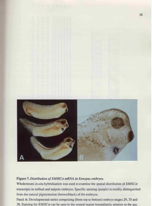
Figure 7 Distribution of XM HCa mRNA in Xenopus embryos... 38
Figure 8 Sequence of a Xenopus MLC2 cD N A ...39
Figure 9 XMLC2 expression during development... 41
Figure 10 XMLC2 expression in the prospective heart region of tailbud embryos... 42
Figure 11 Distribution of XMLC2 mRNA in Xenopus embryos... 44
C h ap ter 4 Figure 12 Induction o f XM HCa expression in animal pole explants... 51
Figure 13 XM HCa expression in explants is induced by activin A but not by bFG F... 53
Figure 14 XM HCa expression is induced in discrete foci within cultured explants 55 Figure 15 Induction of XM HCa in animal pole explants requires a minimumof tissue. 51 Figure 16 Induction of XM HCa and Gsc in both dorsal and ventral caps treated with a high dose of activin...59
Figure 17 Induction of XM HCa results from an early response to activin... .62
Figure 18 Activin induction of XM HCa is not inhibited, at later stages, by exposure to bFGF...63
C h ap ter 5 Figure 19 Histological analysis of a longitudinal section of a stage 40 embryo... 69
Figure 20 Histological analysis of sections of untreated, low-dose activin treated and high-dose activin treated animal pole explants... 70
Figure 21 Detection of both cardiac and skeletal muscle cell types within the embryo... 72
C h ap ter 6
Figure 23 Fate mapping of the heart anlage in Xenopus... 82
Figure 24 Explant dissections of the stage 23 embryo... 83
Figure 25 Heart forming potential of explants of the heart field...84
Figure 26 Comparative analysis of explant conjugates...88-89
Figure 27 Heart forming potency of smaller regions of the heart field... 91
Figure 28 Heart-forming potency of the lateral region in vivo after removal of the
anterior ventral cells... 93
Figure 29 Restriction of the heart field...101
Figure 30 The GM l double-gradient model... 105
C h ap ter 7
Figure 31 Expression patterns of XNkx2.5 and XGATA4 in the late tailbud stage
Tables
Heart Forming Potential of Expiants of the Heart Field
Table 1... 85
Table 2 ... 85
Heart Forming Potential of Explant Conjugates
Table 3 ...90
Restriction of the Heart Morphogenetic Field
T a b le d ... 100
Graphs
Restriction of the Heart Morphogenetic Field
Graph 1... 98
Chapter 1
Embryological origin of vertebrate cardiac mesoderm
Fate mapping studies in mouse, chick and frog embryos have uncovered striking similarities
between the embryological origin of the heart in vertebrates. Both the heart and skeletal
musculature are mesodermal derivatives but have separate embryological origins from the earliest
point they are identifiable. In the mouse embryo at the early streak stage, the cells fated to form
heart are situated adjacent and lateral to the cells destined to form notochord and endoderm
(Lawson and Pedersen, 1992). The skeletal muscle is derived from a more lateral region (fig. li).
In the chick embryo, regions of cells fated to form particular structures appear more dispersed, but
at the early primitive streak stage two regions of cells that later form heart have been identified
(fig. lii). These abut the region of cells of the prospective notochord while the cells of the
prospective somite lie more laterally (Rosenquist, 1966; Rosenquist, 1970; Hatada and Stem,
1994).
The most detailed account of the origins of cardiac muscle has been provided by studies of
amphibian embryos. Fate maps of the 32-cell stage Xenopus embryo indicate that the blastomeres
of the dorsal marginal zone (Cl and C2) most commonly give rise to the heart (Dale and Slack,
1987 and figure liii). At gastrula stages in both Xenopus (Keller, 1975) and the urodele
(Holtfreter, 1938) the two heart primordia are situated either side of the dorsal blastopore lip, deep
and lateral to the cells of the prospective notochord but in a more anterior dorsal position than the
prospective somitic mesoderm, situated laterally (fig. liii). A similar relationship in
embryological origin for both the cardiac and skeletal musculature has recently been confirmed in
another amphibian, Rana pipiens (Saint-Jeannet and Dawid, 1994).
Taken together, these fate mapping studies indicate that in most vertebrates cardiac mesoderm
is «anterior dorsal derivative, despite the final ventral position of the heart whilst the skeletal
musculature is derived from more lateral mesoderm. A notable exception to this is the zebrafish
{Brachydanio rerio). Recent fate mapping of the mid blastula zebrafish embryo has identified a
zone of heart progenitors centred at the future ventral axis (Stainier et ai, 1993; Lee et al.y
1994b).
Vertebrate cardiogenesis
In mouse and chick embryos, formation of a beating heart is a relatively rapid process. In
mouse embryos, the prospective myocardium is first identifiable around 7.5 days p.c^presomite
stage, when cells of the anterior splanchnic mesoderm assume a cuboidal morphology (DeRuiter
et a i, 1992). These cells constitute the cardiogenic plate which fuses to form a horse-shoe shaped
ant. post.
blood islands
allan-lateral
vr
rhfa
M O U SE
Ihta
ps
CHICK
AP
■ECTODERM
/
I
w /^SOM ITE-^
V I BLOOO _ HE>
\
NOTO ...CHORO
PROHEPHROS HEART
D
1 T
-ENDODERM
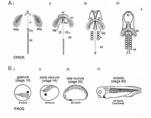
Figure \,Origin o f the cardiac mesoderm in vertebrate embryos
Fate maps of similar stage mouse, chick and Xenopus embryos. The area of cells fated to
form heart in each case lie adjacent and lateral to, the cells of the prospective notochord. (I)
Fate map of the early streak stage mouse showing the approximate regions from which
structures identifiable at the early somite stage are derived. Ant: anterior. Post.: posterior.
Not + end.: notochord and endoderm (From Lawson, 1992). (II) Ventral view of the stage 5
chick embryo. The left and right heart-forming areas (Ihfa and rhfa) lie on either side of the
prospective notochord (n) and Hensen's node (hn) (from Osmond, 1991). (Ill) Fate map of
the stage 6 Xenopus embryo indicating the approximate areas from which various cell types
PS
ht ce
aip
O s
sp
CHICK
B
gastrula (stage 10)
s %
9 hours
FR O G
early neurula
(stage 14) late neurula (stage 20)
16 hours 22 hours
IV
tadpole (stage 33)
A4 hours (heartbeat)
Figure 2. The relative position o f the cardiac mesoderm during development
Panel A: Diagrams showing a ventral view of normal heart development in the chick. At
stage 6 (I) the precardiac mesoderm migrates craniomedially toward the developing
headfold (hf). By stage 7 (II), the precardiac cells meet in the midline over the headfold to
form the cardiogenic crescent (cc). The overlying endoderm invaginates to form the anterior
intestinal portal (aip). At this point the neural folds (nf), first pair of somites (s) and
segmental plates (sp) are visible. At stage 8 (III), the cardiac mesoderm begins to
differentiate into bilateral heart tubes (ht). At stage 9 (IV), the tubes fuse cranially in the
midline and the sinus vinosus (sv) forms. The neural folds have fused along much of their
length, so that a neural tube (nt) is visible.
Panel B: Diagrams showing the heart forming regions (dark shading) and somitic muscle forming (light shading) regions at various stages of Xenopus development, from gastrula
stage (10) until tadpole stage (33). The heart primordia is initially in an anterior-dorsal
position whilst the skeletal muscle progenitors lie more laterally (I). At early neurula stage
(II), the cardiac mesoderm lies in the head region, whilst the somitic mesoderm is begining
to differentiate in two strips along the dorsal midline. During neurulation the cardiac
mesoderm undergoes a ventralward migration to its final anterior-ventral position (III). By
stage 33 (IV), a hean tube is visible in the anterior ventral position behind the cement gland
and the somitic muscle has differentiated into segmented blocks along the length of the
with about 3-5 somites (around 8 days p.c.), the two cardiogenic rudiments have formed open
tubes which are fused at the rostral tip. A rostral to caudal fusion takes place giving rise to a single
midline structure at about 8 days p.c. and soon after the heart assumes its characteristic looping,
"S" shape morphology. By 8.5 days p.c. (6 somites), the heart begins to beat regularly.
In the chick embryo, precardiac mesoderm in the epiblast migrates through the primitive streak
and into the lateral plate mesoderm during gastrulation (Rosenquist and Dehaan, 1966;
Rosenquist, 1970). By stage 5 (Hamburger and Hamilton, 1951), there are two distinct bilateral
heart forming areas located either side of Hensen’s node (Rawles, 1943) (fig. 2Ai). As the
primitive streak begins to regress, the precardiac mesoderm migrates craniomedially toward the
developing headfold and by stage 7 fuses at the midline to form a cardiogenic crescent (cc),
analogous to the horse-shoe shaped region described in mouse. At stage 8 as the neural folds
begin to close, the precardiac mesoderm begins to differentiate into bilateral heart tubes. In what
appears to be a similar event to that in mouse, from stage 9 the two open tubes fuse in a rostral to
caudal progression to form a single heart tube that then loops and commences beating (2Aii-iv).
In mammals and birds, the heart is the first organ system to differentiate and function and, at
this stage of development, it is also the most prominent organ in the embryo. Differentiation markers such as MHCa in mouse (Lyons et al., 1990a) and VMHCl in chick (Litvin et al., 1992)
are detected prior to fusion of the developing heart tube and before the onset of molecular
differentiation of skeletal muscle. Heart muscle differentiation therefore predates skeletal muscle
differentiation in these embryos.
In contrast, heart formation only occurs after skeletal muscle formation in amphibians and fish.
During gastrulation stages in Xenopus, the two heart anlage migrate at the leading edge of the
chordamesoderm to an anterior dorsal position (Gerhart and Keller, 1986) (fig. ^i-ii). At the onset
of neurulation (stage 13 (Nieuwkoop and Faber, 1956)), the prospective heart rudiments begin to
move laterally. In the early neurulae (stage 14), prospective heart mesoderm sits at the anterior
lateral edge of the mesodermal mantle between the edge of the neural plate and the ventral
midline(2Bii). The regions of heart mesoderm migrate ventrally and at stage 16, begin to fuse at
the ventral midline. Fusion is complete by early tailbud stage 20 (2Biii). Overt differentiation is
only morphologically distinct by stage 27, when a rudimentary heart tube forms and a heart beat is
not initiated until the tadpole is free swimming, at stage 34 (Nieuwkoop and Faber, 1956).
Transcripts of cardiac actin are detected in the developing somitic mesoderm from the gastrula
stage (Cascio and Gurdon, 1986; Mohun et al., 1994) indicating the onset of terminal
expression of markers in the skeletal musculature and the first morphological signs of
differentiation of the heart in Xenopus. Similarly in zebrafish, an anti-tropomyosin monoclonal
antibody (mAb CHI) is the earliest marker for the cardiac primordia and stains the heart from the
18 somite stage onwards (Stainier et a l, 1993). All the somitic muscles are already CHI positive
at this stage.
Cardiac development In amphibians
Much of our understanding of vertebrate cardiogenesis has come from studies of amphibian
embryos. Experiments with wild-type and cardiac mutant axolotls (Smith and Armstrong, 1990)
and with Xenopus (Sater and Jacobson, 1989; Sater and Jacobson, 1990a; Nascone and Mercola,
1995) have identified a number of distinct events in cardiogenesis that suggest formation of a
morphologically distinct, functional beating heart in the amphibian is a multistep process.
In Xenopus, removal of the dorsal lip from the early gastrula stage prevents heart formation, as
well as differentiation of other dorso-anterior mesodermal derivatives (Sater and Jacobson,
1990b). Furthermore, in classical "organiser graft" experiments, transplantation of the dorsal lip into the ventral marginal zone of host embryos results in the formation of a secondary axis; in
over half the cases this contains a heart derived from the host mesoderm (Sater and Jacobson,
1990b). Together, these results suggest that the establishment of heart mesoderm is initiated by a
dorsalising signal from the dorsal lip of the blastopore. Nascone and Mercola (Nascone and
Mercola, 1995) have recently examined the contributions of influences from the mesoderm and underlying endoderm in such grafting experiments. They found that heart formation in the
secondary axis only occurred if the grafted tissue contained deep endodermal cells. The dorso-
anterior endoderm alone, however was incapable of inducing heart muscle formation from host
tissue. These results suggest that in addition to a dorsalising signal from the organiser mesoderm a
signal from the endoderm is also required for the induction of cardiac muscle.
Further evidence for a role of underlying endoderm in the formation of heart muscle has come
from studies of urodele amphibia which develop more slowly than anurans such as Xenopus.
Removal of the entire endoderm from neurula stage urodele embryos prevents heart formation
(Jacobson and Sater, 1988). Experiments using explant recombinations of endoderm and
mesoderm have located the heart-inducing capability to the anterior ventral endoderm (Jacobson
and Duncan, 1968; Fullilove, 1970). In addition, this tissue has a "formative effect" on heart
formation since the quality of the hearts that form correlates with how long the heart mesoderm is
Additional evidence for a role of endoderm in later stages of cardiogenesis comes from studies
of a mutant form of the axolotl, Ambystoma mexicanwn (Humphrey, 1972). Embryos
homozygous for the cardiac lethal (cl) mutation form a heart but fail to initiate a heartbeat and
eventually die. The cl mutation disrupts normal myofibrillogenesis but the defect can be rescued
by replacing mutant endoderm with wild-type endoderm at the tailbud stage (Lemanski et a/.,
1979). This suggests that the cl mutation interferes with the ability of endoderm to regulate
relatively late steps in heart morphogenesis.
Analysis of the amphibian heart field
It has long been known that in amphibians the region of cells within the embryo capable of
forming heart is initially larger than the area of cells fated to form the heart in the normal embryo.
As early as 1924, Copenhaver removed the presumptive heart mesoderm from Ambystoma
punctatum embryos at tailbud stages 25-29 and showed that mesoderm surrounding the wound
would undergo heart formation, often resulting in the production of two hearts in the embryo from
lateral mesoderm on either side of the ventral midline. Replacement of the anterior ventral and anterior lateral mesoderm with more dorsal and posterior flank mesoderm prevented heart
formation. Ekman (Ekman, 1925) found that anterior lateral mesoderm was capable of heart
formation when transplanted into the anterior ventral region of post neurula embryos, whereas
more posterior mesoderm was not The region of tissue possessing heart forming potency has been
termed the heart morphogenetic field.
In Xenopus, Sater and Jacobson (Sater and Jacobson, 1990a) described a crude map of the
heart morphogenetic field constructed by culturing ventral and lateral explants from anterior,
midanterior, mid posterior and posterior regions of the stage 20 embryo and scoring for beating heart formation. Each explant included all three germ layers, mesoderm, ectoderm and endoderm.
They found that hearts formed from anterior ventral tissue, which includes the cells fated to form
heart, in 100% of cases. Anterior lateral explants formed hearts in 69% of cases. Hearts did not
form in any of the other explants. These results demonstrated that at the end o f neurulation (stage
20), the heart morphogenetic field includes both anterior lateral and anterior ventral mesoderm and that anterior ventral mesoderm has a slightly stronger potency for heart formation than lateral
mesoderm.
In the same study, Sater and Jacobson also described experiments comparing the heart forming
potency of anterior ventral and anterior lateral explants removed at successive stages from early
tailbud stage 20, through to late tailbud stage 28. Anterior ventral explants from all stages formed
initially high, starting at 69% at stage 20 and increasing to 83% at stage 22. After this stage,
however, the frequency of heart formation in lateral explants begins to decrease steadily so that by
stage 28, no lateral explants formed beating hearts. These results indicate that the lateral
mesoderm within the heart morphogenetic field begins to lose its potency to form heart from stage
22 onwards and has lost it completely by stage 28. The spatial extent of the heart morphogenetic
field therefore changes over time. This loss of heart forming potency in the lateral regions is
described as the restriction of the heart morphogenetic field.
Restriction of the heart morphogenetic field is not due to migration of cells with heart-forming
ability to a more ventral position since grafting experiments demonstrate that anterior lateral
mesodermal cells maintain their relative position in the embryo between stages 20 and 28 (Sater
and Jacobson, 1990a). Neural tissue has been shown to have an inhibitory influence on heart
formation (Jacobson and Duncan, 1968) but this is unlikely to account for the restriction of the
heart field in Xenopus. Removal of the neural crest and neural tube at stage 20 and subsequent
culture of ventral and lateral explants from operated embryos gave the same results as those
obtained with unoperated embryos (Sater and Jacobson, 1990a).
There is some evidence from explant studies that endodermal tissue underlying ventro-lateral
mesoderm plays a role in establishing the extent of the heart field. Explants of lateral mesoderm,
removed at stage 20, form hearts with a significantly higher frequency in the presence of
underlying endoderm. By stage 23, the heart forming frequency of lateral mesoderm is unaffected
by the presence or absence of the endoderm. These results indicate that a supportive influence of
the underlying endoderm can be distinguished between stages 20 and 22 but is lost by stage 23.
Heterochronic explant combinations made with anterior lateral endoderm from embryos at stage
20 to 23 and anterior lateral mesoderm from stage 27 to 28 embryos, rarely formed beating hearts
suggesting that the lateral mesoderm loses its competence to respond to signals from underlying
endoderm during tailbud stages.
Summary
An embryological approach in Xenopus, using the formation of a rhythmically beating tissue as
a marker for cardiac muscle, has identified a number of steps in cardiogenesis; dorsalisation of the
mesoderm, an inductive influence of the underlying endoderm, later formative influences from the
endoderm and restriction of the heart morphogenetic field. A first step to understanding the
molecular basis of these steps would be to isolate a molecular marker for cardiac muscle. Good
candidates are genes expressed during the terminal differentiation of this tissue type, such as those
Contractile apparatus of striated muscle
In Xenopus, mononucleate skeletal muscle cells in the myotomes are formed shortly after
neurulation and are fully functional before innervation. These cells do not initially fuse but remain
mononucleate after innervation until shortly before metamorphosis (Hamilton, 1969; Muntz,
1975) when they become multinucleate by fusion with satellite cells (Muntz, 1975). In contrast,
cardiac muscle cells remain mononucleate throughout development.
At the subcellular level both cardiac and skeletal muscle cells contain numerous myofibrils, the
functional units of contraction. Each myofibril consists of bundles of thin filaments arranged
around thick filaments in a hexagonal pattern. This organisation forms a repeating unit or
sarcomere which gives both cardiac and skeletal myofibrils a characteristic striated appearance.
The structural proteins of muscle
The thick filaments of myofibrils are about 15 nm in diameter and about 1.6 |im long. They lie
parallel to one another in the middle of the sarcomere, thus forming the dark A bands of the
myofibril. Every thick filament consists of hundreds of myosin molecules organised in a staggered
array such that the heads of successive myosin molecules project out of the thick filament in a repeating pattern. The heads occur in pairs, which protude from the thick filaments facing away
from the centre of the filament. These protuding heads are able to make contact with adjacent thin
filaments and form the cross-bridges that are integral to the mechanism of muscle contraction.
Myosin has a complex structure, consisting of two identical heavy chains and four light chains
(two regulatory light chains and two alkali light chains). These six polypeptides are organised into
two globular heads and a long, narrow rod-like tail. The tail consists of the two heavy chains
twisted around each other. At the amino terminus, each of these chains is coiled into a globular
head, with which a pair of each type of light chains is associated. The ATPase activity responsible
for the ATP hydrolysis that drives the contraction process is situated in part of the globular head
domain.
The thin filament is largely composed of actin, tropomyosin and troponin. Actin filaments (F
actin) are polymers of a single polypeptide (G-actin) and each filament consists of two polymers
about 4 nm in diameter twisted into a helix with 13.5 molecules per turn. Smaller amounts of
tropomyosin and troponin are associated with the F-actin strands. Tropomyosin is a long
threadlike protein composed of two subunits which form an a-helical coiled coil, that lies in the
10 and associates with seven actin monomers. Troponin is a complex of three polypeptide chains,
troponin T (TnT), troponin C (TnC) and troponin I (Tnl). TnT binds to tropomyosin and is
thought to be responsible for positioning the complex on the tropomyosin molecule. TnC binds up
to four molecules of calcium and Tnl binds to actin. One troponin complex is associated with each
tropomyosin molecule along the thin filament. Troponin and tropomyosin constitute a calcium-
sensitive switch for contraction in both heart and skeletal muscle. They function to regulate the
availability of myosin binding sites on the actin filament.
When the sarcoplasmic level of calcium is low tropomyosin blocks myosin binding sites on the
actin molecule and cross-bridge formation is prevented. At higher calcium concentrations the
tropomyosin molecules shift their position on the F-actin strand allowing myosin heads to make
contact with binding sites on the actin filaments thereby facilitating contraction. Calcium
sensitivity is conferred by the TnC polypeptide of the troponin complex. Upon binding of
calcium, TnC undergoes a conformational change that is transmitted to the tropomyosin molecule.
C ontractile protein isoforms
Almost all of the proteins that comprise the striated muscle myofibril are members of protein
isoform families. Isoform diversity can be generated by either one or both of two mechanisms.
Gene families may encode individual isoforms as is the case with the myosin heavy chain {MHC)
multi gene family (Nguyen et a/., 1982; Buckingham, 1985; Robbins et o/., 1986). Alternatively, multiple protein isoforms are generated from a single gene as the result of alternative splicing. An
example of this is the troponin T gene which potentially gives rise to as many as 64 different
transcripts encoding distinct isoforms (Breitbart et al.y 1985).
Isoform diversity is thought to underlie the unique physiological characteristics of individual
muscle-fibre types although functional differences between isoforms have only been demonstrated
in a few cases. Indeed some protein isoforms are functionally interchangeable indicating that there
maybe functional redundancy among some genes expressed in the myofibre (Bandman, 1992).
S triated muscle isoforms a - Actin isoforms
In mammals, six isoforms of actin are expressed, each from a single gene; p- and y-actins are
expressed in non-muscle cells, aSM - and ySM-actins are expressed in smooth muscle and cardiac
a-actin and skeletal a-actin are expressed in cardiac muscle and skeletal muscle (Weydert, 1988).
Bovine and rabbit, cardiac and skeletal a-actin sequences are highly conserved, differing in only 4
11
muscle-specific actin genes, encoding distinct protein isoforms have been identified, cardiac (a l),
skeletal (a2) and femoral (a3) (Mohun etaL, 1988).
Cardiac actin is the main isoform found in the adult heart but during embryonic stages in
mouse, rat and Xenopus it is also the predominant form found in the developing somites and is
only down-regulated later in development (Minty et a i, 1982; Mayer et al., 1984; Schwartz et al., 1986; Sassoon et al., 1988). Conversely in rat cardiac muscle, a-skeletal and a-cardiac actin
mRNAs are coexpressed, but after birth a-skeletal actin disappears (Mayer et a i, 1984).
Troponin isoforms
In mammals and chick, the TnC isoforms are encoded by two genes, one is expressed in both
cardiac and slow skeletal muscle while the other is expressed in fast skeletal muscle (Parmacek and Leiden, 1989; Toyota et al., 1989). Based upon common sequence features, it has been
proposed that TnC and other calcium-binding proteins such as the calmodulins and the regulatory
and alkali light chains have evolved from a common ancestor (Collins, 1991). TnC sequences are
highly conserved between distant species, for example there is only a single amino acid difference
between human and rabbit fast skeletal isoforms (Bandman, 1992). The fast TnC isoform can bind
four calcium ions per molecule, while the slow/cardiac isoform can only bind three. This
difference appears to have arisen as a result of a natural mutation in one binding site (Putkey et
a l, 1989).
In mammals and birds,TnX isoforms are encoded by three genes, cardiac, slow skeletal and fast
skeletal (Nikovits et a l, 1986; Koppe et a l, 1989; Murphy et a l, 1991). In rat, the cardiac Tnl
isoform is developmentally regulated, being expressed at low levels in the foetal myocardium and
then markedly increased during the perinatal period (Sabry and Dhoot, 1989; Saggin et a l, 1989).
In the foetal rat and human heart, the slow skeletal isoform represents the predominant transcript.
Recently, the Xenopus Tnl isoform has been cloned and has been shown to be expressed
exclusively in the myocardium at embryonic stages (Drysdale et a l, 1994). Cardiac Tnl has an
additional amino terminal domain compared with skeletal Tnis (Leszyk et a l, 1988) and it is
within this region that phosphorylation of a serine residue results in altered calcium sensitivity
(Solaro e ta l, 1976; Mope e t a l, 1980).
TnT isoforms are similarly encoded by three genes, cardiac, slow skeletal and fast skeletal
(Copper and Ordahl, 1985; Breitbart and Nadal-Ginard, 1986; Gahlman et a l, 1987; Smillie e ta l,
1988). Whilst each of the Tnl and TnC genes code for single proteins, the TnT gene produces
12
The cardiac TnT gene generates isoforms that are developmentally regulated and expressed in a
tissue-specific manner (Copper and Ordahl, 1985; Anderson, 1988). Bovine heart TnT isoforms,
containing amino terminal variations, confer different calcium sensitivities to the ATPase activity
o f the cardiac myosin heads in vitro (Tobacman and Lee, 1987). These results and other data with
fast TnT isoforms, show a correlation between TnT isoform composition and tension generated in
muscle fibres in response to changes in the calcium ion concentration (Schachat et a i, 1987;
Reiser era/., 1992).
T ropomyosin isoforms
Muscle cells express two isoforms, a-tropomyosin and P-tropomyosin which may form homo-
or heterodimers. The
a-p
heterodimer appears to be the favoured form when both isoforms are expressed within the same cell, however the ratio of a /a ,a /p, p/p
forms is predominantly determined by the accumulation of the different transcripts that arise from the two genes. In rat and chick,a-
and P-tropomyosin genes have been characterised and each undergoes alternative splicing to generate skeletal muscle, smooth muscle and non-muscle isoforms (Ruiz-Opazo and Nadal-Ginard, 1987; Wieczorek df a/., 1988; Libri gf a/., 1989; Nadal-Ginard and Mahdavi, 1989;Libri etal.y 1990; Lemonnier gf a/., 1991).
Myosin heavy chain isoforms
The myosin heavy chains (MHC) comprise a multigene family and all MHC isoforms
identified so far are encoded by unique genes which are differentially regulated in muscle cells
(Buckingham, 1985; Buckingham et a/., 1986; Mahdavi et a/., 1987). In mammals, the myosin
heavy chain family consists of at least 7 -1 0 genes (Weydert et a/., 1985), whilst in chick and
Xenopus, studies of genomic southerns have suggested that many MHC genes exist in both
species (W ydroe t a l , 1983; Robbins e t a l , 1986).
In rat and chick, different MHC genes are expressed in embryonic {MHCemb), neonatal
{MHCneo) and adult muscle fibres (Whalen et a l, 1981; Bader et a l, 1982; Winkelman et a l,
1983) and MHC genes also differ in fast (MHCfllA and MHCfllB) and slow fibre types (MHCs)
(Masaki, 1974; Gauthier and Lowey, 1977; Gauthier and Lowey, 1979). Two forms of cardiac MHC, a and p have been characterised in vertebrates and they are distinguished by their
enzymatic properties (Hoh, 1979; Mahdavi et a l, 1984). The a-M HC has a calcium- ATPase
activity three times that of P - MHC (Pope et a l, 1980) and the expression of the two isoforms
varies according to species, development, hormonal state, haemodynamic character and
Other MHC genes appear to be functionally equivalent and it has been suggested that gene
duplication evolved as a means of maintaining adequate MHC levels in a dynamic tissue such as
muscle. Indeed, a characteristic of sarcomeric MHC genes is that they appear to be clustered. The
two cardiac MHC genes are 4 Kb apart in both mouse and human (Mahdavi et û/., 1984). Fast
skeletal MHC genes in mouse and human are also found on a single chromosome within a few
hundred Kb (Weydert et a/., 1985; Saez et al., 1987). Similar observations have also been made
on the chicken embryonic and neonatal fast MHC genes (Gulick et a l, 1987).
Myosin light chain isoforms
In mammals, the alkali light chains are encoded by three genes. Two isoforms, M LC lf and
MLC3f, result from the use of different transcription initiation sites and alternative splicing at the
5' end of the transcript (Nabashima et at., 1984; Robert et al., 1984; Strehler et al., 1985). A
second gene encodes an alkali light chain isoform that is expressed in cardiac ventricle and slow
skeletal muscle (MLClv or M LCls) (Barton et al., 1985), while the third gene encodes an
isoform that is expressed in cardiac atria and embryonic skeletal muscle, MLCIA or MLClemb
(Barton and Buckingham, 1985; Barton et al., 1985).
Similarly, the regulatory light chains are encoded by three unique genes. One is expressed in
cardiac ventricle and slow skeletal muscle (MLC2s or MLC2v), one expressed in cardiac atria (MLC2a) and a third expressed in fast skeletal muscle(MLC2f) (Arnold and Siddiqui, 1979;
Whalen et al., 1982; Kumar etal., 1986).
Since any light chain isoform appears able to combine with any MHC isoform, a large range of
different myosin complexes can be generated (Billeter et a i, 1981; Staron and Pette, 1987). There
is no direct evidence that either type of light chain modulates the ATPase activity of myosin
(Wagner and Giniger, 1981) making it difficult to envisage the functional significance of different
isoforms, however, the light chains are extensively conserved throughout evolution suggesting
they do confer distinct functional properties on the thick filament.
Summary
The isoform diversity of structural muscle protein genes can provide markers for specific
striated muscle fibres and particular muscle types. The expression pattern of individual isoforms
commonly varies with developmental stage. Adult cardiac and skeletal muscles express
overlapping sets of isoforms but can often be distinguished by the presence of a particular
Coexpression of adult muscle-type specific isoforms is common although some embryonic
muscle-type specific isoforms exist. It is therefore more difficult to identify suitable markers to
distinguish cardiac and skeletal muscle in the early embryo.
A specific muscle type is comprised of muscle cells that express a range of isoforms, some
specific to that muscle type and others common amongst different muscle types. This raises an
intriguing problem for the regulation of sarcomeric gene expression in embryos. In particular, h o w v t.
the same genes regulated in distinct cells that give rise to different striated muscles?
Transcriptional control in muscle
Regulation of skeletal muscle differentiation by members of the bHLH subfamily of genes
In skeletal muscle, the MyoD family of proteins play a decisive role in initiating
differentiation. Each of the four proteins (MyoD, Myf5, myogenin, MRF4) are capable of
converting cultured non-muscle cells into the skeletal muscle phenotype. All share approximately
80% homology within a 70-amino acid segment that encompasses a region rich in basic residues
and a motif postulated to adopt a helix-loop-helix (HLH) conformation (Murre et al., 1989). The
HLH motif serves as a dimérisation domain of the HLH proteins which brings together their basic
(b) regions to form a bipartite DNA-binding domain that recognises the dyad symmetrical DNA
sequence CANNTG (N is any nucleotide), known as an E-box (Anthony-Cahill et ai, 1992).
bHLH proteins can homodimerise, but their affinity for DNA is dramatically enhanced by
heterodimerisation with several widely expressed bHLH proteins, collectively referred to as E
proteins, E12 and E47. E-boxes are present in the control regions of most, but not all, skeletal
muscle-specific genes and in some genes coexpressed in both skeletal and cardiac muscle types.
Although E-boxes are important for transcriptional activation of many muscle-specific genes, they
are not by themselves sufficient for high level transcription. Binding sites for other cell type-
restricted and ubiquitous factors within muscle control regions co-operate with the E-box to
confer transcriptional activity in skeletal muscle.
Expression and activity of the myogenic factors is regulated by a number of environmental
influences. When myoblasts are exposed to high concentrations of growth factors, the myogenic
HLH proteins are inactivated such that they cannot activate muscle specific genes and the cells
remain proliferative. Overexpression of the myogenic factors can induce cell cycle withdrawal
and initiate myogenesis even when cells are exposed to high concentrations of mitogens (Lassar et
understood. Both activated protein kinase C and the c AMP signal transduction pathway can
silence the transcriptional activities of HLH proteins (Li etal., 1992; Li and Olson, 1992). In
addition it has been shown that, c-Jun and c-Fos can block muscle gene expression in response to
mitogenic signals (Bengal et al., 1992; Li et al., 1992). The electrical activity within the muscle
cell also appears to have a link with transcriptional activity. The activity of MyoD may be
modulated by calcium/calmodulin inhibition via direct binding with its bHLH partners
(Comeliussen et al., 1991). HLH proteins that lack DNA-binding domains form non-functional
heterodimers with bHLH proteins that cannot bind DNA. One such inhibitory protein. Id
(inhibitor of (differentiation), is expressed at high levels in proliferating skeletal myoblasts and is
down regulated during differentiation (Benezra et al., 1990). The decline in Id expression during
myogenesis may cause release of E12/E47 allowing heterodimerisation with MyoD leading to
activation of muscle-specific genes.
None of the myogenic factors isolated so far, are expressed in cardiac muscle and despite
intensive study, no equivalent bHLH factors have been detected in the heart.
Evidence for cardiac-specific bHLH proteins
Consistent with the idea that MyoD-like bHLH proteins may regulate cardiac transcription is
the presence of E-boxes in the control regions of genes expressed in cardiac muscle. One of the best characterised of these genes is a-cardiac actin, which appears to be regulated in skeletal
muscle by MyoD in combination with serum response factor (SRF) and SPl, which are ubiquitously expressed (Olsen, 1993). Indirect evidence that a-cardiac actin transcription is
regulated by bHLH proteins in heart muscle was provided by the observation that overexpression of Id can block activity of the a-cardiac actin promoter in cardiac myocytes (Sartorelli et al.,
1992). The same study identified sequence-specific DNA-binding proteins in nuclear extracts
from cardiac myocytes that recognise this E-box. Antibodies against E proteins recognise this
binding activity, indicating that it does contain bHLH proteins. Further supportive evidence has
come from a study that showed that an antibody raised against helix 2 of the skeletal muscle bHLH proteins recognises a protein in stage 11 chick heart with E-box binding activity (Litvin et
a l, 1993).
E-box-independent pathways for the control of skeletal and cardiac muscle transcription
What transcription factors are responsible for regulating sarcomeric differentiation in cardiac
muscle cells? An alternative approach to the search for cardiac specific bHLH factors has been the
identification of factors responsible for transcriptional activation of cardiac specific genes.
identified distinct promoter elements responsible for E-box independent cardiac specific
expression and separate elements required for expression in skeletal muscle (Lee et al., 1994a). In
a similar manner, transient assays have provided clear evidence of distinct enhancers for the
control of the cardiac/slow twitch muscle troponin C gene in cardiac and skeletal muscle
(Parmacek er fl/., 1992).
Recent studies have characterised the cis regulatory elements and their corresponding factors,
which are components of the E-box-independent pathway for cardiac muscle-specific expression
of the MLC-2 gene. A conserved 28 bp element (HF-1) confers cardiac specific expression and
two distinct factors have been identified that bind to this regulatory element (Zhu et al., 1991;
Navankasattusas et a i, 1992). HE-la is a ubiquitous factor that binds the core region of the
element. A muscle factor, HE-lb, binds to anAT-rich element in the 3' end of the 28 bp element,
which conforms to a consensus binding site for the muscle enhancer factor-2 (MEF-2) family of
proteins (Zhu et al., 1991). By a number of separate criteria, (gel shift, mutagenesis and
expression studies) H P-lb and MEF-2 binding activities appear to be indistinguishable
(Navankasattusas et al., 1992). Point mutations within the single E-box site of the MLC-2
promoter have no effect on cardiac specific expression however point mutations within the MEF-2
sites significantly reduce promoter activity (Navankasattusas et al., 1992; Lee et al., 1994a).
Several laboratories have recently characterised the muscle factors that bind to the AT-rich
MEF-2 sites within the promoters of muscle-specific genes. These factors appear to be widely
expressed throughout the embryo (Chambers et a i, 1994; Edmondson et al., 1994). MEF-2
proteins belong to the MADS box gene superfamily that include serum response factor (SRF) and
transcription factors found in plants and yeast (Pollock and Treisman, 1991). In mouse, several
distinct MEF2 isoforms, produced by alternat we splicing of transcripts from four distinct but
related genes (denoted a-d), have been clone(^ In Xenopus, two MEF-2-like genes have been
identified, SL-1 and SL2. Both genes are expressed in the somitic mesoderm, however SL-1 is
distinguished from SL-2 in being expressed in the presumptive heart region of the early tailbud
embryo (Chambers et a i, 1994).
Summary
Cell lines have provided an ideal model system with which to study the process of skeletal
muscle differentiation and a means by which key regulators of this process could be identified.
Unfortunately, there is a lack of model systems or cell lines for heart muscle. Mesoderm
mesodermal derivative, the possibility of using this system to set up a model system for cardiac
muscle formation was investigated.
Mesoderm formation in the amphibian
The process of mesoderm formation in Xenopus was clearly identified with experiments by
Nieuwkoop (1969). Cells from the blastula animal pole, when cultured alone, form atypical
epidermis. If the same area of cells is cultured in combination with blastula vegetal pole cells the
ectodermal cells are induced to form a range of mesodermal derivatives, such as muscle and
notochord. These observations led to the suggestion that mesoderm induction occurs when signals
from the vegetal hemisphere (pole) induce overlying equatorial ectodermal cells to form
mesoderm. The type of mesodermal derivatives induced in conjugates depends on the origin of the
vegetal pole cells (Dale et al., 1985). Ventral and lateral vegetal pole cells induce ventral
mesoderm from animal pole cells while only dorsal vegetal cells induce dorsal cell types (Dale and Slack, 1987). Transplantation of the dorsal blastopore lip to the ventral side of a host embryo
at the gastrula stage, leads to the formation of a secondary body axis (Speman and Mangold,
1927). The transplanted tissue acts as an organiser centre, recruiting neighbouring host cells into
various tissues of the secondary axis. The organiser region therefore provides patterning
information to the more lateral mesoderm.
A combination of embryological observations led to the proposal of a "three signal" model for mesoderm formation in Xenopus (fig. 3)(Smith and Slack, 1983; Dale and Slack, 1987). During
oogenesis, differences arise between the animal and vegetal poles of the egg. After fertilisation
cytoplasmic movements result in a sub-division of the vegetal pole into dorso-vegetal (DV) and
ventro-vegetal (VV) inducing centres. During blastula stages, the DV region emits a signal(s) that
induces the organiser region in the dorsal marginal zone (DMZ), while the VV region emits a
ventral mesoderm-inducing signal(s). During gastrulation stages, the organiser produces the third,
graded signal which patterns the lateral mesoderm into regions forming somite (M l), lateral plate
mesoderm (M2) and blood islands (M3). Recently, a fourth signal has been proposed to exist in
the ventral marginal zone that actively ventralises the mesoderm (Sive, 1993).
A significant advance in the understanding of mesoderm formation in the amphibian came with
the discovery of soluble growth factor molecules that were capable of mimicking the inductive
effects of vegetal pole cells on ectodermal animal caps (Sive, 1993). Activin A, a transforming growth factor P (TGFp)-like protein, was shown to be a potent inducer of a full range of
dV vV
S IG N A L S 1 & 2
dV, vV
S IG N A L S
MS M2 Ml
dVy vV
Figure 3. The "three signal" model fo r mesoderm formation in amphibian embryos. Signals from the vegetal cells (V) within the blastula embryo induce animal pole cells to
form mesoderm. The most dorsal vegetal cells (dV) cells induce dorsal, organiser
mesoderm, (O), whilst a second signal from ventral vegetal (vV) cells induces the remaining
mesoderm (M). A third signal, originating from the organiser, establishes the dorsal-ventral
ventral (blood). In contrast, basic fibroblast growth factor (bFGF) is only capable of inducing mesoderm of a lateral and ventral type and does not induce notochord.
Further studies with animal caps and dissociated animal pole cells have indicated that the types
of mesodermal derivatives induced in such animal cap assays is dose-dependent. A high dose of
growth factor induces mesoderm of a dorso-anterior nature while progressively lower doses
induce mesoderm of more lateral and ventral origin (Green et a/., 1990).
By exploiting assays for mesoderm formation developed in Xenopus a number of candidate
dorsalising signals have been isolated such as noggin (Smith and Harland, 1992) and chordin
(Sasai et al., 1994) which are expressed in the DMZ. Additionally genes have been identified (e.g.
Xwnt-8 and BMP-4) that are expressed in the ventral and lateral marginal zone and may play a
role in patterning lateral and ventral mesoderm. These signals may also constitute the proposed
fourth signal to the "three signal" model and constitute an active ventralising centre within the
marginal zone (Sive, 1993).
The animal cap assay provides a means by which mesoderm formation can be induced in vivo.
Furthermore, the range of mesodermal derivatives induced can be controlled by varying the dose
and type of mesoderm inducing factor used. The induction of cardiac muscle has not been previously described with any dose or type of factor but this could be attributable to the lack of
any specific marker for that muscle type. In addition heart muscle differentiation appears to occur relatively late in Xenopus, and in previous studies induced caps may have been harvested at a
point before heart muscle formation is detectable.
Aims of the project
The initial goal of the project was to isolate a molecular marker for cardiac muscle in the
Xenopus embryo that will specifically identify heart muscle tissue from other muscle cells and
tissue types. cDNAs of muscle structural protein genes provided likely candidates since a number
of muscle-type specific isoforms have been previously described. A heart muscle marker would
be used to establish whether cardiac mesoderm could be induced from animal pole cells using
soluble growth factors. In the absence of suitable cell lines induction of heart muscle formation
from animal cap tissue may provide an ideal model system to study heart muscle differentiation
and ultimately isolate genes involved in heart muscle formation and differentiation.
A molecular marker of heart muscle would also be useful in the study of later, formative events
events have used the presence of beating tissue as a score for heart muscle formation. The use of a
molecular marker in such a study would be more sensitive and may reveal new information with
21
Chapter 2;
Chemicals, reagents and media
Oligonucleotides were purchased from the NIMR oligonucleotide synthesis facility.
Unlabelled nucleotides were obtained from Pharmacia, digoxygenin-labelled nucleotides from
Boehringer Mannheim and radiolabelled nucleotides from Amersham. All other chemicals were
supplied by Fisons, Sigma or BDH. Restriction and modifying enzymes were obtained from New
England Biolabs, Boehringer Mannheim and International Biotechnologies Inc., apart from T7,
T3 and SP6 RNA polymerases which were purchased from Promega and T7 DNA polymerase and the Klenow subunit of DNA polymerase I, which were supplied by Pharmacia. TBE
electrophoresis buffer was prepared according to Sambrook et al. (Sambrook, 1989).
Embryo and explant culture
Xenopus laevis embryos were obtained by artificial fertilisation (Smith and Slack, 1983),
dejellied in 2% L-cysteine-HCL (pH 7.8-8.1) and cultured in 3/4 normal amphibian medium
(NAM) (Slack, 1984) until the late blastula stage (stage 8). Embryos were then transfered to 1/10 X NAM for further development. At various developmental stages, embryos were either frozen or
used directly for RNA extraction.
Animal pole explants were dissected from blastulae (stages 8-9) using tungsten needles and
comprised 20-25% of the animal hemisphere. Explants were cultured in 3/4 NAM containing 0.1% BSA (Sigma) and 25 pg/ml Gentamycin (Sigma)), in agarose-coated dishes. Human
recombinant activin A and Xenopus basic FGF were kindly provided by Jim Smith (NIMR).
Explants were induced in groups of 10-20 by including activin A or bFGF in the culture medium. Dose ranges of 1-80 units/ml for activin A and 10-120 units/ml for bFGF were tested, employing
the unit definition described previously (Green et a i, 1992).
Expiants dissected at tailbud stages were incubated on agarose-coated dishes in 3/4
NAM/Gent (3/4 NAM containing 25pg/ml gentamycin). Explant conjugates were made by
carefully stripping away the inner endodermal layer of the explant and placing the two inner surfaces together. The resulting conjugate was held together and allowed to heal under the weight
of a slither of agarose. After approximately Ihr conjugates were carefully transferred to a fresh
agarose coated dish and were maintained in 3/4 NAM/Gent for the rest of the incubation period.
Preparation of RNA
Total RNA from adult Xenopus tissues was isolated by a modification of the AGFC method.
(lOmls/gm). Nucleic acid was then prepared as described previously (Chomczynski and Sacchi,
1987). This was resuspended in 1.5 ml of TE, extracted twice with buffered phenol and
precipitated with an equal volume of 5M LiCl overnight. After centrifugation (lOK, 15 min., Æ ),
the RNA was washed twice with 70% ethanol, dissolved in water and reprecipitated. Poly A(+)
RNA was selected from adult heart RNA using the PolyA Tract system (Promega) following the
manufacturers instructions.
RNA from embryos and embryo fragments was prepared using NETS buffer mini RNA prep,
method (Mohun et al., 1984).
RT-PCR
Vertebrate MHC sequences from the EMBL database were analysed with the Staden Analyseq
computer package. Alignment of the sequences using the UWGCG Pileup computer programme
suggested the following two M//C-specific primers for use in a 3' RACE procedure:
MHC-1: CTGTCCAAGTTCCGCAAGGTGCAG
MHC-2: CAACAAGCTTCGGGCCAAGAGCCG
These were used with oligo dT-adapter (GACTCGAGTCGACATCGAT(n)) and adapter
(GACTCGAGTCGACATCG) primers previously described (Frohman et ai, 1988) to amplify a 3' fragment of the Xenopus MHCa cDNA.
Heart cDNA was synthesised using poly A(+) RNA selected from 50p,g of total heart RNA
using MMLV reverse transcriptase (Gibco BRL) as described by the manufacturers. Synthesis was primed using the oligo dT-adapter primer (2 |iM) in a reaction volume of 20 |il and the
product used directly for PCR amplification. Reactions were carried out in 50|il (Saiki et al.,
1989) using primers at a concentration of 2|iM.
In the first round of amplification, 1-5% of the cDNA reaction was used with the MHC-1 and
oligo dT-adapter primers. In the first cycle, samples were denatured at 95°C (5 min), annealed at
55°C (60 sec) and extended at 72°C (10 min). In the subsequent 29 cycles, the duration of each
step was reduced: dénaturation (40 sec), annealing (60 sec), extension (150 sec). For a second
round of amplification, 1% of the reaction was used with the nested MHC-2 and adapter primers
under similar conditions, except that the annealing temperature was increased to 58°C.
Cloning of XM H C œ
Fragment isolation
The PCR amplified DNA fragments and vector phagemid Bluescript pKS(+) were initially
digested with X hol and Hind III restriction enzymes under the conditions recommended by the
supplier. The resulting fragments were separated by preparative TBE agarose gel electrophoresis.
DNA was isolated from the gel by electrophoresis into a sandwich of Whatman 3MM paper and
dialysis membrane. DNA recovered from the paper by brief microfuge spin was precipitated with
2.5 volumes ethanol.
Ligation and transformation
DNA fragments were ligated into vectors with T4 DNA ligase (New England Biolabs) using
the manufacturers buffer and instructions. Reactions were incubated at room temperature for 1
hour. DNA constructs were introduced into bacterial cells as described by Hanahan (Hanahan, 1985). Bacterial colonies containing recombinant plasmids were selected using ampicillin
antibiotic selection.
Isolation of plasmid DNA
Plasmid clones were propagated in TG-1 cells grown in 2 X TY broth containing ampicillin.
DNA from clones was isolated by the CTAB boiling method (Del Sal et al., 1988) and screened
for inserts of approximately the correct size by restriction analysis. Clones judged positive were
then sequenced using T7 DNA polymerase and collapsed supercoiled DNA as template. The Xenopus M H Ca plasmid obtained in this manner was designated pXM HCa
DNA sequencing
The complete sequence of pXM HCa was determined using the dideoxy method as described
by Zhang et al., (Zhang etal., 1988) except the dénaturation step was carried out at 68“C for 15 minutes to hydrolyse any residual RNA. Nucleotide sequence was determined with unmodified T7 DNA polymerase and a-[32-P]-dATP (800 Ci/mol)
RNAase protection assay
In order to obtain a fragment of the M H Ca cDNA suitable for use as an RNAase protection
assay probe template, embryonic (st42) heart cDNA was amplified using the PCR primer MHC-1
and a third MHC-specific primer (MHC-3: GGCATTCATGTGGTGCTTCTGTGG) derived from Xenopus M H Ca DNA sequence. The PCR product was subcloned into the Eco R V site of the