ANTIOXIDANT EFFECT OF ARTEMISIA VULGARIS LEAF EXTRACTS ON OXIDATIVELY STRESSED PRECISION-CUT LIVER SLICES
6
0
0
Full text
(2) Abdul Majeeth Kamarul Haniya et al. Int. Res. J. Pharm. 2013, 4 (10) the presence or absence of leaf extracts (20 μl) and H2O2 (500 µM) and incubated at 37ºC for 1 hour with mild shaking. After incubation, the mixture was homogenized using a Teflon homogenizer. The homogenate was centrifuged at 1500 rpm for two minutes and the supernatant (20 µl) was used for the analysis of various enzymic and non-enzymic antioxidants. Evaluation of Enzymic Antioxidants The method described by Kakkar et al. (1984)8 was used for the assay of superoxide dismutase (SOD) activity. The activity of catalase (CAT) was assayed by the method of Luck (1974)9. The method proposed by Reddy et al. (1995)10 was adopted for assaying the activity of peroxidase (POX). Glutathione reductase (GR) and glutathione s-transferase. activity was determined by the methods of David and Richard (1983)11 and Habig et al. (1974)12 respectively. Evaluation of Non-Enzymic Antioxidant Levels The non-enzymic antioxidants analyzed in the liver homogenate were ascorbic acid, tocopherol, vitamin A and reduced glutathione. The level of ascorbic acid in Artemisia vulgaris leaves was quantified spectrophotometrically by the method of Roe and Keuther (1943)13. The spectrophotometric method proposed by Rosenberg (1992)14 was adopted to estimate the level of tocopherol in Artemisia vulgaris leaves. Vitamin A was estimated by the method of Bayfield and Cole (1980)15. The amount of reduced glutathione present in the leaf sample was estimated by the method proposed by Moron et al. (1979)16.. Figure 1: Effect of Artemisia vulgaris leaf extracts on superoxide dismutase activity. Figure 2: Effect of Artemisia vulgaris leaf extracts on catalase activity. Page 56.
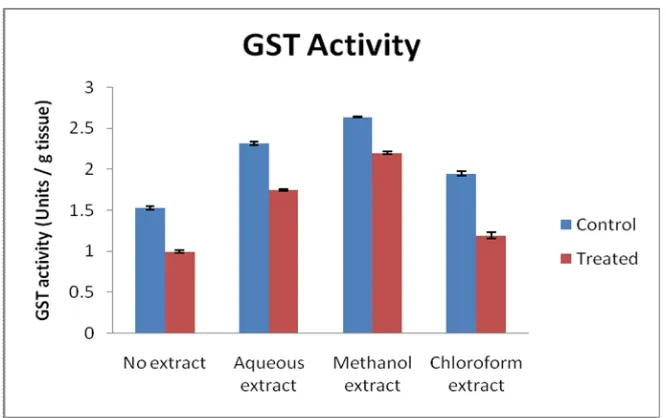
(3) Abdul Majeeth Kamarul Haniya et al. Int. Res. J. Pharm. 2013, 4 (10). Figure 3: Effect of Artemisia vulgaris leaf extracts on peroxidase activity. Figure 4: Effect of Artemisia vulgaris leaf extracts on glutathione reductase activity. Figure 5: Effect of Artemisia vulgaris leaf extracts on glutathione s-transferase activity. Page 57.
(4) Abdul Majeeth Kamarul Haniya et al. Int. Res. J. Pharm. 2013, 4 (10). Figure 6: Effect of Artemisia vulgaris leaf extracts on vitamin C level. Figure 7: Effect of Artemisia vulgaris leaf extracts on vitamin E level. Figure 8: Effect of Artemisia vulgaris leaf extracts on vitamin A level. Page 58.
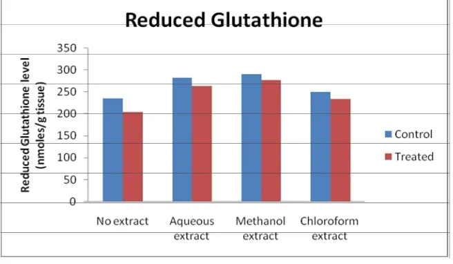
(5) Abdul Majeeth Kamarul Haniya et al. Int. Res. J. Pharm. 2013, 4 (10). Figure 9: Effect of Artemisia vulgaris leaf extracts on reduced glutahione level. RESULTS Enzymic Antioxidant Activity The enzymic antioxidant activities namely SOD, CAT, POD, GR and GST were analyzed in the liver slices. Effect of Artemisia vulgaris leaf extracts on enzymic antioxidant activities in goat liver slices exposed to H2O2 in vitro is graphically represented (Figure 1 to 9). H2O2 exposure caused a significant decrease in SOD activity compared to the control group. The co-treatment with the leaf extracts caused elevation in SOD activity. The maximum activity was observed with the methanolic extract treatment (Figure 1). A decrease in catalase activity was found in H2O2-exposed liver slices when compared to the control group (Figure 2). Coadministration of methanol and aqueous extract with H2O2 caused an increase in catalase activity. The chloroform extract co-administered group showed decreased catalase activity compared to untreated control but the activity was higher than the H2O2-treated group. The activity of peroxidase decreased upon exposure to H2O2 (Figure 3). Treatment with the leaf extracts of Artemisia vulgaris caused an increase in the peroxidase activity compared to the control group. The decrease in peroxidase activity by H2O2 was counteracted by the administration of aqueous and methanol extracts of Artemisia vulgaris leaves. The effect of chloroform extract in peroxidase activity was similar to that of the effect observed for catalase. The glutathione reductase activity increased in all the three extracts in comparison to the control group (Figure 4). Decreased GR activity was found in the slices exposed to H2O2. This effect was reverted by the administration of all the three extracts of Artemisia vulgaris leaves. H2O2 exposure caused a decrease in GST activity (Figure 5). The depletion of GST with the exposure of H2O2 was counteracted by the co-administration of leaf extracts. The methanolic extract showed significantly higher effect than the aqueous and chloroform extracts. Non-Enzymic Antioxidant Levels The non-enzymic antioxidants, namely vitamins C, E, A and reduced glutathione were estimated in the oxidant challenged liver slices with or without the leaf extracts of A. vulgaris. Decreased vitamin C level was found in the H2O2 treated group (Figure 6). However, the treatment of the goat liver slices with the leaf extracts of A. vulgaris reverted the reduction. The methanolic and the aqueous extracts caused an increase in the levels of vitamin C. Among the three extracts. used, the methanolic extract exhibited the maximum protection, followed by the aqueous and chloroform extracts. Similar trend was observed in vitamin E level (Figure 7). Hydrogen peroxide alone caused a marked decline in the levels of vitamin A, while the trend was effectively reverted by the Artemisia vulgaris leaf extracts. Among all the extracts used, the liver slices treated with methanolic extract showed more increase in vitamin A level than the groups treated with the aqueous and chloroform extracts (Figure 8). The oxidant exposure caused a reduction in the levels of GSH when compared to control. The depleting effect of H2O2 treatment was very well counteracted by the administration of the leaf extracts, where the methanolic extract was found to be better than the other two extracts (Figure 9). DISCUSSION Many studies have shown that the administration of herbal extracts can improve the antioxidant status of tissues, both in vivo and in vitro. Liu et al (2009)17 reported that a diet enriched with protandium, a combination of five phytochemicals from medicinal plants, improved SOD activities and suppressed tumor promoter-induced oxidative stress in mice. Gupta et al. (2007)18 have reported that the methanol extract of Oldenlandia umbellate exerts a protective effect on CCl4 - induced hepatic injury by increasing the activity of catalase in rats. Mahesh et al. (2007)19 demonstrated that the administration of an aqueous extract of Terminalia chebula showed marked increase in GPx activity in aged rat brain, which was suggested to be due to the protection of sulphydryl groups in glutathione from oxidative damage. The activities of glutathione reductase and glutathione S-transferase were decreased in CCl4 and Nnitrosodiethylamine injured rat liver, which were significantly preserved by the synergistic effect of silymarin and garlic20. Visavadiya and Narasimhacharya (2009)21 reported that Asparagus racemosus root powder improved the status of antioxidants namely ascorbic acid, SOD and CAT in hypercholesterolemic rats. Soussi et al. (2006)22 demonstrated that the pre treatment with green tea (Camellia sinensis) significantly improved the levels of vitamins E and A in the liver and kidney of rats with ammonium metavanadate-induced toxicity. An aqueous extract of Ocimum sanctum increased the level of GSH in alcohol treated rats (Shetty et al., 2008)23. The outcome of the present study showed that the leaves of A. vulgaris possessed high Page 59.
(6) Abdul Majeeth Kamarul Haniya et al. Int. Res. J. Pharm. 2013, 4 (10) levels of antioxidants which could effectively protect the stress induced by oxidants in goat liver slices. Sreelatha and Padma (2010)24 reported that CCl4 treatment significantly decreased the activities of antioxidant enzymes such as superoxide dismutase, catalase, glutathione peroxidase, glutathione reductase, and glutathione S-transferase. Treatment with Moringa oleifera extract increased the activities of antioxidant enzymes and glutathione content significantly. The activity of enzymic antioxidants and the levels of non enzymic antioxidants which decreased initially by H2O2 treated goat liver slices was found to be increased on treatment with methanolic extract of both the leaves and rhizomes of Curcuma amada (2012)25. The study carried out by Radha and Padma (2011)26 revealed that the methanol and chloroform extracts of Majorana hortensis leaves can improve the antioxidants (enzymic and non-enzymic) status of liver slices exposed to oxidative stress. CONCLUSION In general, groups promoting the 3Rs (refinement, reduction and/or replacement) of animal welfare for biomedical research have overlooked the immediate welfare gains that may be possible using various in vitro systems as alternative models. With this as the focus, precision-cut liver slices were used as an in vitro system that simulate the in vivo conditions prevailing in experimental animals and perhaps, the human system. The precision-cut liver slices provide a system in which the cells are present in their natural environment. This system was employed to evaluate the antioxidant potential rendered by the Artemisia vulgaris leaf extracts against hydrogen peroxide-induced stress in vitro. All the three extracts tested were capable of improving the levels of antioxidants studied to a significant extent. The methanolic extract was found to be most effective, followed by the aqueous and chloroform extracts. Thus, the results confirmed that the Artemisia vulgaris leaf extracts can improve the antioxidant status in oxidatively stressed tissue, which strengthens the antioxidant potential of the plant. REFERENCES 1. Metelitza DI, Karasyova EI. Initiation and inhibition of free-radical processes in biochemical peroxide systems: a review. Appl Biochem Microbiol 2007; 43: 481-505. http://dx.doi.org/10.1134/S0 00368380705002X 2. Valko M, Rhodes CJ, Moncol J, Izakovic M, Mazur M. Free radicals, metals and antioxidants in oxidative stress-induced cancer. Chem Biol Interact 2006; 160: 1-40. http://dx.doi.org/10.1016/j.cbi.2005.12.009 PMid:16430879 3. Choi Y, Jeong HS, Lee J. Antioxidant activity of methanolic extracts from some grains consumed in Korea. Food Chem 2007; 103: 130-138. http://dx.doi.org/10.1016/j.foodchem.2006.08.004 4. Adeolu AA, Florence OJ, Srinivas K, Masika P, Anthony JA. Assessment of the medicinal potential of the methanol extracts of the leaves and stems of Buddleja saligna. Complement Altern Med 2009; 9: 21-28. http://dx.doi.org/10.1186/1472-6882-9-21 PMid:19580647 PMCid:PMC2715372 5. Muanda F, Koné D, Dicko A, Soulimani R, Younos C. Phytochemical composition and antioxidant capacity of three Malian medicinal plant parts. Evid Based Complement Alternat Med 2009; 2011: 1-8. http://dx.doi.org/10.1093/ecam/nep109 PMid:19736222 PMCid:PMC3 136850 6. Walter HL, Memory PF, Elvin L. Medicinal botany, 2nded. New Hersey: John Wiley and Sons; 2003. 7. Bovenkamp MV, Groothuis GMM, Draaisma AL, Merema MT, Bezuijen JI, Gils MJ, et al. Precision-cut liver slices as a new model to. 8. 9. 10. 11. 12. 13. 14. 15. 16.. 17.. 18.. 19. 20.. 21. 22.. 23.. 24.. 25. 26.. study toxicity-induced hepatic stellate cell activation in a physiologic milieu. Toxicol Sci 2005; 85: 632-638. http://dx.doi.org/10.1093 /toxsci/kfi127 PMid:15728706 Kakkar P, Das B, Viswanathan PN. A modified spectrophotometric assay of superoxide dismutase. Indian J Biochem Biophys 1984; 21: 130-162. PMid:6490072 Luck H. Methods in enzymatic analysis. 2nded. New York: Bergmeyer Academic Press; 1974. Reddy KP, Subhani SM, Khan PA, Kumar KB. Effect of light and benzyladenine on dark treated graving rice (Oryza sativa) leaves changes in peroxidase activity. Plant Cell Physiol 1995; 26: 987-994. David H, Richard JS. Glutathione reductase. In: Bergmeyer J, Grab M. editiors. Methods of enzymatic analysis. 3rded. Weinhein Deer Field Beach: Verlag Chemie; 1983. Habig WH, Pabst MJ, Jakoby M. Glutathione transferase: A first enzymatic step in mercapturic acid formation. J Biol Chem 1974; 249: 7130-7139. PMid:4436300 Roe JH, Keuther CA. The determination of ascorbic acid in whole blood and urine through 2, 4-dinitrophenylhydrazine derivative dehydro ascorbic acid. J Biol Chem 1943; 147: 399-407. Rosenberg HR. Chemistry and physiology of the vitamins, Interscience Publisher, New York; 1992. Bayfield RF, Cole ER. Colorimetric estimation of vitamin A with trichloroacetic acid. Meth Enzymol 1980; 37: 189-195. http://dx. doi.org/10.1016/S0076-6879(80)67026-8 Moron MS, Depierre JN, Mannervik VC. Levels of glutathione, glutathione reductase and glutathione S-transferase activities in rat lung and liver. Bioche Biophys Acta 1979; 582: 67-68. http://dx.doi.org/ 10.1016/0304-4165(79)90289-7 Liu J, Gu X, Robbins D, Li G, Shi R, McCord JM, et al. Protandim, a fundamentally new antioxidant approach in chemo prevention using mouse two-stage skin carcinogenesis as a model. Plos One 2009; 4: e5284. http://dx.doi.org/10.1371/journal.pone.0005284 PMid:19384424 PMCid:PMC2668769 Gupta M, Mazumber UK, Thamilselvan V, Manikandan L, Senthilkumar GP, Suresh R, et al. Potential hepatoprotective effect and antioxidant role of methanol extract of Oldenlandia umbellata in carbon tetrachloride induced hepatotoxicity in Wistar rats. Iran J Pharma Therap 2007; 6: 5-9. Mahesh R, Ramesh T, Nagulendran KR, Velavan S, Begum VH. Effect of Terminalia chebula on monoamine oxidase and antioxidant enzyme activities in aged rat brain. Phcog Mag 2007; 3: 241-245. Shaarawy SM, Tohamy AA, Elgendy SM, Elmageed ZYA, Bahnasy A, Mohamed MS, et al. Protective effects of garlic and silymarin on NDEA-induced rats hepatotoxicity. Int J Bio Sci 2009; 5: 549-557. http://dx.doi.org/10.7150/ijbs.5.549 Visavadiya NP, Narasimhacharya AVRL. Asparagus root regulates cholesterol metabolism and improves antioxidant status in hypercholesteremic rats. eCAM 2009; 6: 219-226. Soussi A, Croute F, Soleiharoup JP, Kammoun A, El Feki A. Impact of green tea on oxidative stress induced by ammonium metavanadate exposure in male rats. C R Biol 2006; 329: 775-784. http://dx.doi. org/10.1016/j.crvi.2006.07.004 PMid:17027638 Shetty S, Udupa S, Udupa L. Evaluation of antioxidant and wound healing effects of alcoholic and aqueous extract of Ocimum sanctum Linn in Rats. Ecam 2008; 5: 95-101. PMid:18317555 PMCid:PMC 2249741 Sreelatha S, Padma PR. Protective mechanisms of Moringa oleifera against CCl4-induced oxidative stress in precision-cut liver slices. Forsch Komplementmed 2010; 17: 189-94. http://dx.doi.org/10.1159 /000318606 PMid:20829596 Sivaprabha J, Dharani B, Padma PR, Sumathi S. In vitro prevention of oxidative damage by Curcuma amada in goat liver slices exposed to oxidative stress. J Pharma Res 2012; 5: 1108-1111. Palaniswamy Radha, Padma PR. Antioxidant activity of Majorana hortensis leaves subjected to oxidative stress in an in vitro system. Int Res J Pharma 2011; 6: 153-157.. Cite this article as: Abdul Majeeth Kamarul Haniya and Palghat Raghunathan Padma. Antioxidant effect of Artemisia vulgaris leaf extracts on oxidatively stressed precision-cut liver slices. Int. Res. J. Pharm. 2013; 4(10):55-60 http://dx. doi.org/10.7897/2230-8407.041013. Source of support: Nil, Conflict of interest: None Declared. Page 60.
(7)
Figure


Related documents
Within a period of 11 years, thus far four phases can be distinguished: (1) start-phase: rapid and homogeneous colonization by fouling organisms, still
The relation- ship of the current reading to the current subject under study should be clear, the contributions of the great workers of the past must be
The effectiveness of training was determined by asses- sing recruitment rates to a host RCT [14 – 16, 21, 26, 30], the number of patients approached about trial participa- tion
this study was conducted to determine the pattern of acute poisoning cases and their management at the emergency department of Dessie referral hospital, northeast Ethiopia.. Methods
Previous studies show that there is a growing commitment among companies to include sustainability issues within their annual reports, more and more companies being able to measure
For the primary discrete variables of the footwear conditions: Standard shoe vs Brace condition vs High Top Shoe, a repeated measures analysis of variance was undertaken for the
endoscopy reliably predicted the presence of significant findings throughout the gastrointestinal tract; fecal cal- protectin provided valuable diagnostic ability for signifi-
In fluorimetrischen Assays zeigte sich für die Rac-GTPasen Rac1a, RacC und RacE jeweils ein spontaner GDP/MANT-GDP-Austausch, welcher jedoch nicht durch rekombinan- te Trix-Fragmente
