COMPARATIVE ANALYSIS OF LYMPH NODES POSITIVE AND LYMPH NODE
NEGATIVE BREAST CANCER
1
*Dr. Syed Sajid Hussain Shah, 2Dr. Syedha Farwa Batool and 3Dr. Muhammad Hassan Bashir
1PMDC # 87094-P. 2PMDC # 87798-P. 3PMDC # 87338-P.
Article Received on 13/03/2019 Article Revised on 03/04/2019 Article Accepted on 24/04/2019
INTRODUCTION
Pakistan has the highest incidence of breast cancer (BC) among Asian women.[1] Mean age for BC at the time of diagnosis falls in the 4th & 5th decade and the affected women mostly presented at an advanced stage with loco regional spread.[1-3] More than 95% of malignant tumors of breast are adenocarcinomas with infiltrating ductal carcinoma being the most common type (40% - 80% of all invasive cancers).[1,2]
Axillary lymph node (LN) involvement in BC has diagnostic, prognostic as well as therapeutic significance.[4] In 1978, the Union for International Cancer Control (UICC) and American Joint Committee on Cancer (AJCC) developed TNM (―Tumor, node, metastasis‖) staging system, which is commonly used for BC. It has been periodically updated with the seventh edition completed in 2010.[5] The ‗N‘ in TNM staging (AJCC) denotes axillary lymph node involvement, and groups BC patients into three ‗N‘ stages: N1 (1 to 3 LNs positive), N2 (4 to 9 LNs positive), N3 (more than 9 LNs positive). Isolated tumor cells or minimal tumor involvement is denoted as N0.[5]
Tumor size correlates with number of lymph nodes involved; the larger the tumor size, worst is the
prognosis. Moreover in node-negative BC, tumor size becomes the most significant prognostic factor for decisions regarding adjuvant therapy.[6] Prognostic significance for histologic grading as well as lymphovascular invasion (LVI) is limited to node-negative breast cancers, with borderline tumor sizes.[7]
The presence of ER, PR, or HER2/neu in BC predicts the response of tumor to anti-estrogen (tamoxifen) or Herceptin (trastuzumab) therapy. Gaopande et al. (2018)[8] found triple-negative BC (TNBC) in younger women, with higher grade, larger size, and lymphovascular invasion. He et al. (2018)[9] divided invasive intraductal BC into four different subtypes on the basis of ER, PR and HER2 expression and studied the association between different BC subtypes and axillary lymph node involvement.
These histopathologic features have significant prognostic information for breast cancer[3,8,10] and have been described as predictors for axillary lymph node metastasis.[9,11] However limited data[9,12,13] is available for Asian countries including Pakistan, so significance of these variables needs to be assessed in our population.
ISSN 2455-3301
WJPMR
AND MEDICAL RESEARCH
www.wjpmr.com
*Corresponding Author: Dr. Syed Sajid Hussain Shah PMDC # 87094-P.
ABSTRACT
Hence the purpose of the present study was first to evaluate the clinicopathological features of breast cancer and then to compare these in lymph node positive and negative BC patients from two tertiary care centers in Lahore, Pakistan.
METHODS
A total of 150 BC patients were included in this prospective study using the non-probability sampling technique. Only patients with invasive breast cancer with modified mastectomies and axillary lymph node resection from Fatima Jinnah medical college Lahore and Services Hospital, Lahore were included. The study period was from February 2016 to March 2018 and it was approved by Ethics Review Committee of Fatima Jinnah medical college, Lahore. Informed written consent was obtained from all the patients and confidentiality of their personal details was maintained.
Gross examination was done according to standard College of American Pathologists (CAP) protocol with special emphasis on tumor size, skin, nipple and lymph node involvement and confirmed later with histopathological examination. Histological grading (grade I to III) and staging (stage I to IV) were done according to Modified Scarff-BloomRichardson (SBR) grading system and TNM staging respectively. Estrogen receptor (ER), progesterone receptor (PR) and human epidermal growth factor receptor (HER2/neu) status were determined by immunohistochemistry. In this study, HER2/neu score of 3+ and 2+ was recorded as positive and 1+ as negative.
Information regarding lymph node involvement included total number of lymph nodes recovered, number of lymph nodes showing metastatic deposits and extracapsular involvement. BC cases were stratified by tumor size (T1, <2 cm; T2, 2-5 cm; T3, >5 cm) 3,13 and nodal status (‗0‘ LN; ‗1-3‘ LN; ‗>3‘ LN).3
Data was analyzed in Statistical Package for Social Sciences (SPSS, version 20, IBM). Frequencies and percentages were computed and an association between nodal status and clinicopathological variables was seen by Chi-square and Fisher‘s exact probability tests. Two-tailed p value less than 0.05 was considered statistically significant. Odds ratio (OR) with corresponding 95% confidence interval (CI) predicting nodal metastasis were also estimated.
RESULTS
Tumor characteristics and histopathological features of BC are shown in Table-I. Overall 150 lymph node positive and negative BC patients were included with mean age of 46.7 years ± 11.68 (95% CI 44.8 – 48.6; age range 22 – 76 years). More than half of the patients (66.9%) were younger than 50 years at presentation (mean age 40.2 ±years ±6.82; 95% CI 38.9 – 41.5; range 22 – 50 years). Most of the tumors were invasive ductal
carcinoma (IDC) (140; 93.3%) with only 7 cases (4.7%) of invasive lobular carcinoma (ILC), two cases (1.3%) of metaplastic breast cancer (MBC) and one case (0.67%) of mucinous carcinoma.
There were 37 (24.7%) patients with no LN involvement, 47 (31.4%) with 1-3 LNs and 66 (44%) with >3 LNs involved by tumor. Extracapsular extension was noted in 80 (53.3%) cases. The minimum number of affected lymph nodes recovered per case was one and maximum number was 45 (95% CI 44.3 – 45.7).
According to histological grading and TNM staging, most of the cases were high grade (II & III = 92%) and late stage tumors (II & III = 83.4%) (Table-I). Tumor size ranged from 0.2 cm to 19.5 cm (mean 4.94 ± 3.4; 95% CI 4.4 – 5.5). Univariate analysis revealed that menopausal status (p=0.009), tumor stage (p=0.0001), tumor size (p=0.001), LVI (p=0.0001) and PNI (p=0.007) were significantly associated with nodal status (Table-I). Multivariate analysis was done to compare the different clinicopathological characteristics with lymph node involvement (‗0‘ LN Vs ‗1-3‘ LN & ‗0‘ LN Vs ‗>3‘ LN). Menopausal status, tumor grade and tumor stage, tumor size, LVI, PNI and skin and nipple invasion were significant predictors for ‗>3‘ LN involvement with highest odds ratio reported for tumor stage, tumor size and LVI respectively (Table-II).
DISCUSSION
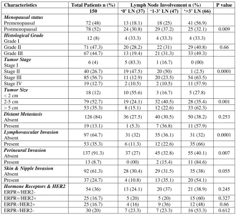
Table I: Patient and tumor characteristics by Lymph node involvement in Breast Cancer.
Characteristics Total Patients n (%) Lymph Node Involvement n (%) P value 150 ‘0’ LN (37) ‘1-3’ LN (47) ‘>3’ LN (66)
Menopausal status
Premenopausal 72 (48) 13 (18.1) 18 (25) 41 (56.9)
Postmenopausal 78 (52) 24 (30.8) 29 (37.2) 25 (32.1) 0.009
Histological Grade
Grade I 12 (8) 4 (33.3) 4 (33.3) 4 (33.3)
Grade II 71 (47.3) 20 (28.2) 22 (31) 29 (40.8) 0.66
Grade III 67 (44.7) 13 (19.4) 21 (31.3) 33 (49.3)
Tumor Stage
Stage I 6 (4) 5 (83.3) 1 (16.7) 0 (00)
Stage II 40 (26.7) 19 (47.5) 20 (50) 1 (2.5) 0.0001
Stage III 85 (56.7) 11 (12.9) 20 (23.5) 54 (63.5)
Stage IV 19 (12.7) 2 (10.5) 2 (10.5) 11 (57.9)
Tumor Size
< 2 cm 18 (12) 10 (55.6) 3 (16.7) 5 (27.8)
2-5 cm 79 (52.7) 19 (24.1) 32 (40.5) 28 (35.4) 0.001
> 5 cm 53 (35.3) 8 (15.1) 12 (22.6) 33 (62.3)
Distant Metastasis
Absent 126 (84) 36 (27.5) 40 (30.5) 50 (38.2) 0.253
Present 19 (13.1) 1 (5.3) 7 (36.8) 11 (57.9)
Lymphovascular Invasion
Absent 97 (64.7) 31 (32) 35 (36.1) 31 (32) 0.0001
Present 53 (35.3) 6 (11.3) 12 (22.6) 35 (66)
Perineural Invasion
Absent 137 (91.3) 37 (27) 45 (32.8) 55 (40.1) 0.007
Present 13 (8.7) 0 (00) 2 (15.4) 11 (84.6)
Skin & Nipple Invasion
Absent 92 (61.3) 28 (30.4) 29 (31.5) 35 (38) 0.055
Present 37 (24.7) 4 (10.8) 13 (35.1) 20 (54.1)
Hormone Receptors & HER2
ERPR+/HER2- 54 (36) 13 (24.1) 20 (37) 21 (38.9) 0.245
ERPR+/HER2+ 25 (16.7) 5 (20) 5 (20) 15 (60) 0.327
ERPR-/HER2+ 25 (16.7) 4 (16) 9 (36) 12 (48) 0.66
ERPR-/HER2- 30 (20) 7 (23.3) 7 (23.3) 16 (53.3) 0.612
LN, lymph node; Vs, versus; n, number of patients; OR, odds ratio; CI, confidence interval; ER, estrogen receptor; PR, progesterone receptor; HER2, human epidermal growth factor receptor 2.
Table II: Odds Ratio (OR) for Clinicopathological Predictors of Nodal Status in BC.
‘1-3’ LN ‘> 3’ LN
Characteristics OR 95% CI P Value OR 95% CI P Value Menopausal Status
Premenopausal Vs Postmenopausal 0.873 0.36-2.14 0.822 0.33 0.14-0.76 0.013 Tumor Size (cm)
2-5 Vs <2 2.95 0.87-9.99 0.136 5.614 1.37-22.98 0.014 >5 Vs <2 5 1.04-24.03 0.072 8.25 2.19-30.96 0.002 >5 Vs <5 1.243 0.45-3.45 0.798 3.625 1.45-9.09 0.006 Tumor Grade
II Vs I 1.467 0.29-7.37 0.702 0.833 0.69-0.99 0.036
III Vs I 2.154 0.41-11.2 0.421 0.765 0.58-0.99 0.01
Tumor Stage
II Vs I 5.263 0.56-49.29 0.193 0.792 0.65-0.97 1
III Vs I 9.091 0.94-87.96 0.066 24.09 2.56-226.9 0.002
IV Vs I 36 1.8-718.7 0.029 0.143 0.02-0.88 0.0001
Present Vs Absent Perineural Invasion
Present Vs Absent 1.044 0.98-1.11 0.501 1.2 1.17-1.34 0.007 Skin & Nipple Invasion
Present Vs Absent 3.138 0.91-10.79 0.094 4 1.23-13.06 0.024 Distant Metastasis
Present Vs Absent 3.063 0.59-15.72 0.287 3.5 0.73-16.74 0.128 Hormone Receptors & HER2
ERPR+/HER2- Vs ERPR-/HER2- 0.733 0.19-2.89 0.734 1.375 0.42-4.56 0.765 ERPR+/HER2+ Vs ERPR-/HER2- 1.25 0.22-7.08 1 0.962 0.24-3.89 1 ERPR-/HER2+ Vs ERPR-/HER2- 0.444 0.09-2.28 0.428 0.909 0.21-4.02 1 ERPR+/HER2+ Vs ERPR+/HER2- 1.591 0.34-7.37 0.703 0.49 0.13-1.79 0.348 ERPR-/HER2+ Vs ERPR+/HER2- 0.606 0.15-2.49 0.728 0.424 0.11-1.69 0.322 ERPR-/HER2+ Vs ERPR+/HER2+ 0.356 0.06-2.08 0.384 0.582 0.12-2.88 0.689
LN, lymph node; CI, confidence interval; ER, estrogen receptor;
PR, progesterone receptor; HER2, human epidermal growth factor receptor 2.
Axillary LN status is an important predictor for BC prognosis. Rates of BC recurrence are higher for patients with LN metastasis (70% risk) as compared to patients diagnosed with LN negative BC (20% - 30% risk). Mammon et al.[10] showed 25% cases as LN negative, 35.6% with 1-3 LNs involved and more than 60% with >4 LNs involved. Aziz-unNisa et al.[3] reported 28.7% of cases with no nodal involvement, 23.3% of cases with 1-3 positive LNs and 48% with >1-3 LNs involved. Our study is in agreement with findings of these researchers with 24.7% LN negative, 31.3% with 1-3 LNs and 44% with >3 positive LNs.
Local studies have reported younger age, higher grade and stage at the time of presentation.[1,2] We also report similar demographic findings as maximum number of high grade (II & III; 92%) and late stage (II & III; 83%) tumors with nodal metastasis were also seen in our patients with mean age of 46.7 years. Age of BC is 40 to 50 years in the developing countries (including Pakistan) at time of presentation, with a mean of 47/48 years.[2,10] and 60 to 70 years in the developed world with a mean of 61 to 63 years.[14,15]
An inverse relationship between age and axillary LN involvement has been reported previously with younger patients (<40 years) having a higher risk for nodal involvement.[13] In our study a statistically significant association was observed between age and LN involvement (‗>3‘ LN) with a lower risk for postmenopausal women (OR 0.33; 95% CI 0.140.76; p < 0.013). However two studies done in Asian populations showed no relationship with age.9,12 Infiltrating ductal carcinoma (IDC) was found to be the most common histological type of BC (93.3%) in our study and other local studies as well.[1,2,10] In contrast developed countries have a frequency of 70% to 80% IDC and 5% to 15% of ILC.16 Lower incidence of ILC in our patients may be attributed to multiple factors that have known protective effect like high parity, birth of first child at an early age, prolonged lactation etc.[16]
Scarff-Bloom-Richardson histological grades reported in literature include 5.1%-33% for grade I, 31%-57% for grade II and 20%-63.6% for grade III.[2,7] Mammon et al.[10] reported 94.7% high grade tumors (grade II and III) in their study. Distribution of histological grades in our study (92% grade II & II) is within the ranges reported in literature, with a lower risk of nodal metastasis for grade I as compared to grade II (OR 0.88) and grade III tumors (OR 0.77).
presenting with nipple and skin invasion). The present study reports 32.7% cases of nipple and skin invasion with statistically significant association with nodal status (p <0.05) and higher risk for metastasis to the LNs (OR 3.14 for ‗1-3‘ LN & 4 for ‗>3‘ LNs).
Previously published data regarding association between ER, PR and Her2/neu reactivity and nodal status gives conflicting reports. Bevilacqua et al.[20] found increased frequency of LN involvement in ER/PR positive cases while Moosavi et al.[13] and Friedman et al.[14] found no statistically significant association between ER/PR/Her2 reactivity and nodal status. Our observations were similar to the latter two studies with no relationship between these two variables. Similarly a study done in Pakistan also showed no significant correlation with lymph node metastasis.[3]
CONCLUSION
Our study confirms BC as a heterogeneous disease with varied clinical, pathological and molecular features, and prognostic behavior. Results of this study reveal statistically significant differences when clinicopathological characteristics were compared with nodal status. However larger prospective studies are required to develop and validate a nomogram for prediction of nodal metastasis in our population.
REFERENCES
1. Naeem M, Khan N, Aman Z, Nasir A, Samad A, Khattak A. Pattern of breast cancer experience at lady reading Lahore, Peshawer. J Ayub Med Coll Abbottabad, 2008; 20: 22-25.
2. Khokher S, Qureshi MU, Riaz M, Akhtar N, Saleem A. Clinicopathologic profile of breast cancer patients in Pakistan: Ten years data of a local Cancer Lahore. Asian Pac J Cancer Prev., 2012; 13: 693-698. doi:10.7314/ APJCP.2012.13.2.693
3. Azizun-Nisa, Bhurgri Y, Raza F, Kayani N. Comparison of ER, PR & HER 2/Neu (C-erb B2) reactivity pattern with histologic grade, tumor size and lymph node status in breast cancer. Asian Pac J Cancer Prev., 2008; 9: 553-556.
4. Layeequr Rahman R, Crawford SL, Siwawa P. Management of axilla in breast cancer - The saga continues. Breast., 2018; 24: 343–353.
5. Edge SB, Byrd DR, Compton CC, Fritz AG, Greene FI, Trotti III A. Editors. Part VII Breast. AJCC Cancer Staging Manual. 7th ed. New York, NY: Springer, 2010; 347-376.
6. Cianfrocca M, Goldstein LJ. Prognostic and predictive factors in early-stage breast cancer. The Oncologist, 2004; 9: 606-616.
7. Le Doussal V, Tubiana-Hulin M, Friedman S, Hacene K, Spyratos F, Brunet M. Prognostic value of histologic grade nuclear components of Scarff-Bloom-Richardson (SBR). An improved score modification based on a multivariate analysis of
1262 invasive ductal breast carcinomas. Cancer, 1989; 64: 1914–1921.
8. Gaopande VL, Joshi SS, Kulkarni MM, Dwivedi SS. A clinicopathologic study of triple negative breast cancer. J Sci Soc., 2018; 42(1): 12-15. doi: 10.4103/0974-5009.149469.
9. He ZY, Wu SG, Yang Q, Sun JY, Li FY, Lin Q, Lin HX. Breast cancer subtype is associated with axillary lymph node metastasis. A retrospective cohort study. Medicine (Baltimore), 2018; 94(48): e2213. doi: 10.1097/MD.0000000000002213 10. Mamoon N, Sharif MA, Mushtaq S, Khadim MT,
Jamal S. Breast carcinoma over three decades in northern Pakistan — are we getting anywhere? J Pak Med Assoc., 2009; 59: 835-838.
11. Greer LT, Rosman M, Charles Mylander W, Liang W, Buras RR Chagpar AB, et al. A prediction model for the presence of axillary lymph node involvement in women with invasive breast cancer: a focus on older women. Breast J., 2014; 20: 147153. doi: 10.1111/tbj.12233.
12. Kuo YL, Chen WC, Yao WJ, Cheng L, Hsu HP, Lai HW, et al. Validation of Memorial Sloan–Kettering Cancer Center nomogram for prediction of non-sentinel lymph node metastasis in non-sentinel lymph node positive breast cancer patients an international comparison. Int J Surg., 2016; 11(7): 538–543. doi:10.1016/j.ijsu.2016.05.005
13. Moosavi SA, Abdirad A, Omranipour R, Hadji M, Razavi AE, Najafi M. Clinicopathological Factors Predicting Nonsentinel Lymph Node Metastasis of Breast Cancer in Iran. Asian Pac J Cancer Prev.,
2014; 15: 7049-7054.
doi:10.7314/APJCP.2014.15.17.7049.
14. Friedman D, Gipponi M, Murelli F, Meszaros P, Solari N, Massa M, et al. Predictive factors of non-sentinel lymph node involvement in patients with invasive breast cancer and sentinel node micrometastases. Anticancer Res., 2016; 33: 4509-4514.
15. American Cancer Society. Cancer Facts and Figures 2018. Atlanta, GA: American Cancer Society, 2018. 16. Li CI, Uribe DJ, Daling JR. Clinical characteristics
of different histologic types of breast cancer. Br J Cancer., 2005; 93: 1046-1052. doi: 10.1038/sj.bjc.6602787.
17. Gilani GM, Kamal S, Akhter AS. A differential study of breast cancer patients in Punjab, Pakistan. J Pak Med Assoc., 2003; 53: 478-480.
18. Guray Durak M, Akansu B, Akin MM, Sevinç AI, Koçdor MA, Saydam S, et al. Factors predicting non-sentinel lymph node involvement in sentinel node positive breast carcinoma. Turk J Pathol, 2011; 27: 189-195. doi: 10.5146/ tjpath.2011.01074. 19. Eldweny H, Alkhaldy K, Alsaleh N, Abdulsamad M,