Effect of Sitting Posture on Development of
Scoliosis in Duchenne Muscular Dystrophy Cases
Pokharel RK,
1Brisco L,
2Mandal M,
2Agrawal JP,
3Dillon D,
4Vitale M,
5Woodland P,
4Jacoby P,
6Downs J
71Department of Orthopaedics, Institute of Medicine Tribhuvan University, Maharajgunj, Kathmandu Nepal, 2Muscular Dystrophy Foundation, Kathmandu, Nepal, 3Department of Medicine, Institute of Medicine Tribhuvan University, Maharajgunj, Kathmandu, Nepal, 4Department of Orthopaedics, Princess Margaret Hospital, Perth, Australia, 5Morgan Stanley Children’s Hospital of New York – Presbyterian, Columbia University Medical Centre, New York, 6Centre for Child Health Research, Telethon Institute for Child Health Research, The University of Western Australia, Perth, Australia, 7School of Physiotherapy and Exercise Science, Curtin University, Perth, Australia.
ABSTRACT
Correspondence:
Dr. Rohit Kumar Pokharel, Department of Orthopaedics (Spine
Unit) Institute of Medicine, Tribhuvan University Teaching Hospital, Kathmandu,
Nepal. Email: [email protected], Phone: +977 1 4410911.
INTRODUCTION
Duchenne Muscular Dystrophy (DMD) is a X- linked recessive disorder due to mutation in the dystrophin gene,1 that cause progressive muscle weakness leading to feet off around 9 to 12 years of age and scoliosis often develops.2 Different treatment options are recommended to prevent scoliosis3 however, it does develop in many boys.4 Surgery is considered for moderate to large curves, ideally before respiratory function deterioration.5, 6
The needs of DMD boys in Nepal are now being acknowledged and rehabilitation strategies used
proactively.7 It has been observed that they have prolonged ambulatory phase,8 and scoliosis is not common even after loss of ambulation.9 Culturally, Nepalese people practice cross legged fl oor sitting. In DMD boys with muscle weakness, this posture may increase lumbar lordosis that has been associated with greater spinal stability.10 In this study, we sought to examine and identify the relationships between cross legged sitting, lumbar posture and scoliosis.
Background:
Scoliosis is a frequent association in boys with Duchenne Muscular Dystrophy when the ability to
walk is lost around nine to 12 years of age. This study assessed the contribution of physical factors including lumbar
posture to scoliosis in non-ambulatory youth with DMD in Nepal.
Methods:
Linear regression was used to assess effects of time since loss of ambulation, muscle strength, functional
severity and lumbar angle as a binary variable on coronal Cobb angle; again logistic regression was used to assess
effects of muscle strength and cross-legged sitting on the presence of a lordotic lumbar posture in 22 non-ambulant
boys and young men.
Results:
The boys and young men had a mean (SD) age of 15.1 (4.0) years, had been non-ambulant for 48.6
(33.8) months and used a median of 3.5 (range 2 to 7) postures a day. The mean Cobb angle was 15.1 (range 0 to
70) degrees. Optimal accuracy in predicting scoliosis was obtained with a lumbar angle of -6° as measured by skin
markers, and both a lumbar angle ≤-6° (P=0.112) and better functional ability (P=0.102) were associated with less
scoliosis. Use of cross-legged sitting postures during the day was associated with a lumbar angle ≤-6° (OR 0.061;
95% CI 0.005 – 0.672; P=0.022).
Conclusions:
Use of cross-legged sitting posture was associated with increase in lumbar lordosis. Higher angle of
lumbar lordosis and better functional ability are associated with lesser degree of scoliosis.
Keywords:
Duchenne Muscular Dystrophy; lumbar lordosis; lumbar posture; Nepal; scoliosis.
METHODS
Twenty fi ve boys and young men with DMD from Nepal at non-ambulant stage who had not received surgical interventions were included in this study, after taking verbal and written consent from the boys and their parents. Relevant information such as age when ability to walk was lost was reported by the primary caregiver or the client himself.
Each case was examined for muscle strength, functional ability, lumbar sitting posture, spinal deformities, posture through the course of the day, as detailed below. Finally, past and current use of glucocorticoid medications was recorded as a binary variable.
1. Muscle strength was assessed with manual muscle testing, each muscle group was graded using the Medical Research Council (0 to 5) scale which has been found reliable in DMD.11 A total muscle strength score was calculated by dividing the sum of the individual muscle group scores by the total possible muscle strength score and expressing as a percentage.
2. Functional abilities were measured with the EK scale, a functional severity scale developed specifi cally for non-ambulatory boys and men with DMD giving a score ranging from 0 to 30, and scores have explained variation in muscle strength, contractures and respiratory capacity.12
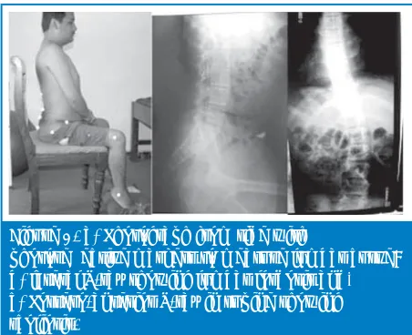
3. Lumbar posture was assessed with a lateral photograph of the lumbar spine taken whilst seated on a fi rm-surfaced stool with hips and knees at 90 degrees fl exion, and feet as fl at as possible.13 Photo-refl ective markers were taped (3M doubled sided tape, Pymble, NSW) to the bony landmarks of the right anterior superior iliac spine, midpoint of the greater trochanter, lateral femoral condyle, lateral malleolus and the spinous processes of T10, L2, L4 and S2. A camera (Canon EOS 500D) was placed on a tripod a distance of 1.5m away and at the level of T8. Sitting posture as tall as possible (with minimal support as necessary for safety) was then photographed. A lumbar angle was calculated as the angle between the intersection of the tangents drawn through the T10/L2 markers and the L4/S2 markers13 and measured with Image J software, a Java image processing program that measures angles between manually marked positions on a digital image (National Institute of Mental Health, Bethesda, Maryland, USA) (Figure 1a). This measure was applicable to in-the-fi eld measurement. A
4. Scoliosis was assessed clinically and an antero-posterior X-ray of the spine was taken in supine. Any lateral curvature in spine was measured as the Cobb angle using standard methods (Figure 1c).
5. Postures over the course of one day were recorded by the primary caregiver on a 24 hour diary card with response categories indicating the type of lying, sitting and standing used during each 15 minute epoch of the day.
This study was approved by the Ethics Committees of the Nepal Health Research Council, Kathmandu, Princess Margaret Hospital and Curtin University, Perth.
Figure 1. a) Photograph from side with
photorefl ective markers to measure lumbar curve; b) lateral X-ray showing lumbar lordosis and, c) Postero-anterior X-ray in supine showing scoliosis.
Linear regression was used to assess predictors of functional severity and the relationship between the radiological and photographic measures of lumbar posture. To determine a cut-off value for the photographically measured lumbar angle with optimal sensitivity and specifi city for predicting scoliosis, a receiver operator characteristic (ROC) analysis was performed with presence of scoliosis defi ned as greater than 10° of Cobb angle. A linear model was calculated to assess predictors of coronal Cobb angle and a logistic regression model was calculated to assess the infl uence of variables on the presence of a lumbar angle ≤-6°. Analysis was conducted with SPSS software version 19 (SPSS, Chicago, Illinois).
RESULTS
(coeff 0.68, P=0.022), increased time since walking was lost (coeff 0.11, P=0.001) and weaker muscle strength (coeff -0.33, P<0.001) predicted poorer EK severity scores. As is usual for DMD, the older boys in our sample were more severely affected refl ecting increasing weakness with time. Five (22.7%) of the group had previously used and 13/22 (59.1%) were currently using glucocorticoid medications.
The DMD boys were awake an average of 16.9 (1.0) hours and adopted a median of 3.5 (range 2 to 7) different postures during the day. Supine lying (n=16; 72.7%), side lying (n=18; 81.8%) and cross legged sitting (n=15; 68.2%) were common postures during the course of their usual day. Other postures included diamond sitting on the fl oor and sitting on a chair (Table 1). No boy sat in a wheelchair with postural supports or was supported for standing.
The postural lumbar angle during stool sitting was on average -3.6 (13.3) degrees, ranging from 29.8 to -17.8, three of the young men needing minimal support to maintain balance for their photograph. The area under the ROC curve was 0.767 (95%CI 0.555-0.978) for lumbar angle as a predictor of the presence of scoliosis (>10°), and optimal accuracy (taking into account sensitivity and specifi city) was obtained with an angle of -6° (Figure 2). The lumbar angle increased with the size of the lordosis as measured by lateral X-ray taken in side
lying (coeffi cient 0.738; 95% CI 0.227-1.249; P=0.007). A photographically derived angle of ≤-6° was observed in 12/22 (54.5%). The mean Cobb angle was 15.1° (range 0 to 70°): four boys aged 11, 16, 17 and 19 years had a coronal Cobb angle ≥30°. Three of 12 who were 15 years or older (25%) and two of eight who were 17 years or older (25%) had a curve ≥30°.
Table 1. Different postures used over the day by 22 non-ambulant boys and young men in Nepal.
Hours/daya
Posture N (%) Mean (SD) Range
Lying Supine 16 (72.7) 2.6 (1.2) 0.8-5.0 Prone lying 6 (27.3) 0.9 (0.5) 0.3-1.5 Side lying 18 (81.8) 3.2 (1.6) 1.5-7.0 Chair
sitting
Wheel chairb
10 (45.5)
6.3 (3.1) 1.5-11.3
Chair with a back
6 (27.3)
4.4 (1.9) 1.8-6.5
Stool 3 (13.6) 2.8 (3.1) 0.8-6.3 Floor
sitting
Diamond 3 (13.6)
4.6 (2.6) 1.5-6.3
Cross legged
15 (68.2)
8.1 (3.5) 2.0-15.5
Long 6 (27.3) 5.9 (5.3) 0.5-12.0
a average and range statistics calculated for those who used the posture; b wheelchair without additional seating supports
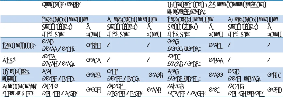
Table 2. Relationships between predictor variables and coronal Cobb angle calculated with linear regression, for all cases (n=22) and without one case who presented with both lumbar kyphosis and lordosis (n=21).
All cases (n=22) Excluding case with lumbar lordosis and kyphosis (n=21)
Univariate model Multivariate model Univariate model Multivariate model Coeffi cienta
(95% CIs)
P value
Coeffi cienta (95% CIs)
P value
Coeffi cienta (95% CIs)
P value
Coeffi cienta (95% CIs)
P value
Time off feet 0.14
(-0.11 - 0.38) 0.258 -
-0.17
(-0.07 – 0.41) 0.159 -
-MRC% -0.51
(-1.18 - 0.17) 0.131 -
--0.43
(-1.12 - 0.26) 0.211 - -EK severity
score
1.19
(-0.26 - 2.64) 0.102 0.86
(-0.65 - 2.37) 0.102 1.17
(-0.25 – 2.59) 0.100 0.74
(-0.69 – 2.17) 0.293 Lumbar angle
(<6° vs ≥ 6°)
-13.90
(-29.52 - 1.72) 0.078
-10.75
(-27.22 - 5.72) 0.112
-16.72
(-31.66 - -1.78) 0.03
-13.87
(-29.78 – 2.05) 0.084
a coeffi cient = change in the outcome for each unit change in the predictor variable.
Age and time since the ability to walk was lost did not predict coronal Cobb angle. The EK severity score (refl ecting functional ability) and a lumbar angle ≤-6° were stronger predictors with the latter being the
Figure 2. Receiver operator curve showing sensitivity and specifi city for lumbar angle predicting the presence of scoliosis >10°. Optimal accuracy was obtained with a lumbar angle of ≥6°.
Figure 3. A lateral photograph of a 15 year old man with a) a lumbar angle 11.9° (≤-6°) and b) scoliosis of Cobb angle 10°.
After adjustment for EK score in a multivariate model, the coeffi cient reduced to -10.7 indicating as would be expected that severity was a confounder. The lumbar spine was purely lordotic or kyphotic in all subjects except for one 16 year old boy with upper lumbar kyphosis, lower lumbar lordosis and marked antero-posterior asymmetry of disc height at L4/5. His lumbar angle refl ected the lower lumbar lordosis measuring -15.4° and his Cobb angle was 40°. Because of co-occurring lordosis and kyphosis in the lumbar spine, linear regression analyses were repeated without this case (Table 2). Cobb angle was not related to past (P=0.778) or current use of glucocorticoid medications (P=0.569).
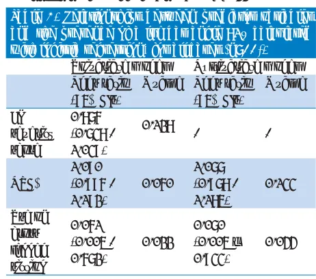
A lower EK severity score (P=0.128), higher MRC%
cross-legged sitting and MRC% in a multivariate logistic regression model, use of cross-legged sitting remained protective for a lumbar angle ≤-6° (P=0.044).
Table 3. Relationships between predictor variables and the presence of a lumbar angle >6° calculated with logistic regression, for all cases (n=22).
Univariate model Multivariate model Odds ratio
(95% CIs)
P value Odds ratio (95% CIs)
P value
EK severity score
0.876 (0.738 - 1.039)
0.128
-
-MRC%
1.090 (0.996 - 1.192)
0.060
1.077 (0.978 - 1.185)
0.133
Use of cross legged sitting
0.061 (0.005 - 0.672)
0.022
0.070 (0.005 – 0.933)
0.044
DISCUSSION
Duchenne Muscular Dystrophy is characterized by progressive muscle weakness which is due to the disease process itself and also secondary to disuse atrophy. Joint contracture and development of deformities including that of spine is due to lack of support from weak muscles.2 Spinal deformity, scoliosis, usually develops after loss of ambulation.4 In DMD boys living in Nepal, we found that a typical day involved time spent in supine and side lying posture, sitting in an unadapted wheelchair and/or sitting in cross legged sitting on the fl oor. We identifi ed a weak relationship between a lordotic lumbar posture in sitting and a less severe scoliosis, with this lumbar posture more likely when cross-legged sitting postures were used during the day. The boys and young men in our sample lost muscle strength and function as would be expected in DMD, but there was only a weak relationship between the size of the Cobb angle and time since they had lost the ability to walk. As would be expected, the relationship between the lumbar sitting posture and Cobb angles was confounded by functional severity but a lumbar angle ≤-6° was nevertheless associated with approximately 11° reduction in Cobb angle. Therefore, use of a variety of posture daily including those that encourage a lordotic lumbar posture could be protective against scoliosis.
associated with delayed onset of scoliosis as well as its slower development, likely related to slower decline in muscle strength.15 Approximately 30% of the UK cohort had received prednisolone according to local protocols13 but a moderate to severe scoliosis nevertheless developed for many. In our sample, past or current glucocorticoid use was not associated with Cobb angle although their duration of use had in many cases been relatively short.
For those in Nepal, economic factors reduce opportunities for the implementation of comprehensive management,2, 3 and we would expect poorer outcomes but the proportion who had developed scoliosis was much smaller than expected. It is interesting also to note a prolonged ambulatory phase among Nepalese DMD boys compared to that of the Japanese boys.8
A variety of postures were used daily including cross legged sitting on the fl oor. This is a widely practiced sitting position in Nepal over long periods of time throughout the day and its practice was associated with maintenance of a lordotic lumbar angle. The ability to sit cross legged on the fl oor with a lumbar lordosis requires adequate range of movement at the lumbar spine and hip joints in order to maintain anterior rotation of the pelvis in combination with fl exion, abduction and external rotation at the hip. With poor muscle strength as seen in DMD, the upper trunk is then held high and balanced over this base of support. This possibly reduces axial rotation of the spine and presumably its contribution to a developing scoliosis. A long term environment that includes practice of cross legged sitting may allow development of the necessary range of motion at the pelvis and hips to achieve this position, even before the boys lose the ability to walk.
With muscle weakness, the spine often develops a long thoracolumbar scoliosis in the fi rst instance that later becomes kyphoscoliotic; and the presence of a lumbar lordosis has been suggested as protective of spinal symmetry in DMD. For example, Oda et al16 found seven of 43 patients with DMD who did not have progressive spinal deformity to have normal sagittal plane alignment and a scoliosis of <30°. In a laboratory study, a lumbar lordosis when sitting on a base tilted 15° was associated with better tolerance of spinal loads in eight non-ambulant boys with DMD.10 For our sample, maintaining a lumbar angle ≤-6° as measured photographically was the strongest univariate predictor of Cobb angle, more so than factors such as time since walking was lost and muscle strength, and also despite the absence of supported seating systems. The effect of lumbar angle was moderated by functional severity in the multivariate model, as would be expected, and this effect was stronger when the case with an unusual
deformity of both lumbar kyphosis and lordosis was excluded. Using multiple postures throughout the day including cross legged sitting may be protective of spinal symmetry, although we recognize that these strategies are not directly applicable to boys in different cultures.
To characterize lumbar lordosis, we used a fi eld measure to examine lumbar posture with skin markers13 and found that a lumbar angle -6° was the cut-point with optimal sensitivity and specifi city for predicting the presence of scoliosis. The boys had lean body shapes and we were confi dent that the skin markers were able to illustrate the posture of the lumbar spine. Data were available to assess the relationship between radiological measurement of lumbar lordosis in side lying and our photographic measure for some of our sample, and the strong relationship was encouraging. We elected to use the photographic measure in our analyses because it was assessed in the sitting position and its in-the-fi eld application. Further study of this measure taking account of any infl uence of BMI would be valuable to understanding its application to other populations.
Our study is the fi rst to report physical assessment of DMD boys in a developing country and our fi ndings are particularly applicable to those living in similar countries. We also feel that assessment of boys living within different environmental circumstances to those participating in most other studies in the literature affords a valuable opportunity to learn much more about factors that can infl uence outcomes. Our recruitment rate was high and we assessed a comprehensive range of variables to describe impairments and activities. However, we also acknowledge the limitations of our study. The sample size was unavoidably small comprising boys and young men of families who lived in the Kathmandu area and were interested to receive interventions for their son by being registered with the Muscular Dystrophy Foundation Nepal. Only cross-sectional data was collected and we would recommend additional longitudinal study to track this potential protective role of sitting habit.
are non-ambulant,3 but specifi c guidelines for the lumbar spine have not been found. Development and testing of seating strategies that allow for greater postural variety including reproduction of a sitting posture with the lumbar angle at approximately -6° could confer additional advantages for non-ambulant boys with DMD.
CONCLUSIONS
Use of cross-legged sitting posture was associated with increase in lumbar lordosis. Higher angle of lumbar lordosis and better functional ability are associated with lesser degree of scoliosis.
ACKNOWLEDGEMENTS
We would like to acknowledge the valuable contributions of the boys and young men with Duchenne Muscular Dystrophy in Nepal and their families. We are also grateful for the support of the staff and Board members of the Muscular Dystrophy Foundation Nepal, the Muscular Dystrophy Child Care Society Nepal and the Muscular Dystrophy Association Nepal.
REFERENCES
1. Manzur AY, Kinali M, Muntoni F. Update on the management of Duchenne muscular dystrophy. Archives of Disease in Childhood. 2008;93(11):986-90.
2. Katharine B, Richard F, David JB, Laura EC, Paula RC, Linda C, et al. Diagnosis and management of Duchenne muscular dystrophy, part 1: diagnosis, and pharmacological and psychosocial management. Lancet neurology. 2010;9(1):77-93.
3. Katharine B, Richard F, David JB, Laura EC, Paula RC, Linda C, et al. Diagnosis and management of Duchenne muscular dystrophy, part 2: implementation of multidisciplinary care. Lancet neurology, 2010;9(2):177-189.
4. Kinali M, Messina S, Mercuri E, Lehovsky J, Edge G, Manzur AY, et al. Management of scoliosis in Duchenne muscular dystrophy. a large 10-year retrospective study. Developmental Medicine and Child Neurology. 2006;48(6):513-8.
5. Karol L.A. Scoliosis in patients with Duchenne muscular dystrophy. Journal of Bone and Joint Surgery. 2007;89S1:155-62. 6. Sussman M. Duchenne muscular dystrophy. Journal of the
American Academy of Orthopaedic Surgeons. 2002;10(2):138-51.
7. Joshi SK. Disability in Nepal. KUMJ. 2004;2(1):3-4.
8. Satoshi M, Tsuyoshi A, Yasuhiro T, Rohit P, Jagdish A, Mukesh M, et al. International comparison of physical activity in Nepalese and Japanese boys with Duchenne muscular dystrophy. Kobegakuin Journal of Rehabilitation Research, 2012;7(2):82-93.
9. Rohit P. Orthopaedic management in Muscular Dystrophy. Souvenir 3rd International seminar on Duchenne Muscular
Dystrophy. 2008;49-52.
10. Kerr TP, Lin JP, Gresty MA, Morley T, Robb SA. Spinal stability is improved by inducing alumbar lordosis in boys with Duchenne Muscular Dystrophy: a pilot study. Gait & posture. 2008;28(1):108-12.
11. Florence J, Mendell JR, Moxley RT, Griggs RC, Brooke MH, Fenichel GM, et al. Intrarater reliability of manual muscle test (Medical Research Council scale) grades in Duchenne's muscular dystrophy. Physical Therapy. 1992;72(2):115-22. 12. Steffensen B, Hyde S, Lyager S, Mattsson E. Validity of the EK
scale: a functional assessment of non-ambulatory individuals with Duchenne muscular dystrophy or spinal muscular atrophy. Physiotherapy research international. 2001;6(3):119-34. 13. O'Sullivan PB, Mitchell T, Bulich P, Waller R, Holte J. The
relationship between posture and back muscle endurance in industrial workers with flexion-related low back pain. Manual therapy. 2006;11(4):264-71.
14. Kinali M, Main M, Eliahoo J, Messina S, Knight RK, Lehovsky J, et al. Predictive factors for the development of scoliosis in Duchenne Muscular Dystrophy. European Journal of Paediatric Neurology. 2007;11(3):160-6.
15. Alman B, Raza SN, Biggar WD. Steroid treatment and the development of scoliosis in males with duchenne muscular dystrophy. Journal of Bone and Joint Surgery (Am). 2004;86-A(3):519-24.