1535-9778/05/$08.00⫹0 doi:10.1128/EC.4.3.577–587.2005
Copyright © 2005, American Society for Microbiology. All Rights Reserved.
Dim1p Is Required for Efficient Splicing and Export of mRNA
Encoding Lid1p, a Component of the Fission Yeast
Anaphase-Promoting Complex
Robert H. Carnahan,
1† Anna Feoktistova,
1Liping Ren,
1Sherry Niessen,
2John R. Yates III,
2and Kathleen L. Gould
1*
Howard Hughes Medical Institute and Department of Cell and Developmental Biology, Vanderbilt University School of Medicine, Nashville, Tennessee,1and The Scripps Research Institute,
La Jolla, California2
Received 18 November 2004/Accepted 24 December 2004
Schizosaccharomyces pombe Dim1p is required for maintaining the steady-state level of the
anaphase-promoting complex or cyclosome (APC/C) component Lid1p and thus for maintaining the steady-state level and activity of the APC/C. To gain further insight into Dim1p function, we have investigated the mechanism
whereby Dim1p influences Lid1p levels. We show thatS. pombecells lacking Dim1p orSaccharomyces cerevisiae
cells lacking its ortholog, Dib1p, are defective in generalized pre-mRNA splicing in vivo, a result consistent with the identification of Dim1p as a component of the purified yeast U4/U6.U5 tri-snRNP complex. Moreover, we find that Dim1p is part of a complex with the splicing factor Prp1p. However, although Dim1p is required for
efficient splicing of lid1ⴙ pre-mRNA, circumventing the necessity for this particular function of Dim1p is
insufficient for restoring normal Lid1p levels. Finally, we provide evidence that Dim1p also participates in the
nuclear export oflid1ⴙmRNA and that it is likely the combined loss of both of these two Dim1p functions which
compromises Lid1p levels in the absence of proper Dim1p function. These data indicate that a mechanism acting at the level of mRNA impacts the functioning of the APC/C, a critical complex in controlling mitotic progression.
The fission yeast Schizosaccharomyces pombe provides an excellent model organism for the analysis of cell cycle regula-tion. In particular, genes involved in the G2/M transition and in
progression through mitosis have been identified and studied extensively. Entry into mitosis depends upon Cdc2p function, the single Cdk in fission yeast. Cdc2p activity depends both upon its association with Cdc13p, a B-type cyclin, and upon the balance between positive and negative regulatory phosphory-lation events (22). Beyond its activation, however, our under-standing of how Cdc2p promotes the events of mitosis is lim-ited.
In an effort to identify downstream targets of Cdc2p function which coordinate entry into mitosis, we had isolated second-site mutations, one of which wasdim1-35, capable of reducing the restrictive temperature of a novel cdc2 mutant, cdc2 -D217N (5, 6). When shifted to restrictive temperature,dim1-35
mutant cells proceed through mitosis in the absence of nuclear division, demonstrating an uncoupling of proper DNA segre-gation from other cell cycle events. In contrast, deletion of
dim1from theS. pombegenome produces a lethal G2arrest.
Lethality is rescued by overexpression of the mouse dim1⫹
homolog, mdim1. Deletion of the Saccharomyces cerevisiae dim1homolog,DIB1, is also lethal. Bothmdim1anddim1⫹are capable of rescuing lethality of thedib1::HIS3mutant.
Inter-estingly, dim1-35 cells display sensitivity to the microtubule-destabilizing drug thiabendazole. In the presence of this drug,
dim1-35cells proceed through mitosis and display a cut (cell untimely torn) phenotype.dim1-35cells also lose minichromo-somes at elevated rates (5). These properties led us to suggest that Dim1p was involved somehow in the entry and transit of
S. pombecells through mitosis.
Dim1p is a highly conserved, essential 17-kDa protein (5). Although structurally it is a member of the thioredoxin super-family (31, 42), the catalytic sites present in thioredoxin are absent in Dim1p, and the biochemical function of Dim1p has yet to be elucidated. In an effort to further understanddim1⫹
function, a synthetic lethal screen was performed with the temperature-sensitivedim1-35mutant, andlid(lethal indim1
-35) mutants were isolated. In a tantalizing connection to cell cycle-regulated proteolysis,lid1⫹was found to encode a com-ponent of the anaphase-promoting complex (APC) or cyclo-some (APC/C) (4).
The APC/C is a ubiquitin ligase required for regulated de-struction of certain proteins during mitosis and G1phase
(re-viewed in reference 41). It is a multisubunit complex that has been conserved throughout evolution. While the majority of subunits are stably associated throughout the cell cycle, the addition of transiently expressed CDC20 protein family mem-bers and posttranslational modifications activate the APC dur-ing mitosis and G1phases (24). InS. cerevisiaeandS. pombe,
13 core APC components have been identified through a com-bination of genetic and biochemical approaches (24, 40, 41).
We reported previously that Dim1p is required for main-taining the steady-state level of the APC component Lid1p and
* Corresponding author. Mailing address: 1161 21st Ave. South, MCN B-2309, Nashville, TN 37232. Phone: (615) 343-9502. Fax: (615) 343-0723. E-mail: [email protected].
† Present address: Department of Cancer Biology, Vanderbilt Uni-versity School of Medicine, Nashville, TN 37232.
577
on September 8, 2020 by guest
http://ec.asm.org/
thus for maintaining the steady-state level and activity of the APC/C (4). We report here the results of our investigation into the mechanism whereby Dim1p influences Lid1p levels. We have found thatS. pombecells lacking Dim1p orS. cerevisiae
cells lacking its ortholog, Dib1p, are defective in pre-mRNA splicing in vivo, a result consistent with the identification of Dim1p as a component of the purified yeast U4/U6.U5 tri-snRNP complex (14, 34). Moreover, we find that Dim1p can be copurified with the splicing factor Prp1p in a complex that is similar in composition to the human B⌬1 complex. Sincelid1⫹
has four introns (4), the decrease in Lid1p levels we observed in the absence of Dim1p function might have been explained simply by defective pre-mRNA splicing. However, we provide evidence that this is not the full explanation for thedim1-35
phenotype and that Dim1p has roles in bothlid1⫹pre-mRNA splicing and the nuclear export oflid1⫹mRNA.
MATERIALS AND METHODS
Yeast methods, strains, and media.S. pombestrains used in this study are listed in Table 1. Strains were grown in yeast extract medium or minimal medium with appropriate supplements (21). Crosses were performed on glutamate me-dium (minimal meme-dium lacking ammonium chloride and containing 0.01 M glutamate [pH 5.6]). Random spore analysis and tetrad analysis were performed as described previously (21). Double mutant strains were constructed and iden-tified by tetrad analysis. Unless otherwise indicated, transformations were per-formed by electroporation (29). Theprp1⫹gene was tagged at its chromosomal locus to encode a C-terminally tandem affinity purification (TAP)-tagged variant (35) by a PCR-mediated strategy as described previously (2). Proper integration of the TAP cassette was confirmed by PCR and immunoblotting.
Construction oflid1ⴙexpression vectors.By using site-directed mutagenesis, NdeI and BamHI restriction sites were placed at the initiating methionine codon and just downstream of the stop codon oflid1⫹in the genomic clone oflid1⫹
(pKG1295), and the four introns were removed from thelid1⫹open reading frame (ORF) by site-directed mutagenesis to make pKG1430, a procedure per-formed by using a Bio-Rad Muta-Gene kit according to manufacturer’s instruc-tions, to create thelid1⌬iallele. The genomic and cDNA versions of thelid1⫹
ORF as NdeI-BamHI fragments were cloned downstream of the thiamine-repressiblenmt1 promoter or its attenuated version,nmt1-41, in the vectors pREP1 (19) and pREP42HA, which add N-terminal hemagglutinin (HA) epitopes (12). All four vectors were able to rescue growth oflid1-6and thelid1
null mutant. Overexpression oflid1⫹from these vectors was achieved by growth in the absence of thiamine, while repression was achieved by growth in the presence of 5g of thiamine/ml.
To introduce an N-terminal HA tag oflid1⌬iat thelid1⫹genomic locus, an Nde1 fragment encoding three copies of the HA epitope was introduced at the NdeI site of pKG1430, and thelid1⫹sequences were subcloned into the yeast expression vector pIRT2 that carries the LEU2 selectable marker to make pKG2259. A diploid strain with the relevant genotypelid1⫹/lid1::ura4⫹leu1-32/ leu1-32was transformed with this vector, and Ura⫹Leu⫹colonies were selected and allowed to sporulate. Haploid progeny that were Ura⫹Leu⫹were isolated, grown to confluence in the absence of selection, and plated onto appropriate medium containing 5-fluoroorotic acid as described previously (10). Colonies that were Ura⫺Leu⫺were then isolated, and the correct replacement of the
lid1::ura4⫹locus with the epitope-tagged version oflid1⌬iwas confirmed by PCR and Southern blotting.
Plasmids and molecular biological techniques.All plasmid manipulations and bacterial transformations were done according to standard techniques (32). Es-sential features of plasmid construction are described. All sequencing of plasmid DNA was performed by using Sequenase 2.0 (USB, Cleveland, Ohio) or Thermo Sequenase (Amersham Life Sciences, Cleveland, Ohio) according to the manu-facturer’s instructions. PCR amplifications were performed by usingTaq poly-merase and Gene Amp reagents (Perkin-Elmer, Norwalk, Conn.),Pfu polymer-ase, BioExact (ISC BIOEXPRESS, Kaysville, Utah), or TaqPlus Precision (Stratagene, La Jolla, Calif.) according to the manufacturer’s instructions. Am-plifications were accomplished by using a PTC-100 programmable thermal con-troller or a PTC-150 minicycler (MJ Research, Watertown, Mass.).
Immunoprecipitations, immunoblots, and sucrose gradient sedimentation.
Protein lysates were made by glass bead disruption of the cell walls in a minimal volume of NP-40 buffer. For denatured lysates, lysed cells were heated to 95°C in sodium dodecyl sulfate (SDS) lysis buffer (10 mM NaPO4[pH 7.4], 1.0% SDS, 1 mM dithiothreitol, 1 mM EDTA, 50 mM NaF, 100M Na3VO4, 4g of leupeptin/ml) for 2 min and extracted with NP-40 buffer (6 mM Na2HPO4, 4 mM NaH2PO4, 1.0% NP-40, 150 mM NaCl, 2 mM EDTA, 50 mM NaF, 100M Na3VO4, 4g of leupeptin/ml) and protease inhibitors. For native lysates, heat-ing in SDS lysis buffer was omitted. For immunoblots, a 1/5 volume of 5⫻sample buffer was added to the extracts. For quantitative immunoblots, denatured and clarified lysates were normalized by bicinchoninic acid assay (Pierce, Rockford, Ill.) so that equal amounts of protein were loaded into each well of 4 to 20% Tris-glycine polyacrylamide gel. Fractionated proteins were transferred onto Immobilon P membranes (Millipore Corp., Bedford, Mass.) and blotted with anti-Cdc5p (1/5,000), anti-Cdc3p (1/500), and anti-Cdc4p (1/500) rabbit poly-clonal antisera and anti-Cdc2p PSTAIR monopoly-clonal antibody (1/5,000) (Sigma, St. Louis, Mo.). 9E10 and 12CA5 mouse monoclonal antibodies (1g/ml) were used to detect Myc- and HA-tagged proteins. Goat anti-rabbit and anti-mouse TABLE 1. S. pombestrains used in this study
Strain designation Genotype Source or reference
KGY28 h⫺972 P. Nurse
KGY69 h⫹975 P. Nurse
KGY246 h⫺leu1-32 ura4-D18 ade6-M210 Lab stock
KGY390 h⫺dim1-35 ura4-D18 leu1-32 Lab stock
KGY396 h⫹dim1-35 leu1-32 Lab stock
KGY1088 h⫺prp1-myc::Kanrura4-D18 leu1-32 ade6-M210 This study
KGY1170 h⫺prp1-TAP::Kanrura4-D18 leu1-32 ade6-M210 This study
KGY1141 h⫺prp2-1 26
KGY1180 h⫹dim1::his3⫹leu1-32::nmt1mdim1⫹his3-D1 ura4-D18 ade6-M210 Lab stock
KGY1302 h⫹lid1-myc::Kanr Lab stock
KGY1305 h⫺lid1-myc::Kanrdim1-35 Lab stock
KGY1336 h⫺lid1-myc::Kanrura4-D18 leu1-32 ade6-M210 Lab stock
KGY1365 h⫹cut9-HA::Kanr Lab stock
KGY1420 h⫺cwf3-myc::Kanrleu1-32 ura4-D18 ade6-M210 his3-D1 Lab stock
KGY1430 h⫺cwf2-myc::Kanrleu1-32 ura4-D18 ade6-M210 his3-D1 Lab stock
KGY1739 h⫹dim1-35 cut9-HA::Kanr This study
KGY1612 h⫺lid1::HA-lid1⌬i dim1-35 leu1-32 ura4-D18 ade6-M21X This study
KGY1858 h⫺dim1-HA::Kanrleu1-32 ura4-D18 ade6-M210 This study
KGY2455 h⫹rae1-1 leu1-32 ura4-D18 This study
KGY3210 h⫺dim1-35 cwf2-myc::Kanrleu1-32 ura4-D18 ade6-M210 his3-D1 This study
KGY3211 h⫺dim1-35 cwf3-myc::Kanrleu1-32 ura4-D18 ade6-M210 his3-D1 This study
KGY3514 h⫺lid1::HA-lid1⌬i leu1-32 ura4-D18 ade6-M21X This study
on September 8, 2020 by guest
http://ec.asm.org/
secondary antibodies (Jackson Immunoresearch Laboratories, Inc.) were used at a 1/25,000 dilution. Proteins were visualized with the ECL⫹detection system (Amersham) by fluorescence scanning (Storm Phosphoimager; Molecular Dy-namics Inc., Sunnyvale, Calif.).35S-labeled lysates were prepared in an identical manner except that cells were grown overnight in minimal medium and then grown for 4 h in the presence of 1 mCi of35S-Trans label (ICN Pharmaceuticals, Costa Mesa, Calif.) prior to lysis.
Immunoprecipitations were performed for 1 h on ice followed by a 30-min incubation with 50l of a 1:1 slurry of protein A-Sepharose (Pharmacia, Pis-cataway, N.J.). Immunoprecipitates were washed six times with NP-40 buffer and then resuspended in sample buffer. Anti-Myc immunoprecipitations were per-formed by using 5g of 9E10 antibody and 5g of rabbit anti-mouse antibody (Cappel; Orfanon Teknika Corp., West Chester, Pa.). Unless otherwise noted, 12CA5 immunoprecipitations were performed by using 20g of 12CA5 which had been coupled to protein A-Sepharose using dimethyl pimelimidate (Sigma). After 1.5 h of incubation, the immunoprecipitates were washed with NP-40 buffer and resuspended in sample buffer.
Proteins were resolved on SDS–6 to 20% polyacrylamide gels. For immuno-blotting, proteins were then transferred by electroblotting onto a polyvinylidene difluoride membrane (Immobilon P; Millipore Corp.). Epitope-tagged proteins were detected with 12CA5 (to detect the HA tag) or 9E10 (to detect the Myc tag) antibody at 2g/ml in Tris-buffered saline followed by alkaline phosphatase-conjugated goat anti-mouse polyclonal antibodies (Sigma). Cdc2p was detected with anti-PSTAIRE antibodies (immunoblots were visualized by using enhanced chemiluminescence [ECL; Amersham]). For visualization of35
S-labeled pro-teins, the protein gels were fixed, treated for fluorography (Amplify; Amersham), dried, and exposed to film.
Protein complexes were obtained by using the TAP strategy as described previously (35), except that the lysates were clarified at 3,000 rpm on a tabletop GS-6R centrifuge in lieu of ultracentrifugation. TAP pellets were subjected to mass spectrometric analyses as described previously (8). Sucrose gradient sedi-mentation analysis was performed exactly as described previously (20), except that gradients were centrifuged for 16 h at 25,000 rpm.
RNA and Northern blots.Total RNA fromS. pombecells was prepared as described previously by Moreno et al. (21). TheS. cerevisiae dib1⌬shutoff strain (KLG1806) was grown under permissive conditions and shifted to restrictive conditions as described previously (5). The control mutants used wereprp3-1
(ts125) (KLG1825) (7, 39), prp18 (ts503) (KLG1229) (39), and cdc28-1N
(KLG1760) (25). Total RNA was prepared from these cells by extraction with hot acidic phenol as described previously (11). To detect mRNAs, total RNA (20
g) was resolved with formaldehyde agarose gels and capillary blotted to Gene-Screen⫹(Dupont-NEN, Boston, Mass.) or Duralon-UV (Stratagene).tf2dRNA was detected by using32P-labeled oligonucleotides complementary to both in-tronic (TFIID I) and exonic (TFIID E) sequences as described previously (17, 26).his3⫹RNAs were detected by using the32P-labeled EcoRV-DraI segment of the genomic clone (10) as a probe. Blots ofS. cerevisiaeRNAs were hybridized with labeled probes fromRP51a,DYN2, andGLC7ORFs or a PCR fragment representing theACT1intron sequences. Blots were exposed to PhosphorImager screens and visualized by using MD Image Quant software version 3.3 (Molec-ular Dynamics).
Microscopy.All microscopy was performed with a Zeiss Axioskop II equipped with az-focus motor drive, and images were captured with an Orca II charge-coupled-device camera (Hamamatsu, Japan). Images were obtained, processed, and analyzed with OpenLab 2.1.3 software (Improvision, Lexington, Mass.).
In situ hybridization.Cells were fixed in suspension with 3.7% formaldehyde for 30 min, washed two times in 0.1 M potassium phosphate, pH 6.5 (K-Pi buffer), washed once in K-Pi buffer plus 1.2 M sorbitol (K-Pi/SORB), and resuspended in 1 ml of K-Pi/SORB. Three microliters of-mercaptoethanol was added, and cells were incubated for a further 10 min. Thirty microliters of Zymolase 20T (10 mg/ml) was then added to cells, and they were incubated for 30 to 60 min with rotation. Cells were then washed three times with K-Pi/SORB, once with K-Pi, once with K-Pi plus 0.1% NP-40, and once with K-Pi. The cells were then resuspended in a solution containing 100l of 50% formamyde, 4⫻
SSC (1⫻SSC is 0.15 M NaCl plus 0.015 M sodium citrate), 1⫻Denhardt’s solution, 125g of tRNA/ml, 10% dextran sulfate, and 500 mg of denatured salmon sperm DNA/ml and incubated for 1 h at 37°C. A 418-pg/ml oligo(dT)50 probe, 3⬘-end labeled with digoxigenin-11-dUTP (Boehringer Mannheim) as previously described (13), was added to the cells, and they were incubated overnight at 37°C while rotating. The cells were then washed for 1 h in 2⫻SSC at room temperature, 1 h in 1⫻SSC at room temperature, 30 min in 0.5⫻SSC at 37°C, 30 min in 0.5⫻SSC at room temperature, 5 min in phosphate-buffered saline containing 1% bovine serum albumin (PBAL) at room temperature, and 1 h in PBAL at room temperature. Cells were resuspended in 50l of PBAL, and
rabbit polyclonal anti-digoxigenin antibody conjugated to fluorescein isothiocya-nate was added at a dilution of 1:25 and then incubated for 3 to 4 h at room temperature. Cells were subsequently washed two to three times in PBAL and mounted onto slides as described previously (13).
Yeast two-hybrid analysis.The yeast two-hybrid system used in this study was described previously (15). The indicated cDNAs were cloned into the bait plas-mid pGBT9 and/or the prey plasplas-mid pGAD424 (Clontech, Palo Alto, Calif.) and sequenced to ensure the absence of PCR-induced mutations and to ensure that the correct reading frame had been retained. To test for protein interactions, both bait and prey plasmids were cotransformed intoS. cerevisiaestrain PJ69-4A.
-Galactosidase reporter enzyme activity in the two-hybrid strains was measured by using the Galacto-Star chemiluminescent reporter assay system according to the manufacturer’s instructions (Tropix Inc., Bedford, Mass.), with the exception that cells were lysed by glass bead disruption. Each sample was measured in triplicate. Reporter assays were recorded on the Mediators PhL luminometer (Aureon Biosystems, Vienna, Austria).
Green RNA.The system for imaging the localization of specific mRNA tran-scripts was adapted from a similar system inS. cerevisiae(a generous gift of Kerry Bloom [3]). The green fluorescent protein (GFP)-fused MS2 coat protein (CP) (CP-GFP) was subcloned by PCR from pCP-GFP into the NdeI/NotI sites of pREP41NT (35), creating the pREP41CP-GFP vector. Primers used to amplify CP-GFP were pCP-GFP-For (5⬘-TAGGCGCGCCCATATGGCTTCTAACTT TACTCAGTTCGTTCTCGTCG-3⬘) and pCP-GFP-Rev (5⬘-TTTCCTTTTGCG GCCGCCCGGGTCGACTTATTTGTATAGTTCATTG-3⬘). To create a host vector for RNA transcripts of interest, an attenuatedadh1promoter (a generous gift of Charlie Albright) was subcloned from pSK utilizing PstI/XhoI and ligated into the pREP2 vector (19) previously digested with PstI/SalI, creating the pRAM vector (pREP withadhmutated). The 154-bp MS2 binding site sequence was excised from pIIIA/MS2-2 by digestion with EcoRI. This fragment contains two tandem copies of the 25-nucleotide MS2 coat protein binding site and a single adjacent SmaI site for cloning sequences of interest. The fragment was first treated with Klenow fragment to create blunt ends and subsequently ligated into the SmaI site of pRAM to create pRAM-MS2. All sequences examined were cloned into the SmaI site of pRAM-MS2, and visualization was accomplished by cotransformation with pREP41CP-GFP and examination of GFP fluorescence in live cells of the strains indicated.
RESULTS
dim1ⴙfunction is required for pre-mRNA splicing.To learn
more about the biochemical function of Dim1p, we examined the biochemical basis for the decreased abundance of the APC/C component Lid1p in the absence of dim1⫹function. Because Lid1p is involved in the process of proteolysis, we began our analysis by examining Lid1p degradation rates in the presence and absence ofdim1⫹function. For this purpose, the
lid1⫹ORF including its four introns was tagged with sequences encoding three copies of the HA epitope at its 5⬘end, and the fusion construct was placed under control of the thiamine-repressible attenuated nmt1 promoter (nmt41). The plasmid expressingHAlid1⫹was able to rescue both thelid1-6andlid1
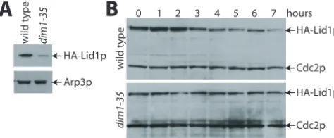
null alleles, and overexpression elicited no obvious phenotype in any background (data not shown).HAlid1⫹was then over-expressed in wild-type anddim1-35cells. Although HA-Lid1p was readily detected in lysates prepared from wild-type cells, it was present at significantly reduced levels indim1-35cells even when the cells were grown at 25°C, a temperature fully per-missive for growth (Fig. 1A). This result reproduces what was observed previously for endogenous Lid1p in the absence of Dim1p function (4).
Despite the lower level of total HA-Lid1p indim1-35cells compared to wild-type cells, we were able to determine the relative half-life of HA-Lid1p in the two strains. After maximal expression was achieved by growing cells in the absence of thiamine, synthesis of the RNA and protein was repressed by adding thiamine and cycloheximide, respectively, to the
on September 8, 2020 by guest
http://ec.asm.org/
dium. The amount of HA-Lid1p that remained was deter-mined at hourly time points by immunoblotting with antibodies to the HA epitope. In wild-type cells, after an initial burst of protein production, the amount of HA-Lid1p declined steadily over the course of the experiment (Fig. 1B, upper panel). Although expressed at significantly reduced levels from the onset, the half-life of HA-Lid1p indim1-35cells appeared even longer than that in wild-type cells (Fig. 1B, lower panel). Thus, increased rates of HA-Lid1p protein degradation could not explain the decreased abundance of Lid1p in the absence of
dim1⫹function.
Based on this result, we then predicted that HA-Lid1p was synthesized at reduced levels in the absence ofdim1⫹function. To test this hypothesis, wild-type anddim1-35cells expressing maximal levels of HA-Lid1p were pulsed for 10 min with35
S-Trans label, and the amount of HA-Lid1p produced was de-termined by immunoprecipitation. Despite similar amounts of
35S incorporation into the strains during a 10-min pulse, there
was significantly less HA-Lid1p produced indim1-35cells than that produced in wild-type cells (data not shown).
We next examined the steady-state level ofHAlid1⫹mRNA produced from thenmt41promoter indim1-35cells relative to wild-type cells by Northern blot analysis. Although the total levels ofnmt41HAlid1⫹RNA production were similar at both permissive and nonpermissive temperatures, the nmt41 HAlid1⫹ RNA was split into two bands in the dim1-35cells (Fig. 2A, strain 3). The faster-migrating band comigrated with thenmt41HAlid1⫹RNA from wild-type cells. Sincelid1⫹ con-tains four introns, it seemed likely that the upper band repre-sented an unspliced form ofnmt41HAlid1⫹RNA. To test this possibility directly, the four introns were removed from the
nmt41HAlid1⫹construct. When the cDNA version oflid1⫹was
FIG. 1. HA-Lid1p cannot be overproduced in the absence of
dim1⫹function. Wild-type anddim1-35cells were transformed with pREP42HAlid1⫹. Transformants were grown at 25°C in the absence of thiamine for 20 h, and samples were collected. A) Total protein lysates were prepared from the samples and resolved by SDS-polyacrylamide gel electrophoresis (PAGE). Following immunoblotting with 12CA5 to detect HA-Lid1p and anti-Arp3p serum (19a) to detect Arp3p as a loading control, proteins were visualized by enhanced chemilumines-cence. B) After maximal induction of pREP42HAlid1⫹ in wild-type (upper panel) anddim1-35(lower panel) cells, 5g of thiamine/ml was added to the medium to prevent further expression from thenmt41
promoter. Equal numbers of cells were collected at hourly intervals, and protein lysates were prepared. Proteins were resolved by SDS-PAGE. HA-Lid1p was detected with 12CA5 antibodies, and Cdc2p, which served as a loading control, was detected with anti-PSTAIR monoclonal antibody. The immunoblot in the lower panel was devel-oped for longer than the immunoblot in the upper panel so that the lower level of HA-Lid1p indim1-35cells could be visualized through-out the course of the experiment.
FIG. 2. Cells lackingdim1⫹function are defective for pre-mRNA splicing. A) Thelid1⫹ORF with (strains 3 and 4) or without (strains 5 and 6) its four introns was introduced into the pREP42HA vector. Wild-type cells (strains 2, 4, and 6) anddim1-35cells (strains 1, 3, and 5) were transformed with vector alone (strains 1 and 2) or the pREP42HAlid1⫹ constructs (strains 3 to 6). Transformants were grown at 25°C in the absence of thiamine for 18 h and then transferred to 36°C for 0 or 4 h. Total RNA was prepared and subjected to Northern analysis with a fragment of thelid1⫹ORF as probe. Note by the absence of bands in 1 and 2 that endogenous levels oflid1⫹RNA are not detected in these exposures. B) Total RNA was purified from wild-type (wt),prp2-1shifted to the nonpermissive temperature for the indicated number of hours, a strain containingdim1::his3⫹and nmt1-T81 dim1⫹integrated at theleu1locus grown in presence of thiamine for the indicated number of hours, anddim1-35cells shifted to the nonpermissive temperature for the indicated number of hours. Twenty micrograms of total RNA from each sample was resolved by electro-phoresis and subjected to Northern blot analysis with oligonucleotide probes complementary to the intron and exon sequences within the
tf2dgene.his3⫹RNAs were detected with a32P-labeled probe derived
from the genomic clone. PC, precursor mRNA, M, mature mRNA. C)
S. cerevisiaecells lackingDIB1are defective in pre-mRNA splicing. Strain KGY1023 was maintained in synthetic medium containing raf-finose and galactose.DIM1expression was repressed by shifting the cells to synthetic medium containing glucose (SD). Aliquots of cells were collected at the number of hours indicated following the shift into synthetic medium containing glucose. Total RNA was also purified from temperature-sensitive mutants prp3-1, prp18 (ts503), and
cdc28-1Nshifted to the restrictive temperature (35.5°C) for the num-ber of hours indicated. Twenty micrograms of total RNA was electro-phoresed and blotted. Northern blots probed with theACT1intron sequence or theDYN2,GLC7, andRP51a ORFs. Note the mature form of GLC7 mRNA does not decline because of the presumed longer half-life of the species.
on September 8, 2020 by guest
http://ec.asm.org/
overexpressed indim1-35cells in parallel with the intron-con-taining form, the upper band was no longer observed (Fig. 2A, strain 5). Thus,dim1-35cells appeared to be defective in the splicing oflid1⫹pre-mRNA.
The failure of dim1-35 cells to splice lid1⫹ mRNA might explain the reduced levels of Lid1p in this strain. To test whether Dim1p was required for general pre-mRNA splicing in vivo, RNA was prepared from wild-type cells,prp2-1(a bona fide pre-mRNA splicing mutant) cells, cells genetically de-pleted fordim1⫹(adim1 null mutant containingnmt1 -regu-latabledim1⫹), anddim1-35cells, and the RNA was subjected to northern analysis using probes directed at two intron-con-taining genes, tf2dand his3⫹. Like prp2-1cells, cells lacking
dim1function accumulated unspliced RNAs (Fig. 2B). We next tested whether theS. cerevisiaeortholog of Dim1p, termed Dib1p, was required for pre-mRNA splicing, since Dib1p had also been implicated in pre-mRNA splicing due to its copurification with the U4/U6.U5 tri-snRNP (14, 34). Pre-viously, in order to investigate the phenotype of cells lacking Dib1p function, we had created a conditional expression strain, KGY1023 (5). Because GAL1-drivenDIB1was not sufficiently repressed by glucose addition, we made use of the ubiquitin-N-degron tagging strategy described previously by Althoefer et al. (1). A plasmid expressing budding yeastDIB1still allowed growth in the presence of glucose. However, a single integrated copy of GALS::UBdim1⫹rescued growth under inducing con-ditions but failed to rescue growth under repressing concon-ditions. BothS. pombe dim1⫹and mouse mDim1 rescue thedib1null mutation (5). KGY1023, which lacks endogenous DIB1and harbors plasmid-borneS. pombe dim1cDNA under control of theGAL1 promoter, arrests growth following 6 h of glucose repression (5). KGY1023 mRNA was compared to that iso-lated from three control strains: (i)prp3-1, a positive control for a defect in the first step of splicing (39); (ii)prp18(ts503), a positive control for a defect in the second step of splicing (39); and (iii)cdc28-1N, a G2arrest (25) control to ensure that
any observed splicing defects were not secondary to cell cycle arrest. We assayed four intron-containing transcripts: ACT1
andRP51a, which are routinely used to analyze splicing defects inprpmutants;DYN2, a transcript inS. cerevisiaethat contains two introns; andGLC7, which encodes a cell cycle-regulated protein. Whendim1expression was repressed, intron-contain-ing forms of all four transcripts steadily accumulated to levels comparable to what was observed in theprpmutants (Fig. 2C). Also, the levels of mature DYN2 and RP51a decreased throughout the time course. These results were comparable to those observed with theprp3-1mutant but distinct from those observed withprp18andcdc28-1N. These data therefore sug-gest thatDIB1is essential, either directly or indirectly, for the first step of pre-mRNA splicing in vivo.
Dim1p copurifies with known splicing factors.To determine
whether Dim1p was a part of the S. pombe U4/U6.U5 tri-snRNP, similar to Dib1p inS. cerevisiae(14, 34), we examined whether it was present in a high-molecular-weight complex by sucrose gradient sedimentation. We found that a percentage of Dim1p sedimented deeper into the gradient than Cdc5p (which runs at approximately 40S) (20) (Fig. 3A), indicating that Dim1p was present in a complex considerably larger than what would be expected from the tri-snRNP (34). The remain-der of Dim1p sedimented near the top of the gradient,
consis-tent with a monomer or small complex (Fig. 3A). To determine if other components of the S. pombe U4/U6.U5 tri-snRNP behaved similarly on sucrose gradients, we modified the S. pombeortholog ofS. cerevisiae PRP6, prp1⫹(also known as
zer1⫹) (38), at its endogenous locus to encode C-terminally Myc13- or TAP-tagged versions of Prp1p (30, 35). Both tagged strains grew normally, suggesting that the epitope did not com-promise the function of Prp1p. By sucrose gradient fraction-ation, Prp1p-Myc13 sedimented deep into the gradient simi-larly to one portion of Dim1p. To determine if the complex that contained Prp1p also contained Dim1p, tandem affinity purification was carried out on two separate occasions from a
prp1-TAPstrain, and the protein composition of a portion of each TAP complex was analyzed by silver staining (Fig. 3B), with the remainder analyzed by multidimensional tandem mass spectrometry (16). Proteins identified from both purifications that were absent from TAP purifications performed on un-tagged cells or from unrelated TAP purifications (data not shown) are listed in Table 2. As a means of comparison, results from the purification of theS. cerevisiaepenta-snRNP complex (34) and theS. pombeCdc5p splicing complex (23) are also shown. In both Prp1p-TAP purifications, Dim1p was present,
FIG. 3. Dim1p copurifies with known splicing factors. (A) Protein lysates ofdim1-HAorprp1-mycstrains were fractionated by sucrose gradient sedimentation. Fractions were collected from the bottom (fraction 1) of the gradients, resolved by SDS-PAGE, and then immu-noblotted with the 12CA5 or 9E10 antibody to detect Dim1p-HA (upper panel) and Prp1p-Myc (lower panel), respectively. The signal on the right-hand portion of the anti-Myc blot is a nonspecific blotch. The positions of sedimentation markers are provided. (B) A silver-stained gel of the purified Prp1p-TAP complex. (C and D) The indi-cated proteins (bait vector/prey vector) were tested by two-hybrid analysis. LEU⫹TRP⫹transformants were tested for growth on selec-tive medium (data not shown) and assayed for-galactosidase activity measured in relative light units.
on September 8, 2020 by guest
http://ec.asm.org/
as were most components of the U4/U6.U5 tri-snRNP and the U2 snRNP. Indeed, the protein composition of this complex is very similar to the recently described human B⌬1 complex that lacks the U1 snRNP and the nineteen complex (18). The amino acid sequence coverage of Prp1p was 60%, and the greatest sequence coverage of copurifying proteins was ob-tained for Dim1p and Prp31p, at 58 and 61%, respectively. While these results are not quantitative, they did raise the possibility that Dim1p interacted with Prp1p directly or indi-rectly through an association with Prp31p, and we tested whether Dim1p was capable of binding either of these two proteins. We found that Dim1p interacted with Prp1p by
two-hybrid analysis using full-length constructs of each protein, but it did not show an interaction with Prp31p (Fig. 3C). In con-trast, Prp1p showed a strong interaction with both Dim1p and Prp31p (Fig. 3C and D), indicating that Prp1p might bind both proteins directly and simultaneously.
The dim1-35mutation selectively affects the production of
Lid1p.A block to pre-mRNA splicing as in thedim1-35mutant
would be expected to affect the levels of most transcripts and their protein products. However, this seemed incongruous for two reasons. First, we had used Cdc2p and Arp3p as loading controls for our immunoblots and observed no difference in their abundance between wild-type anddim1-35cells (4) (Fig.
TABLE 2. Comparison of mass spectrometric results from protein purifications ofS. cerevisiaepenta-snRNP,S. pombeCdc5-TAP, and
S. pombePrp1-TAP
S. pombe S. cerevisiae Human 1a 2a 3a S. pombe S. cerevisiae Human 1a 2a 3a
snRNP core proteins
Smb1p Smb1p SmB/B⬘ X X X
Smd1p Smd1p SmD1 X X X
Cwf9p Smd2p SmD2 X X X
Smd3p Smd3p SmD3 X X X
Sme1 Sme1p SmE1 X ⫺ X
Smf1p Smf1p SmF1 X X X
Smg1p SmG1p SmG1 X X X
Lsm2p Lsm2p LSM2 X ⫺ ⫺
Lsm3p Lsm3p LSM3 X ⫺ ⫺
Lsm4p Lsm4p LSM4 X ⫺ ⫺
Lsm5p Lsm5p LSM5 X ⫺ ⫺
Lsm6p Lsm6p LSM6 X ⫺ ⫺
Lsm7p Lsm7p LSM7 X ⫺ ⫺
Lsm8p Lsm8p LSM8 X ⫺ ⫺
U1 snRNP proteins
U1-70 Snp1p U1-70 X ⫺ ⫺
U1-A Mud1p U1-A X ⫺ ⫺
U1-C Yhc1p U1-C X ⫺ ⫺
——b Prp42p —— X ⫺ ⫺
Prp40p Prp40p —— X ⫺ ⫺
Prp39p Prp39p —— X ⫺ ⫺
SPBC23E6.01c Nam8p —— X ⫺ ⫺
Luc7p Luc7p —— X ⫺ ⫺
—— Snu56p —— X ⫺ ⫺
SPBC24E9.10 Snu71p —— X ⫺ ⫺
U2 snRNP proteins
U2Ap Lea1p U2A⬘ X X X
O13649 Msl1p U2-B⬘ X X X
Sap61p Prp9p SF3a60 X X X
Sap62p Prp11p Sf3a66 X X X
Sap114p Prp21p Sf3a120 X X X
Sap49p Hsh49p SF3b53 X ⫺ X
Sap145p Cus1p SF3b150 X X X
Prp12p Rse1p SF3b130 X X X
Prp10p Hsh155p SF3b160 X X X
Uap2p Cus2p Tat-SF1 X ⫺ ⫺
SPBC29A3.07c —— p14 ⫺ ⫺ X
O94290 1st3p/Snu17p —— X X ⫺
U5 snRNP proteins
Cwf6p Prp8p U5-220 X X X
Brr2p Brr2p U5-200 X X X
Cwf10p Snu114p U5-116 X X X
Prp28p Prp28p U5-100 ⫺ ⫺ ⫺
Cwf17p —— U5-40 ⫺ X X
a
1,S. cerevisiaepenta-snRNP (34); 2,S. pombeCdc5p complex (23); 3,S. pombePrp1p-TAP purification X, protein present;⫺, protein not present.
b
——, no identified homolog.
c
SwissProt or Entrez accession no.
Tri-snRNP proteins
Prp1p Prp6p U5-102 X ⫺ X
Q09856 Prp3p HPR3 X ⫺ X
Q9UTC7 Prp4p HPR4 X ⫺ X
Snu66p Snu66p SART1 X ⫺ X
Prp31p Prp31p PRP31 X ⫺ X
SPAC607.03c Snu13p SNU13 X ⫺ ⫺
—— Snu23p —— X ⫺ ⫺
—— Prp38p —— X ⫺ X
—— Spp381p —— X ⫺ ⫺
Q9USR2 Sad1p Q96RK9 X ⫺ ⫺
Dimlp Diblp U5-15 X ⫺ X
Ntc proteins
Cdc5p Cef1p CDC5L X X –
Prp5p/Cwf1p Prp46p PRL1 X X ⫺
Cwf2p/Prp3p Cwc2p RNPS1 X X ⫺
Cwf3p Syf1p SYF1 X X ⫺
Cwf4p Syf3p CRN1 X X ⫺
Cwf7p Snt309p SPF27 X X ⫺
Cwf8p Prp19p PRP19 X X ⫺
Cwf12p Isy1p ISY1 X X
Cwf13p Prp45p SK1P X X ⫺
—— Ntc20p —— X ⫺ ⫺
O59733 Syf2p GCIP-IP X X ⫺
Second-step factors
Q9Y7Y2 Slu7p SLU7 – X –
Cwf5p Slt11p ECM2 X X ⫺
Prp17p Prp17p PRP17 ⫺ X ⫺
Prp22p Prp22p PRP22 ⫺ X ⫺
Other factors
Cwf11p —— O60306 ⫺ X ⫺
Cwf15p Cwc15p HSPC148 ⫺ X ⫺
Cwf21p —— SRm300 ⫺ X ⫺
Cwf22p Cwc22p M1F4G ⫺ X ⫺
Cwf24p Cwc24p O15541 ⫺ X ⫺
Cwf27p Cwc27p PPIL1 ⫺ X ⫺
O43031 Spp2p Q9BQA8 ⫺ X ⫺
Cwf14p Cwc14p G10 ⫺ X ⫺
Cwf16p Cwc16p Q9BW85 ⫺ X ⫺
Cwf18p —— MGC23918 ⫺ X ⫺
Cwf19p —— —— ⫺ X ⫺
Cwf20p —— —— ⫺ X ⫺
SPCC1281.02c —— SPF30 ⫺ ⫺ X
Ded1p (DEAH box) Ded1p Abstrakt ⫺ ⫺ X
on September 8, 2020 by guest
http://ec.asm.org/
1A), although both thecdc2⫹and arp3⫹primary transcripts contain four introns. Second, it would be difficult to imagine a scenario in which a general block in protein production would generate the dim1-35 mutant phenotype. To examine this question more carefully, we compared the levels of several proteins whose pre-mRNAs contain introns in the dim1-35
mutant and wild-type cells. The levels of Cdc3p (profilin), Cdc4p (a myosin light chain), Cdc5p (a pre-mRNA splicing factor), Arp3p (a component of the Arp2/3 F-actin-nucleating complex), Cwf2p (a pre-mRNA splicing factor), and Cwf3p (a pre-mRNA splicing factor) were not significantly different in thedim1-35mutant relative to wild-type cells using the amount of Cdc2p in the lysates as a loading control (Fig. 4A). Only Lid1p levels were found to be significantly different (Fig. 4A). This was also true if the specific signals were quantitated against total protein loaded onto the gels rather than Cdc2p abundance (data not shown). Thus, thedim1-35mutation se-lectively affects the production of Lid1p among tested proteins. If Dim1p controlled the levels of Lid1p solely by regulating the splicing of lid1⫹ RNA, then production of Lid1p in
dim1-35 cells should be restored to wild-type levels by the removal of the four introns from thelid1⫹ORF. To test this hypothesis, we examined the level of HA-Lid1p produced from thelid1⫹cDNA (lid1⌬i) under control of thenmt41promoter. Unexpectedly, we found that HA-Lid1p levels were still re-duced indim1-35cells relative to that of wild-type cells, al-though the level of Cdc2p did not vary (Fig. 4B, lanes 5 and 6). We then considered the possibility that overproduction of HA-Lid1p from a heterologous promoter was overwhelming the capacity ofdim1-35cells to produce HA-Lid1p. Therefore, we constructed a gene replacement strain. First, we introduced sequences encoding three copies of the HA epitope at the 5⬘ end of the open reading frame of the intron-deleted version of
lid1⫹. Next, the tagged version oflid⌬iwas used to replace the endogenous gene (see Materials and Methods) so that expres-sion would occur from the endogenous lid1 promoter. The
HAlid1⌬istrain was wild type in morphology and growth rate (data not shown). TheHAlid1⌬iallele was then combined with thedim1-35mutation, and endogenous HA-Lid1p levels were examined after a shift to a restrictive temperature. The amount of HA-Lid1p produced indim1-35cells was barely detectable and significantly less than that in wild-type cells (Fig. 4C). Thus, there appears to be a second block to Lid1p production downstream of pre-mRNA splicing indim1-35cells.
Dim1p function is required for efficient pre-mRNA export.
The process we thought to examine next in cells lackingdim1⫹
function was the export of RNA from the nucleus. The local-ization of poly(A)⫹RNA was examined indim1-35anddim1⌬
cells and compared with that in wild-type cells and a bona fide nuclear export mutant,rae1-1(9). In wild-type cells, poly(A)⫹ RNA was not detected in the nucleus at appreciable levels (Fig. 5A). In the dim1-35mutant, poly(A)⫹ RNA could be detected in the nucleus even at permissive temperature, and staining within the nucleus increased during the temperature shift (Fig. 5A). However, nuclear accumulation was neither as complete nor as rapid as that observed inrae1-1cells (Fig. 5A). In the dim1⌬ cells maintained by dim1⫹ expressed from a regulatable thiamine-repressible promoter, nuclear pre-mRNA accumulation was observed concomitantly with the timing of promoter repression (Fig. 5B). Furthermore, this
accumulation paralleled the timing of the loss of pre-mRNA splicing (Fig. 5C).
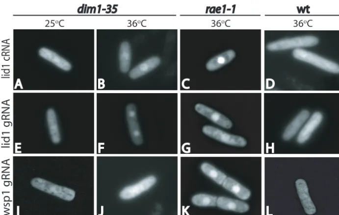
The dim1-35mutation selectively affects the export oflid1
mRNA.One possible explanation for the selective effects of
Dim1p loss of function on Lid1p levels is that the function of Dim1p in either pre-mRNA splicing and/or nuclear mRNA export is specific tolid1transcripts or a subset of transcripts that includeslid1. Since our data indicated a generalized defect of pre-mRNA splicing (Fig. 2), we examined the specificity of thedim1-35mRNA export defect for lid1transcripts. To ad-dress this, we adapted theS. cerevisiae“green RNA” system, developed for live cell monitoring of specific mRNA tran-scripts (3, 33), for use inS. pombe(see Materials and Meth-ods). In wild-type cells, thelid1⫹GFP-labeled mRNA tran-script (gRNA) was not detected in the nucleus at appreciable levels (Fig. 6). However, in thedim1-35mutant,lid1⫹gRNA could be detected in the nucleus at the restrictive temperature (Fig. 6). However, as observed with total mRNA (Fig. 5), nuclear accumulation was not as complete or as rapid as that
FIG. 4. Dim1p affects Lid1p levels independently of pre-mRNA splicing. (A) Cells were grown at permissive temperature (25°C) and shifted to 36°C for 4 h. Protein lysates were prepared and normalized according to protein concentration. Equal amounts of protein from wild-type (wt) (KGY246) anddim1-35(KGY390) cells were blotted with specific antisera for Cdc5p, Cdc3p, Arp3p, Cdc4p, and Cdc2p or from strains that express Myc or HA epitope-tagged proteins (KGY1365, KGY1739, KGY1302, KGY1305, KGY1420, KGY3211, KGY1430, and KGY3210) with 9E10, 12CA5, and Cdc2p antibodies. Immunoblots were analyzed with a Molecular Dynamics Storm Phos-phorImager, and the intensity of each band was normalized against the Cdc2p loading control. Results are presented as means⫾standard deviations (n⫽3 to 10). (B)Wild-type (lanes 1, 3, and 5) anddim1-35
(lanes 2, 4, and 6) cells were transformed with empty plasmid (lanes 1 and 2), the pREP42HAlid1⫹construct containing introns (lanes 3 and 4), or the pREP42HAlid1⫹construct lacking introns (lanes 5 and 6). Following maximal RNA production at 20 h of growth in the absence of thiamine and a shift to 36°C for 4 h (Fig. 2), protein lysates were prepared and resolved by SDS-PAGE. HA-Lid1p (upper panel) and Cdc2p which served as a loading control (lower panel) were detected by immunoblot analysis. (C) Lysates were prepared from wild-type cells (lane 1) or wild-type anddim1-35cells expressingHA-lid1⌬ifrom the endogenouslid1promoter (lanes 2 and 3, respectively) after a shift to 36°C for 4 h. Following separation by SDS-PAGE, HA-Lid1p (up-per panel) and Cdc2p as a loading control (lower panel) were detected by immunoblotting.
on September 8, 2020 by guest
http://ec.asm.org/
observed in rae1-1 cells (Fig. 6). The accumulation of lid1
transcript in dim1-35 is unrelated to the role of Dim1p in splicing, as both wild-type andlid1⌬itranscripts showed com-parable levels of accumulation. Furthermore, no appreciable accumulation of lid1transcript was observed in prp2-1cells, which are strongly inhibited for pre-mRNA splicing, at the restrictive temperature (data not shown). To investigate the specificity of the RNA export defect, we also examined the localization of the unrelatedwsp1transcript. Whilewsp1 tran-scripts with or without introns do accumulate in the nucleus of
rae1-1cells (Fig. 6 and data not shown), which show a block of
generalized mRNA export (9), no significant accumulation of these transcripts was observed indim1-35cells (Fig. 6).
DISCUSSION
In this study, we have investigated the observed dependence of Lid1p protein levels on Dim1p function. Consistent with previous observations that Dim1p orthologs associate with splicing factors (14, 34, 43), we have found that S. pombe
Dim1p is required for efficient pre-mRNA splicing. However, our data indicate that Dim1p’s essential function extends
be-FIG. 5. Localization of poly(A)⫹RNA indim1-35cells. (A) poly(A)⫹RNA was detected by fluorescence in situ hybridization in wild-type,
rae1-1, anddim1-35cells grown at 25°C and shifted to 36°C for the times indicated. (B and C)dim1::his3⫹cells carrying a single integrated copy ofnmt81::mDIM1⫹(KGY1180) were grown in minimal medium lacking thiamine and then shifted to medium containing thiamine for the times indicated. DAPI, 4⬘,6⬘-diamidino-2-phenylindole. (B) poly(A)⫹RNA was detected by fluorescence in situ hybridization. (C) Total RNA from these cells was examined by Northern blot analysis for the accumulation of TFIID pre-mRNA. PC, precursor; M, mature RNA.
on September 8, 2020 by guest
http://ec.asm.org/
yond pre-mRNA splicing to mediating the export of at least certain mRNAs from the nucleus.
While Dim1p orthologs have previously been purified along with the U4/U5.U6 tri-snRNP (14, 34), we purifiedS. pombe
Dim1p with the U4/U5.U6 tri-snRNP component Prp1p (equivalent of Prp6 inS. cerevisiae) in a large splicing complex that appears similar in protein composition to the recently described B⌬1 complex isolated from human splicing extracts (18). Indeed, by sucrose gradient fractionation, we did not detect a smaller Prp1p-containing protein complex. The Prp1p-TAP complex contained U4/U5.U6 tri-snRNP and U2 snRNP components, while it lacked any detectable contribu-tion of the nineteen complex that is a hallmark of the U2,U5,U6 complex that has predominated purification of splicing complexes fromS. pombe(23). While Dim1p was iden-tified within this large protein complex, fractionation of S. pombelysates by sucrose gradient sedimentation indicates that Dim1p/Dib1p exists outside of this splicing complex as well, most likely in smaller complexes or on its own. Thus, our biochemical analyses leave open the possibility that Dim1p/ Dib1p performs functions outside of the U4/U5.U6 tri-snRNP and possibly in processes other than pre-mRNA splicing.
Because Dib1p was initially copurified with a much smaller U4/U5.U6 tri-snRNPS. cerevisiaecomplex, it seems likely that it interacts directly with at least one U4/U5.U6 tri-snRNP component. Indeed, in a genome-wide two-hybrid analysis ofS. cerevisiae protein interactions, Dib1p was found to interact only with Prp6p, and Prp6p interacted only with Dib1p (36). The human homolog of Prp6p has also been shown to interact with the human homolog of Prp31p (18a). We have established the conservation of these interactions by showing thatS. pombe
Dim1p interacts with Prp1p in a two-hybrid assay and that Prp1p interacts with Prp31p. Prp1p contains many TPR re-peats, and it will be interesting to narrow down the domain
responsible for Dim1p and Prp31p interactions in the future. The structure of Dim1p family proteins has been determined by both nuclear magnetic resonance (42) and X-ray crystallog-raphy (31), and it has previously been suggested that the key role of these proteins in splicing complexes might involve bind-ing of RNA via a conserved basic patch on their surfaces (43). However, we have been unable to detect any direct interac-tions between Dim1p and numerous RNA species (data not shown). Therefore, the basic patch region may be critical for a protein-protein interaction with an acidic region of the Prp1p-Prp31p splicing complex.
Since the completion of theS. pombegenome sequence, it has become clear that⬃45% ofS. pombe genes contain in-trons, and therefore, it is unexpected that a mutation in a protein required for general pre-mRNA splicing would have a very specific defect in the metaphase-to-anaphase transition due to inadequate production of a single component of the APC, Lid1p/Apc4p. TheS. pombe APC contains 13 compo-nents, and several of these components are produced from genes containing introns. Of these proteins, however, only Lid1p levels fall significantly in the absence of Dim1p function (4; our unpublished data). This might indicate thatdim1-35is a hypomorphic mutant that allows significant pre-mRNA splic-ing to occur. In this scenario, only the levels of short-lived, low-abundance proteins or RNAs would be expected to change dramatically within a 4-h temperature shift experiment. The scarcity of Lid1p combined with its requirement for APC func-tion might make it an ideal target for regulafunc-tion of the cell cycle via an arrest in proper mRNA processing. Indeed, our data suggest that at least in the case of Dim1p, this regulation is directed rather specifically towards Lid1p. Alternatively, and because we found it difficult to envision thatlid1⫹would sur-face as the single most critical low-abundance message or tar-get, we have entertained possible explanations for thedim1-35 FIG. 6. Localization of individual transcripts indim1-35cells.lid1without (A to D) (lid1cRNA) or with (E to H) (lid1gRNA) introns as well as controlwsp1gRNA (I to L) were generated by placing them downstream of the MS2-CP binding sites in pRAM-MS2. Coexpression of the gRNA expression constructs with pREP41CP-GFP allowed visualization of their localization at either 25°C (A, E, and I) or 36°C (B to D, F to H, J, and K) indim1-35cells (A, B, E, F, I, and J),rae1-1cells (C, G, and K), and wild-type (wt) cells (D, H, and L).
on September 8, 2020 by guest
http://ec.asm.org/
mutant phenotype other than a block to pre-mRNA splicing. Clearly, our biochemical fractionation results showing that a substantial fraction of Dim1p is not a part of a splicing complex and the lid1RNA localization results are compatible with a specific requirement for Dim1p in other steps of pre-mRNA processing. It is also intriguing to us thatprp1anddim1 mu-tants display similar phenotypes. Likedim1-35cells,prp1 mu-tants have been shown to have defects in pre-mRNA splicing, poly(A)⫹RNA nuclear transport, and cell cycle control (26, 27, 28, 37, 38). This finding suggests that a complex containing Dim1p and Prp1p, and perhaps other proteins, might be crit-ical in the transition steps between pre-mRNA splicing and transport of the mature transcript from the nucleus to the cytoplasm. These effects on RNA export are unlikely to be secondary effects related to defects in splicing, as the two defects are detected roughly simultaneously in dim1 mutant cells.
While undertaking these studies, we have generated the first system for real-time imaging of specific RNAs inS. pombe. ThisS. pombegreen RNA system should be of use in future studies to define additional factors involved in RNA processing and export.
ACKNOWLEDGMENTS
We thank Melanie D. Ohi for assistance in tabulating the compar-ative analysis of TAP purifications. We also thank Jeff Flick and Shel-ley Sazer for reagents and protocols used in the in situ hybridization experiments. Finally, we thank Kerry Bloom for reagents and protocols used to adapt the Green RNA system toS. pombe.
This work was supported by NIH grant GM47728 to K.L.G. and NIH grant RR11823-09 to J.R.Y. K.L.G. is an investigator of the Howard Hughes Medical Institute.
REFERENCES
1.Althoefer, H., A. Schleiffer, K. Wassmann, A. Nordheim, and G. Ammerer.
1995. Mcm1 is required to coordinate G2-specific transcription in Saccharo-myces cerevisiae. Mol. Cell. Biol.15:5917–5928.
2.Bahler, J., J. Q. Wu, M. S. Longtine, N. G. Shah, A. McKenzie III, A. B. Steever, A. Wach, P. Philippsen, and J. R. Pringle. 1998. Heterologous modules for efficient and versatile PCR-based gene targeting in Schizosac-charomyces pombe. Yeast14:943–951.
3.Beach, D. L., E. D. Salmon, and K. Bloom.1999. Localization and anchoring of mRNA in budding yeast. Curr. Biol.9:569–578.
4.Berry, L. D., A. Feoktistova, M. D. Wright, and K. L. Gould.1999. The
Schizosaccharomyces pombe dim1⫹gene interacts with the anaphase-promot-ing complex or cyclosome (APC/C) component lid1⫹and is required for APC/C function. Mol. Cell. Biol.19:2535–2546.
5.Berry, L. D., and K. L. Gould.1997. Fission yeast dim1⫹encodes a func-tionally conserved polypeptide essential for mitosis. J. Cell Biol.137:1337– 1354.
6.Berry, L. D., and K. L. Gould.1996. Novel alleles of cdc13 and cdc2 isolated as suppressors of mitotic catastrophe inSchizosaccharomyces pombe. Mol. Gen. Genet.251:635–646.
7.Blanton, S., A. Srinivasan, and B. C. Rymond.1992. PRP38 encodes a yeast protein required for pre-mRNA splicing and maintenance of stable U6 small nuclear RNA levels. Mol. Cell. Biol.12:3939–3947.
8.Boddy, M. N., P. H. Gaillard, W. H. McDonald, P. Shanahan, J. R. Yates III, and P. Russell.2001. Mus81-Eme1 are essential components of a Holliday junction resolvase. Cell107:537–548.
9.Brown, J. A., A. Bharathi, A. Ghosh, W. Whalen, E. Fitzgerald, and R. Dhar.
1995. A mutation in theSchizosaccharomyces pombe rae1gene causes defects in poly(A)⫹RNA export and in the cytoskeleton. J. Biol. Chem.270:7411– 7419.
10.Burke, J. D., and K. L. Gould.1994. Molecular cloning and characterization of theSchizosaccharomyces pombe his3gene for use as a selectable marker. Mol. Gen. Genet.242:169–176.
11.Collart, M. A., and K. Struhl.1993. CDC39, an essential nuclear protein that negatively regulates transcription and differentially affects the constitutive and inducible HIS3 promoters. EMBO J.12:177–186.
12.Craven, R. A., D. J. Griffiths, K. S. Sheldrick, R. E. Randall, I. M. Hagan, and A. M. Carr.1998. Vectors for the expression of tagged proteins in
Schizosaccharomyces pombe. Gene221:59–68.
13.Forrester, W., F. Stutz, M. Rosbash, and M. Wickens.1992. Defects in mRNA 3⬘-end formation, transcription initiation, and mRNA transport as-sociated with the yeast mutation prp20: possible coupling of mRNA pro-cessing and chromatin structure. Genes Dev.6:1914–1926.
14.Gottschalk, A., G. Neubauer, J. Banroques, M. Mann, R. Lu¨hrmann, and P. Fabrizio.1999. Identification by mass spectrometry and functional analysis of novel proteins of the yeast [U4/U6.U5] tri-snRNP. EMBO J.18:4535– 4548.
15.James, P., J. Halladay, and E. A. Craig.1996. Genomic libraries and a host strain designed for highly efficient two-hybrid selection in yeast. Genetics
144:1425–1436.
16.Link, A. J., J. Eng, D. M. Schieltz, E. Carmack, G. J. Mize, D. R. Morris, B. M. Garvik, and J. R. Yates III.1999. Direct analysis of protein complexes using mass spectrometry. Nat. Biotechnol.17:676–682.
17.Lundgren, K., S. Allan, S. Urushiyama, T. Tani, Y. Ohshima, D. Frendewey, and D. Beach.1996. A connection between pre-mRNA splicing and the cell cycle in fission yeast:cdc28⫹is allelic withprp8⫹and encodes an RNA-dependent ATPase/helicase. Mol. Biol. Cell7:1083–1094.
18.Makarova, O. V., E. M. Makarov, H. Urlaub, C. L. Will, M. Gentzel, M. Wilm, and R. Luhrmann.2004. A subset of human 35S U5 proteins, includ-ing Prp19, function prior to catalytic step 1 of splicinclud-ing. EMBO J.23:2381– 2391.
18a.Makarova, O. V., E. M. Makarov, S. Liu, H.-P. Vornlocher, and R. Luhr-mann.2002. Protein 61K, encoded by a gene (PRPF31) linked to autosomal dominant retinitis pigmentosa, is required for U4/U6.U5 tri-snRNP forma-tion and pre-mRNA splicing. EMBO J.21:1148–1157.
19.Maundrell, K. 1993. Thiamine-repressible expression vectors pREP and pRIP for fission yeast. Gene123:127–130.
19a.McCollum, D. A. Feoktistova, M. Morphew, M. K. Balasubramanian, and K. L. Gould.1996. TheSchizosaccharomyces pombeactin related protein, Arp3, is a component of the cortical actin cytoskeleton and interacts with profilin. EMBO J.15:6438–6446.
20.McDonald, W. H., R. Ohi, N. Smelkova, D. Frendewey, and K. L. Gould.
1999. Myb-related fission yeast cdc5p is a component of a 40S snRNP-containing complex and is essential for pre-mRNA splicing. Mol. Cell. Biol.
19:5352–5362.
21.Moreno, S., A. Klar, and P. Nurse.1991. Molecular genetic analysis of fission yeastSchizosaccharomyces pombe. Methods Enzymol.194:795–823. 22.Morgan, D. O.1997. Cyclin-dependent kinases: engines, clocks, and
micro-processors. Annu. Rev. Cell Dev. Biol.13:261–291.
23.Ohi, M. D., A. J. Link, L. Ren, J. L. Jennings, W. H. McDonald, and K. L. Gould.2002. Proteomics analysis reveals stable multiprotein complexes in both fission and budding yeasts containing Myb-related Cdc5p/Cef1p, novel pre-mRNA splicing factors, and snRNAs. Mol. Cell. Biol.22:2011–2024. 24.Peters, J.-M.2002. The anaphase-promoting complex: proteolysis in mitosis
and beyond. Mol. Cell9:931–943.
25.Piggot, J. R., R. Rai, and B. L. A. Carter.1982. A bifunctional gene product involved in two phases of the yeast cell cycle. Nature298:391–393. 26.Potashkin, J., and D. Frendewey.1989. Splicing of the U6 RNA precursor is
impaired in fission yeast pre-mRNA splicing mutants. Nucleic Acids Res.
17:7821–7831.
27.Potashkin, J., D. Kim, M. Fons, T. Humphrey, and D. Frendewey.1998. Cell-division-cycle defects associated with fission yeast pre-mRNA splicing mutants. Curr. Genet.34:153–163.
28.Potashkin, J., R. Li, and D. Frendewey.1989. Pre-mRNA splicing mutants of
Schizosaccharomyces pombe. EMBO J.8:551–559.
29.Prentice, H. L.1992. High efficiency transformation ofSchizosaccharomyces pombeby electroporation. Nucleic Acids Res.20:621.
30.Puig, O., F. Caspary, G. Rigaut, B. Rutz, E. Bouveret, E. Bragado-Nilsson, M. Wilm, and B. Se´raphin.2001. The tandem affinity purification (TAP) method: a general procedure of protein complex purification. Methods24:
218–229.
31.Reuter, K., S. Nottrott, P. Fabrizio, R. Luhrmann, and R. Ficner.1999. Identification, characterization and crystal structure analysis of the human spliceosomal U5 snRNP-specific 15 kD protein. J. Mol. Biol.294:515–525. 32.Sambrook, J., E. F. Fritsch, and T. Maniatis.1989. Molecular cloning: a
laboratory manual, 2nd ed. Cold Spring Harbor Laboratory Press, Cold Spring Harbor, N.Y.
33.SenGupta, D. J., B. Zhang, B. Kraemer, P. Pochart, S. Fields, and M. Wickens.1996. A three-hybrid system to detect RNA-protein interactions in vivo. Proc. Natl. Acad. Sci. USA93:8496–8501.
34.Stevens, S. W., and J. Abelson.1999. Purification of the yeast U4/U6.U5 small nuclear ribonucleoprotein particle and identification of its proteins. Proc. Natl. Acad. Sci. USA96:7226–7231.
35.Tasto, J. J., R. H. Carnahan, W. H. McDonald, and K. L. Gould.2001. Vectors and gene targeting modules for tandem affinity purification in
Schizosaccharomyces pombe. Yeast18:657–662.
36.Uetz, P., L. Giot, G. Cagney, T. A. Mansfield, R. S. Judson, J. R. Knight, D. Lockshon, V. Narayan, M. Srinivasan, P. Pochart, A. Qureshi-Emili, Y. Li, B. Godwin, D. Conover, T. Kalbfleisch, G. Vijayadamodar, M. Yang, M. Johnston, S. Fields, and J. M. Rothberg.2000. A comprehensive analysis of
on September 8, 2020 by guest
http://ec.asm.org/
protein-protein interactions in Saccharomyces cerevisiae. Nature403:623– 627.
37.Urushiyama, S., T. Tani, and Y. Ohshima.1996. Isolation of novel pre-mRNA splicing mutants ofSchizosaccharomyces pombe. Mol. Gen. Genet.
253:118–127.
38.Urushiyama, S., T. Tani, and Y. Ohshima.1997. Theprp1⫹gene required for pre-mRNA splicing inSchizosaccharomyces pombeencodes a protein that contains TPR motifs and is similar to Prp6p of budding yeast. Genetics
147:101–115.
39.Vijayraghavan, U., M. Company, and J. Abelson.1989. Isolation and char-acterization of pre-mRNA splicing mutants ofSaccharomyces cerevisiae. Genes Dev.3:1206–1216.
40.Yoon, H. J., A. Feoktistova, B. A. Wolfe, J. L. Jennings, A. J. Link, and K. L.
Gould.2002. Proteomics analysis identifies new components of the fission and budding yeast anaphase-promoting complexes. Curr. Biol.
12:2048–2054.
41.Zachariae, W., and K. Nasmyth.1999. Whose end is destruction: cell division and the anaphase-promoting complex. Genes Dev.13:2039–2058. 42.Zhang, J., C. Kong, H. Xie, P. S. McPherson, S. Grinstein, and W. S.
Trimble.1999. Phosphatidylinositol polyphosphate binding to the mamma-lian septin H5 is modulated by GTP. Curr. Biol.9:1458–1467.
43.Zhang, Y.-Z., T. Lindblom, A. Chang, M. Sudol, A. E. Sluder, and E. A. Golemis.2000. Evidence that Dim1 associates with proteins involved in pre-mRNA splicing, and delineation of residues essential for Dim1 interac-tions with hnRNP F and Npw38/PQBP-1. Gene257:33–43.