1535-9778/05/$08.00⫹0 doi:10.1128/EC.4.7.1308–1316.2005
Copyright © 2005, American Society for Microbiology. All Rights Reserved.
Galactomannoproteins of
Aspergillus fumigatus
W. Morelle,
1† M. Bernard,
1J.-P. Debeaupuis,
1M. Buitrago,
1‡
M. Tabouret,
2and J.-P. Latge
´
1*
Unite´ des Aspergillus, Institut Pasteur, 25, rue du Docteur Roux, 75724 Paris
Cedex 15,1and Bio-Rad, Clinical Microbiology Division,
59114 Steenvoorde,2France
Received 3 February 2005/Accepted 18 May 2005
Galactofuranose-containing molecules have been repeatedly shown to be important antigens among human fungal pathogens, includingAspergillus fumigatus. Immunogenic galactofuran determinants have been poorly characterized chemically, however. We reported here the characterization of two glycoproteins ofA.fumigatus
with an N-glycan containing galactofuranose. These proteins are a phospholipase C and a phytase. Chemical characterization of the N-glycan indicates that it is a mixture of Hex5-13HexNAc2oligosaccharides, the major
molecular species corresponding to Hex6-8HexNAc2. The N-glycan contained one galactofuranose unit that was
in a terminal nonreducing position attached to the 2 position of Man. This single terminal nonreducing galactofuranose is essential for the immunoreactivity of the N-glycans assessed either with a monoclonal antibody that recognizes a tetra--1,5-galactofuran chain of galactomannan or withAspergillus-infected patient sera.
Galactomannan (GM) is an essential molecule in the life of the human opportunistic fungal pathogenAspergillus fumiga-tus. It is a polysaccharide that is an important structural com-ponent of the cell wall ofA. fumigatus(10). GM is secreted during growth in culture (15) and has also been shown to exist in a glycosylphosphatidylinositol membrane-bound form (C. Costachel et al., unpublished data). The extracellular, mem-brane- or cell wall-linked GMs have a similar chemical orga-nization composed of a linear␣-mannan core with a repeating tetramannose unit (2M-6M-2M-2M) with side chains of -1,5-galactofuranosyl residues with an average degree of polymer-ization of 4 attached to the␣-1,2-linked mannose residues of the mannan chain (10, 15). GM is also produced in the host and is a well-knownA.fumigatusantigen. Antibodies directed against this polysaccharide have been detected in patients with aspergilloma and in experimentally infected animals or in rab-bits or mice hyperimmunized with total extracts ofA.fumigatus
(15, 26). In addition, it has been repeatedly shown that this molecule circulates in the biological fluids of patients with invasive aspergillosis. Indeed, serological diagnosis of this life-threatening fungal infection remains based on the detection of GM in the serum, urine, or bronchoalveolar lavage samples of infected patients (18, 29, 31) since heavily immunocompro-mised patients at risk for invasive aspergillosis are not able to mount an antibody response against A. fumigatus. MAbs or polyclonal antibodies directed against GM have been used in the search for circulating antigens. The only commercial kit available for detection of GM is a sandwich enzyme-linked
immunosorbent assay (ELISA) based on the use of a rat mono-clonal antibody (MAb), EB-A2, that reacts specifically with the galactofuranose (Galf)-containing moiety of the GM. In vitro, the best inhibition was obtained with a tetra- -1,5-galacto-furanosoligosaccharide, suggesting that this oligosaccharide was the epitope recognized by the MAb (30).
Glycoproteins reacting positively with anti-galactofuran MAb EB-A2 have been identified in culture filtrates or myce-lial extracts by Western blotting experiments (11, 30). These results suggested thatA.fumigatuscan secrete both a polysac-charide GM and glycoproteins bearing a galactofuran moiety. Recently, an ␣-galactosidase with a Galf-rich N-glycan was identified inAspergillus niger. Analysis of the N-glycan moiety of the␣-galactosidase showed that it was composed of a mix-ture of Hex7-26HexNAc2substituted with up to three-Galf
residues (36).
In this work, we have isolated and characterized two major galactomannoproteins (GMPs) secreted byA. fumigatusin a glucose-peptone-based medium. Biochemical and molecular analyses have shown that these GMPs were a phospholipase C (PLC) and a phytase. The chemical structure of the N-glycan has been characterized and shown to contain Galf in a terminal nonreducing position.
MATERIALS AND METHODS
Strains and standard culture conditions.A.fumigatusstrain CBS 144-89 was grown in Sabouraud’s liquid medium (2% glucose, 1% mycopeptone; Biokar, Beauvais, France). Cultures were performed in a 15-liter fermentor in Sab-ouraud’s liquid medium for 24 h at 25°C as previously described (15). Paracoc-cidioides brasiliensisstrain B339 was grown in YPD at 37°C as previously de-scribed (2).
Preparation of protein extracts and protein purification.A culture filtrate from a 15-liter fermentor was precipitated using 4 volumes of 100% ethanol overnight at 4°C. After centrifugation (5,000⫻g, 10 min), the precipitate was resuspended in 100 ml of 50 mM ammonium acetate, ultrasonicated, and cen-trifuged and the supernatant was dialyzed against a 10 mM sodium acetate buffer (pH 6.2). Sample was loaded onto a Mono S HR 5/5 column (Pharmacia, Uppsala, Sweden) and eluted with a linear NaCl gradient (0 to 500 mM in 45
* Corresponding author. Mailing address: Unite´ desAspergillus, In-stitut Pasteur, 25, rue du Docteur Roux, 75724 Paris Cedex 15, France. Phone: 33-140613518. Fax: 33-140613519. E-mail: jplatge@pasteur.fr. † Present address: Unite´ Mixte de Recherche CNRS/USTL 8576, Glycobiologie Structurale et Fonctionnelle, IFR 118, Universite´ des Sciences et Technologies de Lille 1, 59655 Villeneuve d’Ascq Cedex, France.
‡ Present address: Instituto Carlos III, Madrid, Spain.
1308
on September 8, 2020 by guest
http://ec.asm.org/
min) at a flow rate of 0.8 ml/min. Fractions were tested by Western blot assays with the Bio-Rad anti-GM EB-A2 MAb. Positive fractions were further purified by gel filtration chromatography on a Superdex 75 HR 10/30 column (Pharma-cia) in the same acetate buffer supplemented with 120 mM NaCl at a flow rate of 0.4 ml/min. The last purification step was performed on the same Mono S HR 5/5 cation-exchange chromatography column equilibrated with acetate buffer (10 mM at pH 5.7). After dialysis against this buffer, positive anti-GM fractions obtained from the gel filtration column were loaded onto the column and eluted with a linear gradient of NaCl (100 to 250 mM in 30 min) at a flow rate of 0.5 ml/min. Purified proteins were kept at 4°C in the elution buffer.
To obtain the gp43 antigen, culture supernatants from a 5-day-old culture of
P.brasiliensiswere inactivated with 0.02% thimerosal for 2 h at 35°C and pre-cipitated with 4 volumes of 100% ethanol. The precipitate was resuspended in 10 mM sodium acetate buffer (pH 5.0) and dialyzed against the same buffer.
Protein analysis.Protein samples were analyzed by sodium dodecyl sulfate (SDS)-polyacrylamide gel electrophoresis (PAGE) (14) using 4% stacking and 10% separating gels. Proteins were stained with Coomassie blue or electrotrans-ferred to nitrocellulose overnight at 30 V in 50 mM Tris HCl (pH 8.0) buffer containing 200 mM glycine and 20% ethanol. Blots were immunolabeled with anti-GM MAb EB-A2 (30). The MAb was used at a 1:10,000 dilution (0.27g immunoglobulin M [IgM]/ml). Immunolabeling used a peroxidase anti-rat IgG (heavy and light chains) conjugate diluted 1:1,000 and the ECL detection method of Amersham. Internal peptide sequencing was performed by J. D’Alayer (Pla-teau Technique d’Analyse et de Microse´quenc¸age des Prote´ines, Institut Pas-teur) on an Applied Biosystems 470 apparatus as described earlier (4). To de-N-glycosylate the protein, 100g of the protein in 100l of 20 mM of NH4HCO3with 2.5l of a mixture of 10% SDS and 10% mercaptoethanol was
boiled for 10 min. After cooling, 100l of 20 mM NH4HCO3, 10l of NP-40
(10%), and 2l of peptide:N-glycanase (PNGase) F (10 U) were added and the mixture was incubated overnight at 37°C. Molecular shifts associated with N-deglycosylation were followed by SDS-PAGE after Coomassie blue staining of the protein.
Sequence analysis.Peptide sequences were used to screen the genome se-quence published in The Institute for Genomic Research (TIGR) database (http://tigrblast.tigr.org//usr/local/db/euk/private/aspergillus/annotation_dbs/ASP .pep) (17 October 2003). The position of the introns in the database was con-firmed after amplification of cDNA by PCR using primers flanking the putative intron identified after analysis of the genomic sequence. For example, in GMP1 a unique intron was identified and the primers employed to verify the presence of the intron were 5⬘-GGCTTCGTTCATGAGCAG-3⬘and 5⬘-GCTGGAAGA TGGACCGCT-3⬘.
Analysis of PLC and phytase activities.Demonstration of the PLC activity was performed using14C-radiolabeled phosphatidylcholine (PC) following a
modifi-cation of the method of Johansen et al. (12). Five micrograms of enzyme was mixed in 100l of 125 mM Tris buffer (pH 6.0) containing 0.1% deoxycholate with 10 g of PC containing 0.045 Ci 14C-labeled PC (L-3-PC, 1,2-di[1-14C]palmitoyl; Amersham). The reaction mixture was sonicated for 1 min in a
bath sonicator and incubated overnight at 37°C. A 10-l sample of the reaction mixture was applied to a thin-layer chromatography (TLC) plate (Silica Gel 60 F254; Merck). Plates were eluted with petroleum ether-ethyl ether-acetic acid
(50/50/1, vol/vol/vol). TLC plates were analyzed with a Typhoon phosphorimager (Molecular Dynamics). To quantify the PLC activity, a colorimetric assay was carried out using a modified version of the protocol of Kurioka and Matsuda (13).p-Nitrophenyl PC (NPCC) was employed as the substrate at 2.5 mM incubated with 10g of protein in 100 mM sodium acetate buffer (pH 5.0). The reaction mixture was incubated for 10 min at 37°C, and the assay was stopped with 1 ml of 6% Na2CO3. The optical density (OD) was measured at 410 nm. For Kmdetermination, concentrations of 2.5 to 100 mM NPCC were used. Quanti-fication of the lysis of different PLC substrates was done basically as described by Preuss et al. (22). Briefly, mixtures of unlabeled (5g) and radiolabeled (0.02
Ci) substrates in 2l were added to 150l of a 150 mM Tris HCl buffer (pH 6.2) containing 5g protein and 0.1% sodium deoxycholate. The radioactive substrates tested and their corresponding unlabeled substrates wereL -3-PC-1,2-di[1-14
C]palmitoyl (Amersham), L -3-phosphatidylethanolamine-1-palmitoyl-2-[1-14C]linoleoyl (PE; Amersham), sphingomyelin [choline-methyl-14C] (SM;
NEN), andL-3-phosphatidyl[2-3
H]inositol (PI; Amersham). After a 1-h incuba-tion at 37°C, 500l of a chloroform-butanol-concentrated hydrochloric acid (10/10/6) solution and 750l distilled water were added to the 150-l reaction mixture. After vortexing, two phases were obtained. The radioactivity present in the upper aqueous phase containing the digested lipid and the lower chlorofor-mic phase containing the undigested phospholipid was quantified.
Phytase activity was determined as previously described (21) using phytic acid (Sigma) as the substrate at 5 mM in 100 mM sodium acetate buffer (pH 5.0). The
reaction mixture was incubated at 37°C. The phosphate released was quantified by the method of Ames (3).
Glycan isolation.Purified GMP was dialyzed against 50 mM ammonium hy-drogen carbonate at 4°C for 48 h. After lyophilization, the protein sample was dissolved in 1 ml of 600 mM Tris-HCl (pH 8.5) and reduced with 100g dithiothreitol (DTT)/mg protein (fourfold molar excess of DTT over the number of S-S bridges). The sample was flushed with argon and incubated at 37°C for 2 h. After addition of 500g of iodoacetic acid per mg protein (5 M excess over DTT), the sample was flushed again with argon and incubated at room temper-ature overnight in darkness. The sample was then dialyzed for 48 h against 50 mM ammonium hydrogen carbonate at 4°C and lyophilized. The reduced car-boxymethylated protein was digested withL
-1-tosylamide-2-phenylethylchlorom-ethylketone bovine pancreas trypsin (EC 3.4.21.4; Sigma) with an enzyme-to-substrate ratio of 1:50 (by mass), and the mixture was incubated for 5 h at 37°C in 50 mM ammonium bicarbonate buffer (pH 8.4). The reaction was terminated by boiling for 5 min before lyophilization.
An aliquot of the tryptic digest of the reduced-carboxymethylated protein was injected onto a Sep-Pak C18cartridge (Waters Ltd.) and the aqueous eluent
tested for reactivity with EB-A2. It was verified for each protein tested that this aqueous C18Sep-Pak eluate always gave a negative reaction with the ELISA
since a positive reaction would indicate a noncovalent binding of galactofuran molecules that would have contaminated the protein during the extraction and purification procedures.
After running this control, PNGase F (EC 3.2.2.18; Roche Molecular Bio-chemicals) digestion was carried out in ammonium bicarbonate buffer (50 mM, pH 8.4) for 16 h at 37°C using 6 U of the enzyme per mg of tryptic digest. The reaction was terminated by lyophilization, and the products were purified on a C18Sep-Pak cartridge to separate the N-glycans from the peptides. After
con-ditioning the C18Sep-Pak cartridge by sequential washing with methanol (5 ml),
1-propanol (5 ml), and 5% acetic acid (2⫻5 ml), the sample was loaded onto the Sep-Pak cartridge and eluted stepwise with 3 ml of 5% acetic acid, 2 ml of 20% 1-propanol, 2 ml of 40% 1-propanol, 2 ml of 60% 1-propanol, and finally 2 ml of 1-propanol.
Sep-Pak-purified N-glycans were digested with 0.5 U␣-mannosidase (from jack bean; EC 3.2.1.24; Roche Molecular Biochemicals) in 200l of 50 mM ammonium acetate buffer (pH 4.5) for 48 h at 37°C. The reaction was terminated by boiling for 5 min before lyophilization. Since McConville et al. (20) have shown that hydrofluoric acid (HF) treatment only hydrolyzed glycosidic bonds involving residues in the furanose configuration and left the other linkages unbroken, N-glycans were also treated with 100l of 48% HF (Aldrich) at 0°C for 48 h. HF was removed under a stream of nitrogen for 1 h (28). Mannosidase-and HF-treated N-glycans were purified on a Sep-Pak cartridge as described above and permethylated for matrix-assisted laser desorption (MALD)-mass spectrometry (MS) and gas chromatography (GC)-MS analysis. When permethy-lated, N-glycans were eluted from the Sep-Pak cartridge with 50% acetonitrile.
Reactivity of N-glycans to anti-galactomannan MAb.To verify the presence or absence of Galf residues in the different fractions containing N-glycans, a com-mercial sandwich ELISA developed for the detection of GM in blood samples was used (PlateliaAspergillus; Bio-Rad) (29). The untreated galactomannopro-tein at a concentration of 10 to 50 ng was employed as a positive control. Equivalent amounts of N-glycans after different treatments were used. For each sample, 1/10 and 1/100 dilutions were tested. ELISA was performed as recom-mended by the manufacturer, except that the samples to be analyzed were used without boiling in the extraction buffer.
Carbohydrate composition of N-glycan.Monosaccharides from Sep-Pak-puri-fied N-glycans were analyzed by gas-liquid chromatography as alditol acetates obtained after hydrolysis (4 N trifluoroacetic acid, 100°C, 4 h), reduction, and peracetylation (27). Derivatized monosaccharides were separated and quantified on a DB5 capillary column (25 m by 0.32 mm; SGE) using a Delsi 200 apparatus (carrier gas, 0.7 bar helium; temperature program, 120 to 180°C at 2°C/min and 180 to 240°C at 4°C/min).
MALD-MS and GC-MS analyses of N-glycans.Permethylation using the so-dium hydroxide procedure was performed according to Ciucanu and Kerek (7). After derivatization, the reaction products were purified on a C18 Sep-Pak
cartridge according to Dell et al. (9). Partially methylated alditol acetates were prepared from permethylated samples for GC-MS linkage analysis as described previously (1). Gas-liquid chromatography-MS analyses were recorded using an Automass II 30 quadrupolar mass spectrometer interfaced with a Carlo Erba 8000 Top gas chromatograph (Finnigan, Argenteuil, France). Electron ionization spectra were recorded using an ionization energy of 70 eV. The gas chromato-graph was equipped with a CP-Sil 5CB/MS capillary column (25m by 0.32 mm; Chrompak), and helium was supplied at a flow rate of 2 ml/min. The partially methylated alditol acetates were dissolved in methanol prior to on-column
on September 8, 2020 by guest
http://ec.asm.org/
jection at 120°C. The GC oven was held at 120°C for 1 min before increasing to 200°C at 2°C/min and then to 240°C at 15°C/min.
MALD ionization-time of flight analysis of N-glycans.Reflectron spectra were performed on a VOYAGER DE STR Pro instrument (Perseptive Biosystem, Framingham, MA). Desorption and ionization were obtained with a pulsed UV laser beam (nitrogen laser, ⫽337 nm). Irradiance was used slightly above the threshold of ion detection. Ion spectra resulted from positive ion mode analysis. Acceleration and reflector voltages were set up as follows: target voltage, 20 kV; first grid at 66% of target voltage; and delayed extraction at 200 ns. Spectra were obtained by accumulation of 100 shots with calibration according to the manu-facturer’s recommendations. Sample were prepared by mixing directly on the target 1l of oligosaccharide solution (about 25 pmol) and 1l of 2.5-dihy-droxybenzoic acid matrix solution (10 mg/ml dissolved in CH3OH-H2O [50:50,
vol/vol]).
Antigenicity testing. Native or de-N-glycosylated GMPs were blotted onto nitrocellulose as described above. After blocking, blots were incubated with a pool of 17 sera from different aspergilloma patients and a pool of 7 control sera (kind gift of P. Recco, Toulouse, France). Human sera were diluted 1/1,000 and labeled with an anti-human peroxidase conjugate diluted 1/1,000. Peroxidase binding was verified with ECL chemiluminescence (Amersham) as described by the manufacturer. Quantification of the humoral response was done in an ELISA format. GMP1, GMP2, and GM (15) were coated onto ELISA plates (Greiner reference no. 762070) at a concentration of 2g/ml in 0.1 M carbonate buffer (pH 9.0). After coating, the wells were washed with phosphate-buffered saline (PBS)–0.1% Tween 20. Patient sera were diluted 1/500 in PBS containing 0.05% Tween 20 and 1% bovine serum albumin. The ELISA contained classically the following steps: incubation for 1 h at 37°C with 24 individual control and patient sera; five washings with PBS–0.05% Tween 20; incubation with a secondary anti-IgG (heavy and light chains) antibody conjugated to peroxidase (Sigma); and five washings and incubation withO-phenylenediamine dihydrochloride (OPD) for OD readings. Experiments were done in duplicate and repeated at least once. Statistical analysis of the data was done using the JMP software (SAS, Cary, NC). Variance analysis was used for mean comparisons and for bivariate analysis of the distribution of one continuous variable to another one.
RESULTS
Identification of GMPs.The two major proteins secreted in the culture filtrate (GMP1 and -2) that reacted with the anti-galactofuran antibody were purified. These proteins bound to the cation-exchange Mono S column. GMP1 was eluted at 140 to 180 mM NaCl at pH 6.2. The GMP1-containing fractions were collected, pooled, and further purified by gel filtration. On Superdex 75, GMP1 was found in fractions of 55 to 60 kDa. These fractions were pooled, dialyzed, and subjected to a sec-ond cation-exchange chromatography at pH 5.7. GMP1 was eluted at 150 to 160 mM NaCl. GMP2 was eluted initially from the cation-exchange Mono S column with a 320 to 340 mM NaCl solution at pH 6.2. A further purification step with the fractions containing GMP2 was realized on a Superdex 75 gel filtration column. GMP2 was recovered in the 55- to 65-kDa-containing fractions.
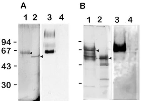
Figure 1 shows the apparentMrof the native and degly-cosylated GMPs. The molecular mass shift resulting from N-deglycosylation was between 10 and 15 kDa, indicating the presence of small N-glycans. N-deglycosylation was always as-sociated with the loss of the reactivity with the anti-GM MAb. Structural characterization of N-glycan of GMP1. (i) Native glycans released by PNGase F.GC analysis showed that the mixture of PNGase F-released oligosaccharides contained mannose, galactose, andN-acetylglucosamine in a molar ratio of 6.4:2.3:2.
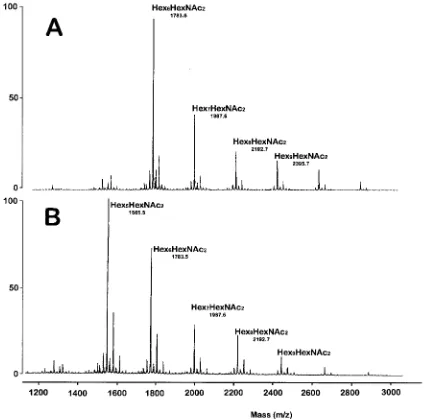
Data from MALD-MS of permethylated PNGase F-released glycans eluting in the 50% (vol/vol) aqueous acetonitrile frac-tion from a C18Sep-Pak cartridge are shown in Fig. 2A. The
data indicated that the glycoprotein contained glycans with a
Hex5-13HexNAc2 composition. The major molecular ion
ob-served corresponded to Hex6HexNAc2 (m/z 1,783). ELISAs
showed that the mixture of native PNGase-released oligosac-charides reacted positively with the anti-GM EB-A2 MAb (data not shown). These experiments indicated the presence of Galf residues in the mixture of N-glycans.
GC-MS analysis of partially methylated alditol acetates gen-erated from the permethylated glycans is shown in Table 1. Key features of these data are as follows. (i) 3,6-Man, 2,6-Man, 2-Man, 6-Man, and 4-GlcNAc are components of the N-glycan core; (ii) Man and Gal are the nonreducing sugars; (iii) the fragmentation pattern of terminal Gal is consistent with a terminally linked hexofuranose sugar; and (iv) no 4-Gal was detected, suggesting that no chains of galactofuran are present in the N-glycan.
HF treatment and ␣-mannosidase digestion of N-glycans released by PNGase F.GC-MS data showed that a 48-h treat-ment at 0°C with 48% HF removed all terminal Galf residues from the N-glycans. In addition, HF-treated N-glycans did not react with the anti-galactofuran MAb, even though a concen-tration of HF-treated N-glycans 100 times higher than un-treated N-glycans was tested by ELISA (data not shown). The MALD-MS spectrum of permethylated HF-treated N-glycans eluting in the 50% (vol/vol) aqueous acetonitrile fraction from a C18 Sep-Pak cartridge is shown in Fig. 2. The overall
MALD-MS pattern of the HF-treated glycan could be roughly superimposed on the native glycan if one hexose unit is re-moved (compare Fig. 2A and B). After HF treatment remov-ing Galf residues, the oligosaccharides had an average size of 6.2 hexose units (calculated as follows: number of hexose units of each oligosaccharide species⫻% of each oligosaccharide species/100), whereas the native oligosaccharide showed an average size of 7.1 hexose units. These data suggested the presence of one galactofuranosyl unit per average oligosaccha-ride. However, the molecular ion atm/z1,783 (Hex6HexNAc2)
was present in a higher concentration than the Hex7HexNAc2
before HF treatment, suggesting that another isomeric
struc-FIG. 1. Analysis of native (lanes 1 and 3) and deglycosylated (lanes 2 and 4) forms of GMP1 (A) and GMP2 (B) on a 10% SDS-PAGE gel stained with Coomassie blue (lanes 1 and 2) or transferred onto ni-trocellulose and revealed by Western blot assay with the EB-A2 anti-galactofuran MAb (lanes 3 and 4). Arrowheads indicate GMP1 (A) and GMP2 (B) before and after deglycosylation. The values on the left are molecular sizes in kilodaltons.
on September 8, 2020 by guest
http://ec.asm.org/
FIG. 2. MALD-MS spectra of permethylated N-glycans fromA.fumigatusGMP1. The N-glycans were released from tryptic glycopeptides by digestion with PNGase F, separated from peptides by Sep-Pak purification, and permethylated. The derivatized glycans were purified by Sep-Pak, and the 50% (vol/vol) aqueous acetonitrile fraction was screened by MALD-MS. (A) Intact N-glycans. (B) N-glycans treated with HF. Major ions are indicated.
TABLE 1. GC-MS analysis of partially methylated alditol acetates obtained from the PNGase F-released N-glycans of GMP1a
Retention
time (min) Characteristic fragment ions Assignment
Relative abundance
No treatment
After HF treatment
26.48 102, 118, 129, 145, 161, 162, 205 Terminal mannose 1.00 1.00
26.82 89, 102, 118, 162, 205, 278 Terminal galactose 0.31 ND
30.99 129, 130, 161, 190 2-Linked mannose 0.76 0.41
32.03 99, 102, 118, 129, 162, 189, 233 6-Linked mannose 0.17 0.14
36.10 129, 130, 189, 190 2,6-Linked mannose 0.28 0.23
36.62 118, 129, 189, 234 3,6-Linked mannose 0.25 0.24
41.25 117, 159, 233 4-Linked GlcNAc 0.48 0.52
aThe 50% acetonitrile fractions from Sep-Pak purifications of permethylated glycans were hydrolyzed, reduced, acetylated, and analyzed by GC-MS. ND, not detected.
on September 8, 2020 by guest
http://ec.asm.org/
ture of Hex6HexNAc2without Galf residues was also present
in the original native mixture of N-glycans.
GC-MS data showed that after HF treatment, 2-linked Man was the only linked mannose residue which decreased signifi-cantly (Table 1), suggesting that terminal Galf residues were attached to the 2 position of Man prior to HF treatment. This result was in agreement with Western blot data showing the absence of reactivity of a culture filtrate ofP.brasiliensiswith EB-A2 (data not shown). This extract contained the major glycoprotein gp43 that was characterized by the presence of
-D-Galf linked in-1,6 to one of the mannosyl residues of the N-glycan mannan core (2).
When native glycan pools were subjected to␣-mannosidase digestion prior to HF treatment, the reactivity of the resulting N-glycans digested with ␣-mannosidase against the anti-GM MAb was identical to the reactivity of the native N-glycans, indicating that this exomannosidase treatment did not remove any Galf residues (data not shown). When the HF-treated N-glycans were digested with ␣-mannosidase, they were trimmed to HexHexNAc2(m/z763), Hex2HexNAc2(m/z967),
and Hex3HexNAc2(m/z1,171).
The chemical and immunochemical data obtained above showed that GMP1 ofA.fumigatuscontained a heterogeneous mixture of N-glycans (Hex5-13HexNAc2). The N-glycans
react-ing with the anti-GM MAb contained only one Galf residue at the terminal nonreducing position attached to the 2 position of the mannose residues.
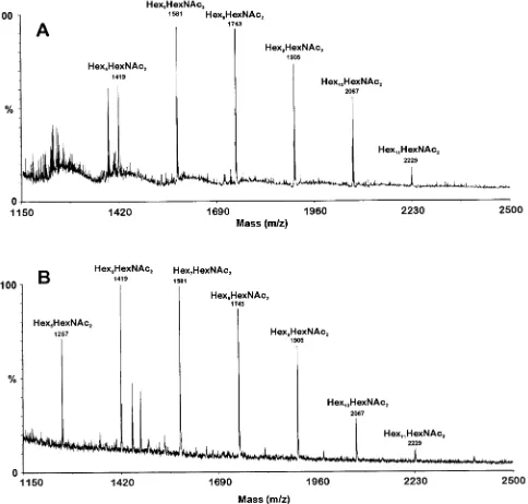
Structural characterization of N-glycan of GMP2.N-glycans of GMP2 were released by PNGase treatment as described for GMP1. ELISAs showed that the mixture of native PNGase-released oligosaccharides reacted positively with the anti-ga-lactofuran EB-A2 MAb (data not shown). These experiments indicated the presence of Galf residues in the mixture of N-glycans. The MALD-MS data indicated that the glycoprotein contained glycans with a Hex6-11HexNAc2 composition. The
major native molecular ions observed corresponded to Hex7HexNAc2(m/z1,581) and Hex8HexNAc2(m/z1,743),
in-dicating that GMP2 N-glycans were similar to GMP1 N-gly-cans in size (Fig. 3). After HF treatment removing Galf resi-dues, the oligosaccharides had an average size of 7.2 hexose units whereas the native oligosaccharides showed an average size of 8.0 hexose units. These data suggested the presence of one galactofuranosyl unit per average oligosaccharide. GC-MS data showed that the Galf was only present at the nonreducing end of the mannan chains since only terminal Galf units were seen and no internal Galf side chains were identified (data not shown). GC-MS data also confirmed the conclusions obtained with GMP1 since the 2-Man concentration diminished after HF treatment of the native oligosaccharides (data not shown). Characterization of GMPs. (i) GMP1.The internal peptide sequence (NDPDHAYGNNIE) obtained from the purified GMP1 belongs to a protein encoded by an open reading frame (ORF) of 1,418 bp with an intron with a size of 50 bp in positions 562 to 612 (confirmed by sequencing the 270 bp obtained by PCR surrounding the intron with cDNA as a template). The ORF (70.m15708 in the TIGR database [http: //tigrblast.tigr.org//usr/local/db/euk/private/aspergillus/annotation _dbs/ASP.pep]) encoded a putative protein of 456 amino acids with five predicted N-glycosylation sites, a predicted molecular mass of 49.8 kDa, and a pI of 6.12. A stretch of hydrophobic
amino acids was seen at the amino terminus, and a putative signal peptide at the NH2terminus was predicted by the
pro-gram SignalP (http://www.cbs.dtu.dk/services/SignalP) and in agreement with the (⫺3⫺1) rule of Von Heijne (35). The putative cleavage site was between A18 and I19.
BLAST analysis of the deduced amino sequence of theA.
fumigatusprotein showed a high degree of similarity with
bac-terial PLCs (47% and 38% similarities with Mycobacterium
tuberculosisandPseudomonas aeruginosaPLCs) and a putative
PLC fromArabidopsis thaliana(37% similarity). No homolo-gies were seen with other fungal phospholipases present in the database, but BLAST against the TIGRA.fumigatusdatabase, however, showed the presence of two other homologous se-quences (clones 65.m07340 and 52.m03766 in the TIGR data-base).
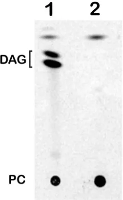
TLC analysis showed that upon incubation of the purified GMP1 with14C-labeled PC, diacylglycerol was released,
show-ing that GMP1 had PLC activity (Fig. 4). The enzymatic ca-pacity to degrade different PLC substrates (PC, PE, SM, and PI) was investigated. Under the experimental conditions tested, the most efficiently degraded substrate was SM. PE was not cleaved. At the same substrate concentration, the amounts of radioactivity released from PC and PI were, respectively, 71 and 31% of the amount released from SM. Using NPCC as a substrate, the enzyme had aKmvalue of 3.85⫾0.7 mM, with a specific activity for the batch analyzed of 30⫾8 pmol/min/
g.
(ii) GMP2.An internal peptide sequence (KALARSVVPF IRASGS) obtained from the 70-kDa GMP2 protein matched PhyAp, a secreted phytase fromA.fumigatus(16, 21, 24, 34). The mature PHYA-encoded enzyme consisted of 439 amino acids with a theoreticalMrof 48,270 (GenBank accession no.
U59804 [58.m07761 in the TIGR database]). The protein pos-sessed seven putative N-glycosylation sites. It had high homol-ogies with other phytases or acid phosphatases: 66%, 61%, and 48% identity to phytases ofA.niger,A.terreus, and
Mycelioph-thora thermophila, respectively (21). In the genome ofA.
fu-migatus, it belong to a cluster of three phytase genes, including
the ORFs 52.m04104 and 66.m04644, and it also had homol-ogies with two acid phosphatase genes, 69.m15283 and 56.m02370. The phytase activity of the GMP2 isolated from the culture filtrate was shown as described in Materials and Meth-ods by the release of phosphate from phytic acid. The enzy-matic activity was not further analyzed since this activity has been extensively studied previously (6, 21, 38). Using phytic acid as a substrate, the batch of enzyme analyzed had a specific activity of 100⫾50 pmol/min/g.
Antigenicity of the GMPs.The two GMPs were recognized in Western blot assays by sera from patients with aspergilloma. After N-deglycosylation, they were no longer labeled with pa-tient antibodies, indicating that the immunogenic moiety of these GMPs was the N-glycan whereas the peptide moiety of these two glycoproteins was not antigenic (Fig. 5A). ELISA data showed that the GMPs were differentially recognized by patient and control sera (P⬍0.01 for both antigens and a df of 23) (Fig. 5B). In addition, a significant linear fit was seen between the OD values obtained with the different antigens and GM in ELISA (data not shown); this result confirmed the exclusive antigenicity of the N-glycans of the phytase and phos-pholipase molecules.
on September 8, 2020 by guest
http://ec.asm.org/
DISCUSSION
Galf-containing molecules have been repeatedly shown to be important antigens among human fungal pathogens. This is true forAspergillusspecies but also inP. brasiliensis(2),
Spo-rothrix schenckii, and dermatophytes (17, 33). The major
exo-cellular antigens ofP.brasiliensisused to detect the presence of antibodies in patients with paracoccidioidomycosis is a 43-kDa glycoprotein which contains an N-linked oligosaccharide ter-minated by a-D-galactofuranosyl residue 136 linked to the mannosyl residues. However, in contrast to A. fumigatus
GMPs, the antigenicity of gp43 was due to the peptide moiety rather than to the Galf-containing N-glycan.
Galf is not present in the human host, and galactofuran determinants could play a role in the host immune reactions duringAspergillusinfections. Antibodies directed against the galactofuran moiety of the GM have already been identified in immunocompetent patients infected withA. fumigatus(15; J. Sarfati et al., unpublished data). In addition, molecules bearing Galf could help the host to recognize the fungus as non-self and induce cytokine synthesis to activate cellular immunity. This issue has not been investigated yet. Molecules recognized
FIG. 3. MALD-MS spectra of N-glycans fromA.fumigatusGMP2. The N-glycans were released from tryptic glycopeptides by digestion with PNGase F, separated from peptides by Sep-Pak purification, and analyzed without derivatization. (A) Intact N-glycans. (B) N-glycans treated with HF. Major ions are indicated.
on September 8, 2020 by guest
http://ec.asm.org/
by the EB-A2 MAb circulate in the biological fluids of patients with invasive aspergillosis. Indeed, serological diagnosis of this life-threatening fungal infection remains based on the detec-tion of GM in the biological fluids of infected patients (18, 29, 31). Initially, it was thought that only polysaccharides were released and circulated during growth in the host. The findings described here show that glycoproteins could also circulate during infection and that the so-called “circulating antigen” is not a single molecule but a family of molecules for which expression could be modulated by the immediate fungal envi-ronment.
The characterization of the N-glycan of the A. fumigatus
GMPs is in agreement with the general composition of
oligo-saccharides ofAspergillus glycoproteins (19). (i) An average oligosaccharide size of Hex5-10GlcNAc2 has been reported.
Large mannan chains (Hex50-200) present in Saccharomyces
cerevisiaeand other yeasts have never been identified in
fila-mentous fungi. (ii) Galf has been shown in severalAspergillus
glycoproteins such as the␣-glucosidase and␣-galactosidase of
A. niger or the-galactosidase of A. oryzae (32, 36, 37). (ii)
When present, Galf is at the nonreducing terminal position. (iv) N-glycan with or without Galf has also been shown to be present simultaneously on the same glycoprotein. Galactose residues found at the nonreducing end of N-glycans can serve as a stop signal for further mannose addition, similar to the role proposed for␣-1,3 mannose inS.cerevisiae(8).
The structure of the N-glycan of the PLC of A. fumigatus
looked similar to the N-glycan of␣-galactosidase A fromA.
niger. The mannan oligosaccharides of the N-glycan of the
␣-galactosidase ofA.nigerwere of a slightly higher molecular weight, with most abundant components being Hex10-15
Hex-NAc2whereas they were Hex6-8HexNAc2inA.fumigatusPLC
and phytase, respectively. The chemical organization of the N-glycan has not been precisely determined since N-glycans of
A.fumigatusandA.nigerwere analyzed as a mixture due to the
limited amounts of purified protein. Data obtained with dif-ferent GMP batches have shown that the oligosaccharide mix-tures obtained are often heterogeneous. For example, treat-ment of native N-glycans from GMP1 with ␣-mannosidase often identified a population of N-glycans without Galf resi-dues that were trimmed to HexHexNAc2 (m/z 763),
Hex2HexNAc2 (m/z 967), and Hex3HexNAc2 (m/z 1,171) by
the ␣-mannosidase treatment. Other batches contained two Galf units that were in the terminal nonreducing position, as shown by GC-MS analysis (data not shown). These variations could be due to the action of a galactofuranosidase that could trim the terminal reducing galactose of the exocellular pro-teins. Exogalactofuranosidases were shown to be secreted by variousAspergillusspecies (23, 36). Studies with bothA.niger
and A. fumigatushave shown that N-glycans from these two
species shared a common essential features: Galf is present as single residues linked to C-2 of the nonreducing terminal man-nose residues of N-glycan. The structure of the Galf-containing N-glycans ofA.fumigatusis shown in Fig. 6.
The recognition of N-glycans of theseAspergillus glycopro-teins by MAb EB-A2 questioned the previously established identity of the epitope recognized by this MAb. Using chemi-cally synthesized galactofurans of different sizes, we showed
FIG. 4. TLC analysis of products released by hydrolysis of PC with GMP1 PLC activity (lane 1). Incubation with GMP1 negative control (heat-inactivated GMP1), lane 2. DAG, diacylglycerol.
FIG. 5. (A) Western blot assays showing the lack of reactivity of patient sera with the deglycosylated forms of phytase (lanes 2) and PLC (lane 4). Respective native proteins are shown in adjacent lanes 1 and 3. (B) OD values obtained by ELISA with native forms of phytase and PLC applied to microtiter plates and incubated with sera from aspergilloma (Asp) and control sera. The values on the left of panel A are molecular sizes in kilodaltons.
FIG. 6. Putative structure of the N-glycans of the GMPs. In the core, the presence or absence of a mannose residue is indicated by parentheses. Side chains of different sizes, with or without a galactose residue at the nonreducing end, are shown on the left and can be linked to any of the mannose residues marked with an asterisk.
on September 8, 2020 by guest
http://ec.asm.org/
that the best-recognized oligosaccharide was a tetra- -1,5-ga-lactofuran (30). The absence of -1,5-galactofuran chains in the N-glycans ofAspergillusGMPs suggests that an oligoman-nan substituted with a Galf on its terminal nonreducing end can be also recognized by this MAb. Although the Galf-Man epitope from the GMP N-glycan has not been identified yet, our data showed that the linkage between the Galf and the oligomannan is critical: a 1,2 linkage allows IgM binding, whereas a 1,6 linkage, as in gp43 of P. brasiliensis, prevents antigen recognition.
Besides being a main component of members of fungal gen-era such asAspergillusandPenicillium, Galf is also present in the cell walls of mycobacteria, in the lipopolysaccharide O antigens of a variety of gram-negative bacteria, and in cell envelope components of eukaryotic parasites. The broad dis-tribution of Galf in critical structures of pathogenic microor-ganisms and its absence in higher eukaryotes make the biosyn-thetic pathway of Galf an attractive target for the development of new antimicrobial drugs. The first enzyme involved in this biosynthetic pathway has been well characterized in the genus
Mycobacterium. It is a UDP-galactopyranose mutase that
con-verts UDP-galactopyranose to UDP-Galf (25). This enzyme has not been identified inA. fumigatus, and no homologous sequence was found in the last release of the A. fumigatus
TIGR database. The first step of the Galf pathway inA.
fu-migatusremains unknown, as well as the enzyme(s) responsible
for the elongation of the galactofuran side chains or the trans-ferases adding the Galf at the terminal nonreducing end of the mannan of the N-glycan.
A third GMP was isolated from the culture filtrate of A.
fumigatus(GMP3) (data not shown). GMP3 was eluted with
GMP2 during the first step of purification and further purified on the cation-exchange Mono S column at pH 6.0 at 250 mM NaCl. Although its N-glycan could not be analyzed due to the very small amount of protein purified, this 85-kDa protein was a GMP since de-N-glycosylation abolished the reactivity of GMP3 with the anti-galactofuran MAb. Two internal peptide sequences (FPVLGHQMTHSMD and ALGQLDDTLII VTA) obtained from this protein matched the singleton 59.m08496 of the TIGR database. This protein had high ho-mologies with alkaline phosphatases (62% with alkaline phos-phatase ofNeurospora crassa). It possessed seven putative sites of N-glycosylation and an N-glycan of 12 kDa (estimated by SDS-PAGE after de-N-glycosylation). The alkaline phospha-tase activity was confirmed usingp-nitrophenylphosphate as a substrate in a 100 mM Tris-HCl (pH 8.0) buffer (10 ng protein was incubated for 30 min at 37°C with 200g PNPP, and the assay was stopped by addition of 3 volumes of 6% Na2CO3)
(data not shown).
The three GMPs ofA.fumigatusstudied here are secreted enzymes that can be an essential part of a phosphate-scaveng-ing system. For example, the main function of the PLC ofA.
fumigatuscould be to retrieve phosphate from phospholipids.
Such a role has been shown for theP.aeruginosaPLC that is regulated at the transcriptional level by Pi. InA.fumigatus, the PLC is also submitted to phosphate repression: it is absent in two defined media containing a high concentration of phos-phate (73 mM) and repressed in a Sabouraud medium supple-mented with 75 mM phosphate (data not shown). The other two enzymes, alkaline phosphatase and phytase, are directly
responsible for phosphate retrieval from the external medium.
A. fumigatus growth is highly dependent on free phosphate
availability to grow: a concentration of around 1 mM phos-phate is found in serum, andA.fumigatusrequires 10 times the amount available in human biological fluids for optimal growth (unpublished data). The results presented here and the iden-tification of an acid phosphatase as the major cell wall protein (5) suggest that phosphate availability and scavenging may be a significant determinant ofA. fumigatusgrowth in the host. Strategies to interfere with such pathways could lead to useful therapies.
REFERENCES
1.Albersheim, P., D. J. Nevins, P. D. English, and A. Karr.1967. A method for the analysis of sugars in plant cell-wall polysaccharides by gas-liquid chro-matography. Carbohydr. Res.5:340–345.
2.Almeida, I. C., D. C. Neville, A. Mehlert, A. Treumann, M. A. Ferguson, J. O. Previato, and L. R. Travassos.1996. Structure of the N-linked oligosaccha-ride of the main diagnostic antigen of the pathogenic fungusParacoccidioides brasiliensis. Glycobiology6:507–515.
3.Ames, B. N.1966. Assay of inorganic phosphate and phosphatases. Methods Enzymol.8:115–118.
4.Beauvais, A., M. Monod, J. P. Debeaupuis, M. Diaquin, H. Kobayashi, and J.-P. Latge´.1997. Biochemical and antigenic characterization of a new dipep-tidyl-peptidase isolated fromAspergillus fumigatus. J. Biol. Chem.272:6238– 6244.
5.Bernard, M., I. Mouyna, G. Dubreucq, J.-P. Debeaupuis, T. Fontaine, C. Vorgias, C. Fuglsang, and J.-P. Latge´.2002. Characterization of a cell-wall acid phosphatase (PhoAp) inAspergillus fumigatus. Microbiology148:2819– 2829.
6.Brugger, R., C. Simoes Nunes, D. Hug, K. Vogel, P. Guggenbuhl, F. Mas-carello, S. Augem, M. Wyss, A. P. van Loon, and L. Pasamontes.2004. Characteristics of fungal phytases fromAspergillus fumigatusandSartorya fumigata. Appl. Microbiol. Biotechnol.63:383–389.
7.Ciucanu, I., and F. Kerek.1984. A simple and rapid method for the perm-ethylation of carbohydrates. Carbohydr. Res.131:201–217.
8.Dean, N.1999. Asparagine-linked glycosylation in the yeast Golgi. Biochem. Biophys.1426:309–322.
9.Dell, A., A. J. Reason, K. H. Khoo, M. Panico, R. A. McDowell, and H. R. Morris.1994. Mass spectrometry of carbohydrate-containing biopolymers. Methods Enzymol.230:108–132.
10.Fontaine, T., C. Simenel, G. Dubreucq, O. Adam, M. Delepierre, J. Lemoine, C. E. Vorgias, M. Diaquin, and J.-P. Latge´.2000. Molecular organization of the alkali-insoluble fraction ofAspergillus fumigatuscell wall. J. Biol. Chem.
275:27594–27607.
11.Haido, R. M. T., M. H. Silva, R. Ejzemberg, E. A. Leitao, V. M. Hearn, G. V. Evans, and E. Barreto Bergter.1998. Analysis of peptidogalactomannans from the mycelial surface ofAspergillus fumigatus. Med. Mycol.36:313–321. 12.Johansen, K. A., R. E. Gill, and M. L. Vasil.1996. Biochemical and molec-ular analysis of phospholipase C and phospholipase D activity in mycobac-teria. Infect. Immun.64:3259–3266.
13.Kurioka, S., and M. Matsuda.1976. Phospholipase C assay usingp -nitro-phenylphosphorylcholine together with sorbitol and its application to study-ing the metal and detergent requirement of the enzyme. Anal. Biochem.
75:281–289.
14.Laemmli, U. K.1970. Cleavage of structural proteins during the assembly of the head of bacteriophage T4. Nature227:680–685.
15.Latge´, J.-P., H. Kobayashi, J.-P. Debeaupuis, M. Diaquin, J. Sarfati, J. M. Wieruszeski, E. Parra, and B. Fournet.1994. Chemical and immunological characterization of the galactomannan secreted by Aspergillus fumigatus. Infect. Immun.62:5424–5433.
16.Liu, Q., Q. Huang, X. G. Lei, and Q. Hao.2004. Crystallographic snapshots ofAspergillus fumigatusphytase, revealing its enzymatic dynamics. Structure
12:1575–1583.
17.Lloyd, K. O.1970. Isolation, characterization and partial structure of peptido galactomannans from the yeast form of Cladosporium wernechii. Biochem-istry9:3446–3453.
18.Maertens, J., J. Verhaegen, H. Demuynck, P. Brock, G. Verhoef, P. Vanden-berghe, J. Van Eldere, L. Verbist, and M. Boogaerts.1999. Autopsy-con-trolled prospective evaluation of serial screening for circulating galactoman-nan by a sandwich enzyme-liked immunosorbent assay for hematological patients at risk for invasive aspergillosis. J. Clin. Microbiol.37:3223–3228. 19.Maras, M., I. van Die, R. Contreras, and C. A. van den Hondel.1999.
Filamentous fungi as production organisms for glycoproteins of bio-medical interest. Glycoconj. J.16:99–107.
20.McConville, M. J., S. W. Homans, J. E. Thomas-Oates, A. Dell, and A. Bacic.
1990. Structures of the glycoinositolphospholipids from Leishmania major. A
on September 8, 2020 by guest
http://ec.asm.org/
family of novel galactofuranose-containing glycolipids. J. Biol. Chem.265:
7385–7394.
21.Pasamontes, L., M. Haiker, M. Wyss, M. Tessier, and A. P. van Loon.1997. Gene cloning, purification, and characterization of a heat-stable phytase from the fungusAspergillus fumigatus. Appl. Environ. Microbiol.63:1696– 1700.
22.Preuss, I., I. Kaiser, and U. Gehring.2001. Molecular characterization of a phosphatidylcholine-hydrolyzing phospholipase C. Eur. J. Biochem.
268:5081–5091.
23.Rietschel-Berst, M., N. H. Jentoft, P. D. Rick, C. Pletcher, F. Fang, and J. E. Gander.1977. Extracellular exo--D-galactofuranosidase from Penicillium
charlesii. J. Biol. Chem.252:3219–3226.
24.Rodriguez, E., E. J. Mullaney, and X. G. Lei. 2000. Expression of the
Aspergillus fumigatusphytase gene in Pichia pastoris and characterization of the recombinant enzyme. Biochem. Biophys. Res. Commun.268:373–378. 25.Sanders, D. A., A. G. Staines, S. A. McMahon, M. R. McNeil, C. Whitfield,
and J. H. Naismith.2001. UDP-galactopyranose mutase has a novel struc-ture and mechanism. Nat. Struct. Biol.8:858–863.
26.Sarfati, J., D. Boucias, and J.-P. Latge´.1995. Antigens ofAspergillus fumiga-tusproducedin vivo. J. Med. Vet. Mycol.33:9–14.
27.Sawardeker, J. S., J. H. Sloneker, and A. Jeanes.1965. Quantitative deter-mination of monosaccharides as their alditol acetates by gas liquid chroma-tography. Anal. Biochem.37:1602–1604.
28.Schneider, P., and M. A. Ferguson. 1995. Microscale analysis of glyco-sylphosphatidylinositol structures. Methods Enzymol.250:614–630. 29.Stynen, D., A. Goris, J. Sarfati, and J.-P. Latge´.1995. A new sensitive
sandwich enzyme-linked immunosorbent assay to detect galactofuran in pa-tients with invasive aspergillosis. J. Clin. Microbiol.33:497–500.
30.Stynen, D., J. Sarfati, A. Goris, M. C. Pre´vost, M. Lesourd, H. Kamphuis, V. Darras, and J.-P. Latge´.1992. Rat monoclonal antibodies against galacto-mannan. Infect. Immun.60:2237–2245.
31.Sulahian, A., F. Boutboul, P. Ribaud, T. Leblanc, C. Lacroix, and F. Der-ouin.2001. Value of antigen detection using an enzyme immunoassay in the diagnosis and prediction of invasive aspergillosis in two adult and pediatric hematology units during a 4-year prospective study. Cancer91:311–318. 32.Takayanagi, T., A. Kimura, S. Chiba, and K. Ajisaka.1994. Novel structures
of N-linked high-mannose type oligosaccharides containing ␣-D -galacto-furanosyl linkages inAspergillus niger␣-D-glucosidase. Carbohydr. Res.256:
149–158.
33.Travassos, L. R.1985.Sporothrix schenckii, p. 121–156.InP. J. Szaniszlo and J. L. Harris (ed.), Fungal dimorphism, with emphasis on fungi pathogenic for humans. Plenum Press, New York, N.Y.
34.Ullah, A. H., K. Sethumadhavan, X. G. Lei, and E. J. Mullaney.2000. Biochemical characterization of cloned Aspergillus fumigatus phytase (phyA). Biochem. Biophys. Res. Commun.275:279–285.
35.Von Heijne, G.1986. A new method for predicting signal sequence cleavage sites. Nucleic Acids Res.14:4683–4690.
36.Wallis, G. L., R. L. Easton, K. Jolly, F. W. Hemming, and J. F. Peberdy.2001. Galactofuranoic-oligomannose N-linked glycans of␣-galactosidase A from
Aspergillus niger. Eur. J. Biochem.268:4134–4143.
37.Wallis, G. L., F. W. Hemming, and J. F. Peberdy.2001. An extracellular
-galactofuranosidase fromAspergillus nigerand its use as a tool for glyco-conjugate analysis. Biochim. Biophys. Acta1525:19–28.
38.Wyss, M., L. Pasamontes, A. Friedlein, R. Remy, M. Tessier, A. Kronen-berger, A. Middendorf, M. Lehmann, L. Schnoebelen, U. RothlisKronen-berger, E. Kusznir, G. Wahl, F. Muller, H. W. Lahm, K. Vogel, and A. P. van Loon.
1999. Biophysical characterization of fungal phytases (myo-inositol hexakisphosphate phosphohydrolases): molecular size, glycosylation pattern, and engineering of proteolytic resistance. Appl. Environ. Microbiol.65:359– 366.