Iran J Public Health, Vol. 48, No.9, Sep 2019, pp.1723-1731
Original Article
Differential Expression Pattern of Epithelial Mesenchymal
Transition Gens: AXL, GAS6, Claudin-1, and Cofilin-1, in
Different Stages of Epithelial Ovarian Cancer
Elham HASSANI
1, Mahmood SHEKARI KHANIANI
1, Mojtaba SAFFARI
2, Amirnader
EMAMI RAZAVI
3, *Reza SHIRKOOHI
2, *Sima MANSOORI
DERAKHSHAN
1,41. Department of Medical Genetics, Faculty of Medicine, Tabriz University of Medical Sciences, Tabriz, Iran 2. Cancer Biology Research Center, Cancer Institute of Iran, Tehran University of Medical Sciences, Tehran, Iran
3. Iran National Tumor Bank, Cancer Institute of Iran, Tehran University of Medical Sciences, Tehran, Iran 4. Immunology Research Center, Tabriz University of Medical Sciences, Tabriz, Iran
*Corresponding Authors: Email: [email protected]
(Received 04 Jul 2018; accepted 11 Aug 2018)
Introduction
Ovarian cancer is fifth reason in cancer depend-ent diseases among females (1). Epithelial ovarian cancer (EOC), correlated with 90% of all ovarian cancers, is the most prevalent and fatal form of
gynecological cancer in developed countries (2). In Iran, ovarian cancer is the 8th prevalent cancer type (3). The usual therapy for ovarian cancer contains reduction of cells after platinum chemo-Abstract
Background: Epithelial ovarian cancer (EOC), is the fatal form of gynecological cancer. Almost 70% of ovari-an covari-ancer patients are detected at ovari-an advovari-anced stage (III-IV) with metastases. Epithelial‑mesenchymal transi-tion (EMT) is a critical process associated with metastasis. This study investigated the expression levels of
AXL, GAS6, Claudin-1, and Cofilin-1, as genes involved in EMT in relation to clinicopathologic features in
ovarian cancer patients.
Methods: In this descriptive study, 78 ovarian epithelial cancer patients were enrolled. Samples were provided by the Iran National Tumor Bank, founded by the Cancer Institute of Tehran University of Medical Sciences in 2017. The expression levels of AXL, GAS6, Claudin-1, and Cofilin-1 genes were investigated in a fresh, frozen tumor sample and normal adjacent tissue by real-time PCR (RT-PCR).
Results: Findings showed a significant relationship between the overexpression of AXL and TNM staging (P=0.03). The expression level of GAS6 decreased in more advanced stages (P=0.01). There is a negative rela-tionship between Cofilin-1 expression level and TNM staging (P=0.002). Claudin-1 expression level was higher in low stages compared with that in high stages (P=0.01). There was no relationship between gene expression levels of target genes with size and grade of the tumor.
Conclusion: Given the importance of these genes in EMT, alteration in their expression pattern can contrib-ute to the progression of the disease and distant metastasis of cancer cells. Additionally, knowing the alteration pattern of these genes expression can help to better understanding and prediction of the prognosis of EOC.
therapy. Despite an initial response to the treat-ment, due to the presence of chemotherapy-resistant residual tumor cells, many patients will be finally affected by metastasis and eventually will die (4). Since ovarian cancer develops with-out any apparent symptoms (5), almost 70% of ovarian cancer patients are detected at an ad-vanced stage (III-IV) with metastases, of which only 45% survive five years after initial diagnosis (6). Probable reasons for this poor prognosis in-clude lack of screening instruments for early-stage diagnosis, the nonspecific symptoms, and drug resistance in advanced disease. Metastasis in cancer is the most challenging problem in clinical issue (7). Metastasis in cancer is the cause of 90% of all cancer-associated die, since the biologic and physical factors that define the final form and heterogenicity of tumors with metastasis are not yet well understood (8). In fact, 90% of all ovari-an covari-ancers are EOC that originates from the ovarian surface epithelium (OSE). The transfor-mation of epithelial cells towards motile mesen-chymal state is a pivotal process associated with metastasis named epithelial-to-mesenchymal transition (EMT) (9).
There are three different forms of EMT; Type 3 happen in cancer (metastasis) (10). In cancerous epithelial cells, cell junctions and apical-basal po-larity are disappeared, cell cytoskeleton is rear-ranged, signaling programs that establish cell shape and alter gene expression program are changed, during EMT. These alterations result in enhanced cell movement and gain of invasive manifestation by cancer cells (11). Thus, attempts to identify molecular targets regulating ovarian metastasis are critical for early detection and treatment of EOC (12). With respect to the steps of EMT, which are essential for metastasis, the researchers of the present study studied a number of genes reported to have a role in EMT in dif-ferent cancers, such as Claudin-1, Cofilin-1, AXL, and GAS6.
Tight Junctions have vital roles in controlling paracellular transport and in keeping cell polarity (13). Claudins are a member of cellular binding molecules that are ingredients of tight junctions, which have crucial roles in cell polarity and cell
transport (14). The Claudins are a group of over 20 proteins that expression is tissue-specific (15). Different pieces of findings offer that Claudin-1 is a crucial ingredient of TJs and is directly have a role in the fence task of TJs. Claudin-1 is located at 3q28-q29 (16). As is known, destroy of the cell-cell junction is an important phase in EMT; in this regard, claudin-1 has capability to directly induce EMT via its interaction with specified EMT-associated transcription factors and signal-ing pathways (17).
Numerous pieces of evidence suggest the im-portant function of the cytoskeleton in the EMT (9). Actin cytoskeleton and its interaction proteins are remodeled and are the motive power for cell migrate and invasiveness of malignant cancer cells (18). Cofilin (CFL), is an F-actin-binding protein disassembled actin filaments and is necessary for reorganize cell cytoskeleton and the formation of filopodia, controls cell migration. (19). Cofilin-1 is located at 11q13. High expression of Cofilin has linked with the invasiveness of different cancer cells (20), such as breast cancer, human prostate cancer (19), and malignant astrocytoma (20). AXL is one of a Receptor tyrosine kinase family member and GAS6 has a high affinity for AXL (12). The AXL gene located on chromosome 19q13.2 (21). GAS6 is located at 13q34. AXL signaling promotes cellular invasion, migration, proliferation, and survival (12). AXL expression was associated with EMT (21). In metastatic ovarian tumor cells, AXL expression is dramati-cally increased and accumulating of these cells to form colony is noticeably depend on the GAS6/AXL signaling pathway (12).
as well as the relationship within the expression rate of four target genes by statistical tests.
Materials and Methods
The biological materials were provided by the Iran National Tumor Bank, founded by the Can-cer Institute of Tehran University of Medical Sci-ences, for cancer research. Fresh frozen tumor and normal adjacent tissue of seventy-eight pa-tients with epithelial ovarian cancer who under-went surgery at the Cancer Institute of Iran were examined in this study. All samples were trans-ferred from Tumor bank and stored at -80 ºC for further investigations. Sample data including pa-tient history, histology, clinical, and paraclinical data were recorded.
All participants had assigned written informed con-sent. This descriptive study was approved by Ethics Committee of Tabriz University of Medical Science with TBZMED.REC.1394.1143 Code of ethics. Total RNA was extracted from homogenized 50µg of tumors and normal tissues, using tripure reagent (Roche, Mannheim, Germany) according to the manufacturer’s instructions. Quantity and quality of prepared RNA were examined by Nanodrop spectrophotometer ND-1000 UV-Vis (Nano-Drop Technology) and running the RNA
in agarose gel. The cDNA was synthesized from 1ug of total RNA, using TaKaRa cDNA synthe-sis kit (TaKaRa, Japan) with a random hexamer as a reaction primer in a final volume of 20 ul following manufacturer’s protocol. The cDNA quality was confirmed by the amplification of housekeeping gene glyceraldehyde3-phosphate dehydrogenase (GAPDH).
Quantitative determination of mRNA levels of target genes was performed in duplicate using SYBR Green I Premix (Takara, Japan) and 1ul of cDNA according to the manufacturer’s instruc-tion in 40 cycles of amplificainstruc-tion. The reacinstruc-tion conditions were 10 min of denaturation and en-zyme activation at 95 °C followed by denatura-tion at 95 °C for 10 sec, annealing at 55 °C for 30 sec, and extension and florescence acquiring at 72 °C for 15 sec. The Rotor-Gene TM 6000 ma-chine (Corbett Life ScienceTM, Germany) was used for this purpose. GAPDH was used as an internal control to normalize the target genes. CT was determined and then dCT was calculated by normalizing with the housekeeping gene. The used Oligonucleotide primers are listed in Table 1 Expression of four target genes (i.e., AXL, GAS6, Claudin-1, and Cofilin-1) was investigated in all tissues by using quantitative real-time re-verse transcription PCR (qRT-PCR).
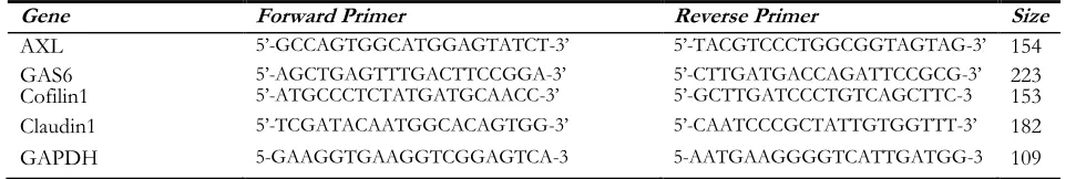
Table 1: Primer’s sequences
Gene Forward Primer Reverse Primer Size
AXL 5’-GCCAGTGGCATGGAGTATCT-3’ 5’-TACGTCCCTGGCGGTAGTAG-3’ 154
GAS6 5’-AGCTGAGTTTGACTTCCGGA-3’ 5’-CTTGATGACCAGATTCCGCG-3’ 223
Cofilin1 5’-ATGCCCTCTATGATGCAACC-3’ 5’-GCTTGATCCCTGTCAGCTTC-3 153
Claudin1 5’-TCGATACAATGGCACAGTGG-3’ 5’-CAATCCCGCTATTGTGGTTT-3’ 182
GAPDH 5-GAAGGTGAAGGTCGGAGTCA-3 5-AATGAAGGGGTCATTGATGG-3 109
GAPDH was used to normalize the gene expres-sion. Then, the correlation with the expression level of target genes and Clinicopathologic Fea-tures (i.e., stage, grade, age, size) of ovarian can-cer patients was investigated.
Statistical procedures
SPSS software ver. 16 (Chicago, IL, USA) was used for statistical evaluation. The qRT-PCR data analyzed normality via the Kolmogorov–Smirnov
method which failed (P<0.05); therefore, nonpar-ametric methods (Mann–Whitney test, Kruskal– Wallis, and Spearman’s rank correlation) were used for statistical analyses. P-value <0.05 were considered as significant.
Results
clinicopatho-logic feature. According to the International Fed-eration of Gynecology and Obstetrics (FIGO) classification, ovarian cancer has divided into
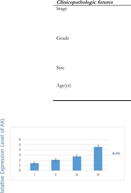
four stages (i.e., I/II/III/IV). Results showed a significant correlation between the overexpres-sion of AXL (P=0.03) and TNM staging (Fig. 1).
Table 2: Number of patient in different considered clinicopathologic feature
Clinicopathologic futures Number of patients
Stage
I 23
II 21
III 21
Iv 13
Grade
I 20
II 20
III 21
IV 17
Size
≤7 10
>7 68
Age(yr)
≤50 44
>50 34
Fig. 1: Expression of AXL in different stages
The differences between the stages I/III (P=0.01) and I/IV (P=0.03) were statistically sig-nificant. Figure 2 indicates the correlation be-tween the expression level of GAS6 and TNM staging of disease. The expression level of GAS6 decreased in more advanced stages (P=0.01).
Fig. 2: Expression of GAS6 in different stages
Fig. 3: Expression of Cofilin1 in different stages
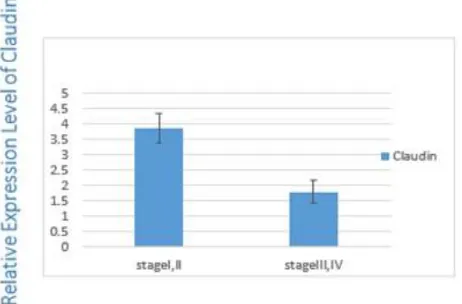
This correlation is significant between the stage I/III (0.002), I/IV (0.001) and II/IV (0.03). There was a statistically significant difference be-tween the expression level of Claudin-1 and TNM staging (P=0.01) (Fig. 4). Claudin-1 expres-sion level was higher in low stages (stage I and II) compared with high stages (stage III and IV) (P=0.01) (Fig. 4).
Fig. 4: Claudin1 expression in low and high stage
There was no relationship between gene expres-sion levels of AXL (P=0.6), GAS6 (P=0.5), Cofil-in-1 (P=0.2) and ClaudCofil-in-1 (P=0.3) with grades of tumor.
Size of tumor was another considered clinico-pathologic feature classified into two groups of >7cm and 7cm. Expression levels of AXL and GAS6 were different in two major tumor size groups, although not statistically significant (P=0.7). Moreover, despite the overexpression of Cofilin-1 and Claudin-1 genes, the researchers did not find any significant relationship between tumor size and the expression of Cofilin-1 (P=0.2) or Claudin-1 (P=0.6).
In line with this finding, the age-specific rates of EOC, FSH, and LH/hCG enhance cell prolifera-tion in primary human OSE and ovarian carci-noma cell lines (22). In this study, patients’ age range was within 15-83. Since the median onset age of ovarian cancer in Iran is estimated to be around 49-50 yr (3), patients were categorized into two age groups of 50 and >50. There is no significant relationship between AXL (P=0.8), GAS6 (P=0.1), Claudin-1 (P=0.5), and Cofilin-1 (P=0.9) gene expression levels and age in this study.
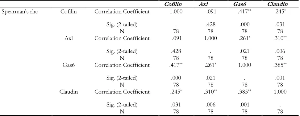
The correlation between target genes expression levels was evaluated in all patients. Cofilin1 and AXL have positive correlations with claudin1 and GAS6. And, GAS6 has positive correlations with AXL, Cofilin-1, and Claudin-1. Claudin-1 showed positive correlations with AXL, GAS6, and Cofil-in-1 (Table 3).
Discussion
In this study, the expression levels of GAS6, AXL, Cofilin-1, Claudin-1, as genes involved in EMT, and their relationship with clinicopatho-logical features include; age at diagnosis, TNM staging, size and grade of tumor were investigated in EOC patients.
Table 3: Correlations between target genes
Cofilin Axl Gas6 Claudin
Spearman's rho Cofilin Correlation Coefficient 1.000 -.091 .417** .245*
Sig. (2-tailed) . .428 .000 .031
N 78 78 78 78
Axl Correlation Coefficient -.091 1.000 .261* .310**
Sig. (2-tailed) .428 . .021 .006
N 78 78 78 78
Gas6 Correlation Coefficient .417** .261* 1.000 .385**
Sig. (2-tailed) .000 .021 . .001
N 78 78 78 78
Claudin Correlation Coefficient .245* .310** .385** 1.000
Sig. (2-tailed) .031 .006 .001 .
N 78 78 78 78
Women diagnosed at an early stage have a much higher survival rate than those diagnosed at a lat-er stage; unfortunately, only 15% of patients with ovarian cancer are diagnosed in early stages and metastasis in tumor is the main problem in EOC (24). Therefore, fined tumor markers in order to improve diagnosis methods is a great challenge in the treatment of EOC (25). A crucial phenome-non correlates with the metastasis is EMT (9). Induction of transcription factors by signaling pathways such as TGFβ and (BMP), Notch, Wnt –β-catenin, Hedgehog, and receptor tyrosine ki-nases, changes the gene expression to promote loss of cell-junctions, cause the change in cyto-skeleton and alteration from epithelial to the mesenchymal form (26). As the EMT associated genes are numerous, the researchers of the pre-sent study selected several up- regulated genes to demonstrate their potential applications as diag-nostic markers (5).
In this study, results indicate that high expression of Cofilin-1 in patient sample tissues correlates with decreasing stage of ovarian cancer. In de-tailed statistical analysis, the differences between stages I/III, I/IV, and II/IV were significant. In another word, the expression level of cofilin-1 in the ovarian tumor was elevated in low stages (I, II) compared to that in high stages (III, IV). The existing pieces of evidence suggest the cru-cial function of the cytoskeleton in EMT (9).
Ac-tin and associated proteins are changed and are necessary power to cell migration. The aggres-siveness in neoplastic cancer cells correlates with the change in the regulation in cell migration (18). The Cofilin action has a crucial role in actin polymerization and constitution of cell mem-brane prominence for cell migration. The up-regulation of Cofilin is correlated with the aggres-sive phenotype of several tumor cells (20). Im-portantly, cofilin-1 play a role in multidrug sistance in pancreatic cancer, and platinum re-sistance in human lung adenocarcinoma cell lines, ovarian cancer, and tumor biopsies (27). Cofilin-1 expression level was significantly increased in breast cancer samples at TNM stages T0, T1 and T2 (based on tumor size (T stage)). There was only a slight increase that was not statistically sig-nificant at TNM stage T3 (28). Up-regulation of Cofilin-1 expression causes the progression of ovarian cancer (18). Aberrant cofilin1 expression was involved in cell invasion.
Tight junctions have a significant role in keeping the cell polarity and also affect cellular transport (15). The claudins are necessary to the build-up tight junctions (TJs) in the epithelial and endo-thelial cell (13). Despite the role of claudins in the formation of mechanical cell adhesion at the site where epithelial and endothelial cells junc togeth-er, also, can recruit proteins involved in cell sig-naling. Thus Claudins are involved in the regula-tion of cell proliferaregula-tion, differentiaregula-tion, and con-sequent neoplastic transformation (29). Claudin-1 had a role in malignant progression of EMT (19). Moreover, had the potency to induction of EMT by interaction with signaling pathways and de-termined transcription factors (17). Tumorigene-sis may result from the Mislocalization of claudin proteins (30). Claudins have a role in cancer pro-gress via the interaction with different extracellu-lar matrix molecules. In cloning research, claudin-1 increase matrix metalloproteinase-2 (MMP-2) activities by contact with membrane-type matrix metalloproteinase-1 (MT1-MMPs). This could increase the invasion ability of cancer cells by the decay of circumambient extracellular matrix in-gredients, such as basement membrane, the clau-din-1 overexpression in OSCC (oral squamous cell carcinoma) is correlated with, advanced stage and grade of the tumor (31). Claudin-1 up-regulated in metastasis tissues of colon cancer, by Mislocalization to the cytoplasm and cell nucleus (30). Claudin-1 expression decreased in breast cancers, while its high expression was proved in thyroid, urothelial, gastric and cervical tumors (31). The simplest define suggest claudin1 ex-pression cause aggressiveness in a tissue-dependent behavior.
In this study, the AXL and GAS6 gene expression differences between tumor stages were statistically significant. GAS6 mRNA in ovarian tumor tissues was elevated more in stage I, II or low stages than stages III, IV. However, in the same tumor sample with overexpression of GAS6 (as a ligand), the ex-pression of AXL (as a receptor) in stage I, II was low-er than its expression in stages III, IV (high stages). In the normal state, expression of Axl is negative-ly affected by GAS6. Although, under hypoxic condition, like tumors, hypoxia inhibits
down-regulation of AXL by GAS6, consequently, tu-mor progression toward EMT may occur in can-cers (7). AXL signaling activates with GAS6 in autocrine or paracrine form. Stress state in the tumor environment has a crucial role in the in-duction of GAS6/AXL signaling (32). In cells with prostate cancer, Axl expression in a hypoxic state not affected by the GAS6 so, it may be available for induction of an EMT-like condition that drives progression of metastatic condition (7). GAS6 is overexpressed in ovarian tumors. Moreover, GAS6 is overexpressed in glioblasto-ma and gastric cancers (25). Metastatic ovarian tumor cells are critically associated with the GAS6/AXL signaling pathway for metastatic colonize (12). Axl is overexpressed in pancreatic adenocarcinoma in stage II samples (7). The find-ings of the present research confirm the results of the recent studies about overexpression of GAS6 and AXL in tumor tissues.
Conclusion
The present study indicates the up-regulation of AXL and down-regulation of GAS6, Coflin-1, and Claudin-1 in the more advanced stages of EOC. Given the importance of these genes in EMT process, alteration in these gene’s expres-sion levels during the progresexpres-sion of disease may contribute to invasive behavior of cancer cells and distant metastasis of the tumor. Additionally, knowing the alteration pattern of these genes ex-pression can help to better understanding and prediction of the prognosis of EOC.
Ethical considerations
Ethical issues (Including plagiarism, informed consent, misconduct, data fabrication and/or fal-sification, double publication and/or submission, redundancy, etc.) have been completely observed by the authors.
Acknowledgements
Conflict of interest
The authors declare that there is no conflict of interests.
References
1. Tiwari A, Hadley JA, Hendricks GL et al (2013). Characterization of ascites-derived ovarian tumor cells from spontaneously occurring ovarian tumors of the chicken: evidence for E-cadherin upregulation. PLoS One, 8:e57582. 2. Lau MT, So WK, Leung PC (2013). Fibroblast
growth factor 2 induces E-cadherin down-regulation via PI3K/Akt/mTOR and MAPK/ERK signaling in ovarian cancer cells. PLoS One, 8:e59083.
3. Arab M, Khayamzadeh M, Tehranian A et al (2010). Incidence rate of ovarian cancer in Iran in comparison with developed countries. Indian J Cancer, 47:322-7.
4. Lynch HT, Casey MJ, Snyder CL et al (2009). Hereditary ovarian carcinoma: heterogeneity, molecular genetics, pathology, and management. Mol Oncol, 3:97-137.
5. Shih Ie M, Davidson B (2009). Pathogenesis of ovarian cancer: clues from selected overexpressed genes. Future Oncol, 5:1641-57. 6. Jiang L, Wang H, Li J et al (2014). Up-regulated
FASN expression promotes transcoelomic metastasis of ovarian cancer cell through epithelial-mesenchymal transition. Int J Mol Sci, 15:11539-54.
7. Mishra A, Wang J, Shiozawa Y et al (2012). Hypoxia stabilizes GAS6/Axl signaling in metastatic prostate cancer. Mol Cancer Res, 10:703-12.
8. Rizvi I, Gurkan UA, Tasoglu S et al (2013). Flow induces epithelial-mesenchymal transition, cellular heterogeneity and biomarker modulation in 3D ovarian cancer nodules. Proc Natl Acad Sci U S A, 110:E1974-83. 9. Sun BO, Fang Y, Li Z et al (2015). Role of
cellular cytoskeleton in epithelial-mesenchymal transition process during cancer progression. Biomed Rep, 3:603-610.
10. Shirkoohi R (2013). Epithelial mesenchymal transition from a natural gestational orchestration to a bizarre cancer disturbance. Cancer Sci, 104:28-35.
11. Lamouille S, Xu J, Derynck R (2014). Molecular mechanisms of epithelial-mesenchymal transition. Nat Rev Mol Cell Biol, 15:178-96. 12. Rankin EB, Fuh KC, Taylor TE et al (2010).
AXL is an essential factor and therapeutic target for metastatic ovarian cancer. Cancer Res, 70:7570-9.
13. Hewitt KJ, Agarwal R, Morin PJ (2006). The claudin gene family: expression in normal and neoplastic tissues. BMC Cancer, 6:186.
14. Boylan KL, Misemer B, De Rycke MS et al (2011). Claudin 4 Is differentially expressed between ovarian cancer subtypes and plays a role in spheroid formation. Int J Mol Sci, 12:1334-58.
15. Litkouhi B, Kwong J, Lo CM et al (2007). Claudin-4 overexpression in epithelial ovarian cancer is associated with hypomethylation and is a potential target for modulation of tight junction barrier function using a C-terminal fragment of Clostridium perfringens enterotoxin. Neoplasia, 9:304-14.
16. Bhat AA, Sharma A, Pope J et al (2012). Caudal homeobox protein Cdx-2 cooperates with Wnt pathway to regulate claudin-1 expression in colon cancer cells. PLoS One, 7:e37174. 17. Zhou B, Moodie A, Blanchard AA et al (2015).
Claudin 1 in Breast Cancer: New Insights. J Clin Med, 4:1960-76.
18. Zhou J, Wang Y, Fei J, Zhang W (2012). Expression of cofilin 1 is positively correlated with the differentiation of human epithelial ovarian cancer. Oncol Lett, 4:1187-1190. 19. Collazo J, Zhu B, Larkin S et al (2014). Cofilin
drives cell-invasive and metastatic responses to TGF-β in prostate cancer. Cancer Res, 74:2362-2373.
20. Nagai S, Moreno O, Smith CA et al (2011). Role of the cofilin activity cycle in astrocytoma migration and invasion. Genes Cancer, 2:859-69.
21. Wu X, Liu X, Koul S et al (2014). AXL kinase as a novel target for cancer therapy. Oncotarget, 5:9546-63.
23. Sharifian A, Pourhoseingholi MA, Norouzinia M, Vahedi M (2014). Ovarian cancer in Iranian women, a trend analysis of mortality and incidence. Asian Pac J Cancer Prev, 15:10787-90.
24. Alliance Ocn. OVARIAN CANCER Statistics 2014-2015. www.ovariancancer.org
25. Buehler M, Tse B, Leboucq A et al (2013). Meta-analysis of microarray data identifies GAS6 expression as an independent predictor of poor survival in ovarian cancer. Biomed Res Int, 2013:238284.
26. Gonzalez DM, Medici D (2014). Signaling mechanisms of the epithelial-mesenchymal transition. Sci Signal, 7:re8.
27. Becker M, De Bastiani MA, Muller CB et al (2014). High cofilin-1 levels correlate with cisplatin resistance in lung adenocarcinomas. Tumour Biol, 35:1233-8.
28. Zhang Y, Tong X (2010). Expression of the actin-binding proteins indicates that cofilin and fascin are related to breast tumour size. J Int Med Res, 38:1042-8.
29. English DP, Santin AD (2013). Claudins overexpression in ovarian cancer: potential targets for Clostridium Perfringens Enterotoxin (CPE) based diagnosis and therapy. Int J Mol Sci, 14:10412-37.
30. Kwon MJ (2013). Emerging roles of claudins in human cancer. Int J Mol Sci, 14:18148-80. 31. Sappayatosok K, Phattarataratip E (2015).
Overexpression of Claudin-1 is Associated with Advanced Clinical Stage and Invasive Pathologic Characteristics of Oral Squamous Cell Carcinoma. Head Neck Pathol, 9:173-80. 32. Rankin EB, Giaccia AJ (2016). The Receptor