Emperor International Journal of Finance and Management Research [EIJFMR] Page 11
PREPARATION OF MgO
NANOPARTICLES AND ITS
CHARACTERIZATION
Paper Recceived 8th Aug 2017 Paper Accepted 20th Aug 2017 Paper Published 30th Aug 2017 Reviewers: Dr. M.Kumaresan
Miss.D.YOGALAKSHMI M.Phil Research Scholar, PRIST University,
Thanjavur Campus, Vallam, Thanjavur, Tamil Nadu, India.
Dr. B. ASHOKKUMAR Research Supervisor,Assistant Professor Department of Chemistry, PRIST University Thanjavur Campus, Vallam, Thanjavur,Tamil Nadu
Abstract
The study on “Preparation of MgO nanoparticles and its characterization”is carried out by a clear study of Nanochemistry and nanoparticles. The unique properties of nanomaterials created an interest among the researchers to devise simple and inexpensive techniques for preparation of nanostructures which have technical importance. The importance of nanoparticles in industrial and environmental fields urges us to prepareand analyseMgO nanoparticles. MgO nanoparticles are synthesized by quick precipitation route and a systematic study of the structural and morphological properties were then carried by using the images from XRD, TEM and SEMtechniques.Further preparation of MgO nanoparticlesby quick precipitation route is concluded as a promising, economic, efficient method.
Keywords— Nanoparticles, MgO Nanoparticles,
Quick Precipitation Method, XRD, SEM, TEM.
I. INTRODUCTION
Nanoparticles have one dimension that measures 100 nanometers or less. The properties of many conventional materials change when formed from nanoparticles. This is typically because nanoparticles have a greater surface area per weight than larger particles which causes them to be more reactive to
some other molecules. Nanoparticles are used, or being evaluated for use, in many fields like medicine, manufacturing, materials, environment, energy and electronics; so it finds a vast application.
Mgo Nanoparticles
Magnesium oxide (MgO) is widely used in the chemical industry as a scrubber for air pollutant gases (CO2, NOX, SOX), as a catalyst support,ceramics and petrochemical products. Itdisplaysa rock salt structure like oxides of other alkaline earth metals. The non-polar (100) face is by far the most stable surface and particles of MgO usually exhibits a cubic shape. For example, when Mg metal is burned in air or oxygen, the MgO smoke particles that are formed are almost perfect cubes having (100) faces. Special procedures to prepare MgO nanoparticles exhibiting (110) and (111) faces have been partially successful, but in general they tend to facet to surfaces containing (100) planes.
Emperor International Journal of Finance and Management Research [EIJFMR] Page 12 is charge neutral (a net dipole moment exists). Thus,
MgO nanoparticles exhibiting (111) faces are intrinsically unstable and should undergo a structural transformation. Highly porous (~ 90%), high surface area (~ 1000 m2/g), thermally stable (1200 K) crystalline films of magnesium oxide nanostructures were prepared using a novel ballistic deposition technique (a collimated atomic beam of Mg was deposited on a silica support under a background pressure of O2). The films consisted of a tilted array of porous nanoscale crystalline filaments.
Surprisingly, the individual filaments exhibited a high degree of crystallographic order with respect to each other. The films had chemical binding sites analogous to those of MgO (100) surfaces. However, the fraction of chemically active, high energy binding sites was greatly enhanced on the nanoporous film. Such properties make these materials attractive for applications as sensors and heterogeneous catalysts.. General Methods in Synthesis of Nano Particles
Presently nano sized metal or semiconductor metal particles are investigated for their size dependent properties and possibility of arranging them in micro assembles. Different methods are used for the synthesis of nanoparticles like, Molecular beam Epitaxy, Chemical vapour decomposition, Thermal decomposition in organic solvents, Reduction by ionizing radiation, Chemical reduction (or) photo reduction in reverse micelles, Chemical reduction with or without stabilizing polymers.
Nanoparticles basically can be synthesized by the two basic approaches as of Top-downor Bottom-upmethod.
Bottom-Up Method
This approach is used for the synthesis of nanoparticles by building organic and inorganic structure atom by atom (or) molecule by molecule from atomic or molecular species via chemical
reaction, biotemplates (or) biological methods by using bacteria or fungi etc., Most of the manufacturers are interested in the ability to control,Particle size, Particle shape, Size distribution, Particle composition, Degree of particle agglomeration.
Top-Down Method
This method starts with bulk materials and then breaks it into smaller pieces by mechanical, chemical (or) matching and etching techniques etc. The top-down method mainly involves physical methods. They are PhysicalVapour Synthesis (PVS), Nano Arc Synthesis (NAS), SolGel method, Co-precipitation, Hydrothermal Synthesis.
Materials
Magnesium nitrate (MgNO3), Sodium bicarbonate (NaHCO3), Poly(NVinyl-2-pyrrolidone) (PVP) and Sodium hydroxide (NaOH) are used. All the reagents are analytical grade and without further purification before utilization.
II. EXPERIMENTAL PROCEDURE
Emperor International Journal of Finance and Management Research [EIJFMR] Page 13 slowly added into the above resulting solution under
stirring. The whole process was carriedout under constant vigorous stirring condition (130 RPM). After the addition of surfactant and precipitating agent the constituent mixture was allowed to stir for 3hours without altering any parameters. After the completion of this whole reaction process, very finely powdered white precipitate MgO was settled at the bottom of the flask. Then the fine powder was separated carefully from the supernatant liquid under vaccum pressure using Buckner funnel. The whole precipitation was washed thoroughly with the helpof doubly distilled water to make the precipitate free from tracer of foreign elements. Then the MgOnanoparticles were obtained by controlled calcination process using muffle furnace for 3hrs at 3500 °C.
Materials Characterization
The sample was characterized by X-ray powder diffraction (XRD) using ‘P Analytical X’ per PRO X-ray diffractometer, Scanning electron microscope (SEM) images were taken with a HITACHI Model S3000H.Transmission electron microscope (TEM) images were taken on a FEI Tecnai G2 20 STWIN High resolution transmission electron microscope. Particle size was analysed using HORIBA LA 910 a laser scattering particle size distribution analyzer range of the system is from 0.02 μm to 1,000 μm in 20 seconds. The UV data was obtained using JASCO V530 UVVisible double beam spectrophotometer.
III. RESULTS AND DISCUSSION X-ray diffraction studies
Nanoparticles and nanoparticle systems play an important role in the recent explosive nanoscience research initiatives. One of the main objectives of nanoscience research is to develop novel synthesis routesthat produce nanoparticles with controlled sizes and shapes. For example, it is critical to control the size and the shape of the metal, alloy or oxide nanoparticles in order to develop nanostructured
heterogeneous catalysts with desired performances. Characterization of the nature of the nanoparticles and their sizes andsize distributions becomes critical in achieving this challenging goal.
X-ray diffraction peak profile analysis is a powerful tool for characterizing the microstructures of crystalline materials. Analysis of the diffraction peak broadening, caused by the finite domain size as well as the strain field, can provide the size distribution of nanoparticles or nanograins of interest. When the nanoparticles become much smaller, however, the surface effects become dominant, the lattice constants become less welldefined, and the shape effects of the individual nanoparticles become non negligible. Themass of the Bragg diffraction peak (in radians)B Linewidth at half maximum, after a correction for the instrumental broadening, an average value of the plate shaped MgOnanoparticles were found to be 20nm whereas the height of the crystallites was 50 ± 10 nm. The sharpness ofthe diffraction peaks indicates that the size of the crystallites is about 50nm.
Emperor International Journal of Finance and Management Research [EIJFMR] Page 14
Fig. 1 XRD pattern of Magnesium Oxide Nanoparticles
Scanning electron microscopy (SEM)
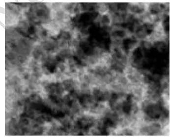
Electron microscopy techniques, on the other hand, can provide information on the size, shape, and location of the individual nanoparticles. It is a formidable task, however, to determine statistically meaningful data on the size distribution of nanoparticles in industrial heterogeneous catalysts, especially when the sizes of the nanoparticles vary significantly with their spatial distribution. The SEM images of the MgO samples are shown in (Fig. 2). It can be seen from the MgO particles exclusively consists of hexagonal lamellarlike structures and the sizes of MgO are 50±10 nm length or width and about 20 nm thickness.
Fig. 2 SEM image of Magnesium Oxide Nanoparticles
Transmission Electron Microscopy
TEM techniques, however, can provide useful information on very small particles, amorphous or highly disordered nanoparticles or particles that contain many incoherent domains. In this work,
experimental data obtained from TEM and XRD techniques will be compared and the pros and cons of the various techniques for determining the size distribution of nanoparticles and nanoparticle systems as well as the challenges and great opportunities in particle size determination will be similar to that of the calculated size from X-ray diffraction. The Sherrer equation was applied to estimate a crystalline size and the size of MgO nanoparticles are about 20nm thickness. On close examination of TEM diffraction pattern of MgO nanoparticles has polycrystalline structure.
Besides that uniform distribution suggests that it has good homogeneities morphology. TEM image of a single platelet of MgO sample, (Fig.3) in which the MgOnanocrystallites exhibit hexagonal shape and very homogeneous crystal structures without any observable pores
Fig. 3 TTEM image of Magnesium Oxide Nanoparticles
IV. CONCLUSION
Emperor International Journal of Finance and Management Research [EIJFMR] Page 15 the applications of MgOnanomaterials in the fields
such as highdensed ceramics, additives in bactericide, refractory, and superconductor products. In addition, the method adopted can be easily extended to synthesis the rod, wire, and tubelike MgO nanostructures. The XRD and TEM results indicate that the obtained MgO has crystalline structure. SEM and TEM images, it can be seen that the sizes of MgO
are 50±10 nm length or width and about 20 nm thickness and the structure is nonporous respectively. Through the present study, it was confirmed that the use of magnesium nitrate with PVP as surfactant would be an excellent and effective method to prepare MgO nanoparticles.
REFERENCE
[1] 1. G. Schmid, Nanoparticles: From theory to applications, WileyVCH Weinheim, 2004.
2. H. Itoh, S. Utamapanya, J. V. Stark, K. J. Klabunde and J. R. Schlup, Chem. Mater., 1993, 5, 71.
3. M. Harcata, Date, M. Appl. Catal. A, 2001, 222, 427.
4. C. George Schatz and et al, J. Phys. Chem. B 2003, 107, 668677.