University of Pennsylvania
ScholarlyCommons
Publicly Accessible Penn Dissertations
2018
The Development Of Intrinsically Fluorescent
Unnatural Amino Acids For In Vivo Incorporation
Into Proteins
Itthipol Sungwienwong
University of Pennsylvania, itthipol@sas.upenn.edu
Follow this and additional works at:https://repository.upenn.edu/edissertations Part of theChemistry Commons
This paper is posted at ScholarlyCommons.https://repository.upenn.edu/edissertations/3037 For more information, please contactrepository@pobox.upenn.edu.
Recommended Citation
Sungwienwong, Itthipol, "The Development Of Intrinsically Fluorescent Unnatural Amino Acids For In Vivo Incorporation Into Proteins" (2018).Publicly Accessible Penn Dissertations. 3037.
The Development Of Intrinsically Fluorescent Unnatural Amino Acids
For In Vivo Incorporation Into Proteins
Abstract
The amino acid acridon-2-ylalanine (Acd) can be a valuable probe of protein dynamics either alone or as part of a Förster resonance energy transfer (FRET) or photo-induced electron transfer (eT) probe pair. We have previously reported the genetic incorporation of Acd by an aminoacyl tRNA synthetase (RS). However, this RS, developed from a library of permissive RSs, also incorporates N-phenyl-amino-phenylalanine (Npf), a trace byproduct of one Acd synthetic route. We have performed negative selections in the presence of Npf and analyzed the selectivity of the resulting AcdRSs by in vivo protein expression and detailed kinetic analyses of the purified RSs. We find that selection conferred a ~50-fold increase in selectivity for Acd over Npf,
eliminating incorporation of Npf contaminants, and allowing one to use a high yielding Acd synthetic route for improved overall expression of Acd-containing proteins. More generally, our report also provides a cautionary tale on the use of permissive RSs, as well as a strategy for improving selectivity for the target amino acid.
In spite of its utility for studying proteins by fluorescence spectroscopy, Acd can potentially be improved by making it longer wavelength or brighter. We reported the synthesis of Acd core derivatives and their photophysical characterization. We also performed ab initio calculations of the absorption and emission spectra of Acd derivatives, which agree well with experimental measurements. The amino acid
aminoacridonylalanine (Aad) was synthesized in forms appropriate for genetic incorporation and peptide synthesis. We show that Aad is a superior FRET acceptor to Acd in a peptide cleavage assay, and that Aad can be activated by an aminoacyl tRNA synthetase for genetic incorporation. Together, these results show that we can use computation to design enhanced Acd derivatives which can be used in peptides and proteins.
Finally, the Aad synthesis has been improved and it will be further tested in vivo incorporations into proteins, and alkylated Aad core analogs show improved brightness making their use as amino acids promising.
Degree Type Dissertation
Degree Name
Doctor of Philosophy (PhD)
Graduate Group Chemistry
First Advisor Ernest J. Petersson
Subject Categories Chemistry
THE DEVELOPMENT OF INTRINSICALLY FLUORESCENT UNNATURAL AMINO ACIDS
FOR IN VIVO INCORPORATION INTO PROTEINS
Itthipol Sungwienwong
A DISSERTATION
in
Chemistry
Presented to the Faculties of the University of Pennsylvania
in
Partial Fulfillment of the Requirements for the
Degree of Doctor of Philosophy
2018
Supervisor of Dissertation
________________________
E. James Petersson
Associate Professor of Chemistry
Graduate Group Chairperson
_________________________
David W. Christianson, Roy and Diana Vagelos Professor of Chemistry and Chemical Biology
Dissertation Committee:
David M. Chenoweth, Associate Professor of Chemistry
Tobias Baumgart, Professor of Chemistry
THE DEVELOPMENT OF INTRINSICALLY FLUORESCENT UNNATURAL AMINO ACIDS
FOR IN VIVO INCORPORATION INTO PROTEINS
COPYRIGHT
2018
iii
ACKNOWLEDGEMENTS
First and foremost, I would like to thank my advisor, Professor E. James Petersson
for providing me with an opportunity to join the laboratory and for his generous guidance
throughout my graduate career. He has supported me not only in scientific research but
also in general life. I am deeply appreciative of his mentorship. Without him, I would not
be able to achieve success in my career. I would also like to thank my dissertation
committee members; Professor David M. Chenoweth, Professor Tobias Baumgart, and
Professor Ivan Dmochowski for their kind support, helpful feedback, and their
encouragement throughout these years.
I would like to acknowledge all members of the Petersson group and alumni, who
have been great colleagues and friends. It was my pleasure to work with these people; Dr.
Lee Speight, Dr. Rebecca Wissner, Dr. Solongo Ziraldo, Dr. Yanxin Wang, Dr. Xing Chen,
Dr. Chris Walters, Dr. Yun Huang, Dr. Conor Haney, Dr. Naoya Ieda, Miklos Szantai-Kis,
John Ferrie, Anand Muthusamy,Taylor Barrett, Joomyung Jun, Chunxiao Liu, Buyan Pan,
Christina Cleveland, E. Keith Keenan, Tiberiu Mihaila, Jimin Yoon. Furthermore, I would
like to thank my collaborators for their helpful support; Professor Rahul Kohli and Zachary
Hostetler (Department of Medicine, Perelman School of Medicine, University of
Pennsylvania), Professor Ryan Mehl (Oregon State University), Professor John Perona
(Portland State University), and Professor Jeffery Saven (Penn Chemistry).
Finally, I would like to acknowledge my family and friends in Thailand. I am quite
iv
degree. I would like to thank the Thai community at the University of Pennsylvania and
Drexel University for mental support and invaluable friendship. I would like to make
special thanks to Nisalak Trongsiriwat, Parawee Kasitinon, and Dr.Weerapha
Panaddasirisuk for their priceless companionship. And last, I need to thank my scholarship
support from the Royal Thai government and my future workplace (Srinakharinwirot
University, Bangkok, Thailand) that provided me with the great opportunity to accomplish
v ABSTRACT
THE DEVELOPMENT OF INTRINSICALLY FLUORESCENT UNNATURAL AMINO ACIDS
FOR IN VIVO INCORPORATION INTO PROTEINS
Itthipol Sungwienwong
Professor E. James Petersson
The amino acid acridon-2-ylalanine (Acd) can be a valuable probe of protein
dynamics either alone or as part of a Förster resonance energy transfer (FRET) or
photo-induced electron transfer (eT) probe pair. We have previously reported the genetic
incorporation of Acd by an aminoacyl tRNA synthetase (RS). However, this RS, developed
from a library of permissive RSs, also incorporates N-phenyl-amino-phenylalanine (Npf),
a trace byproduct of one Acd synthetic route. We have performed negative selections in
the presence of Npf and analyzed the selectivity of the resulting AcdRSs by in vivo protein
expression and detailed kinetic analyses of the purified RSs. We find that selection
conferred a ~50-fold increase in selectivity for Acd over Npf, eliminating incorporation of
Npf contaminants, and allowing one to use a high yielding Acd synthetic route for
improved overall expression of Acd-containing proteins. More generally, our report also
provides a cautionary tale on the use of permissive RSs, as well as a strategy for improving
selectivity for the target amino acid.
In spite of its utility for studying proteins by fluorescence spectroscopy, Acd can
potentially be improved by making it longer wavelength or brighter. We reported the
vi
performed ab initio calculations of the absorption and emission spectra of Acd derivatives,
which agree well with experimental measurements. The amino acid
aminoacridonylalanine (Aad) was synthesized in forms appropriate for genetic
incorporation and peptide synthesis. We show that Aad is a superior FRET acceptor to
Acd in a peptide cleavage assay, and that Aad can be activated by an aminoacyl tRNA
synthetase for genetic incorporation. Together, these results show that we can use
computation to design enhanced Acd derivatives which can be used in peptides and
proteins.
Finally, the Aad synthesis has been improved and it will be further tested in vivo
incorporations into proteins, and alkylated Aad core analogs show improved brightness
vii
TABLE OF CONTENTS
ACKNOWLEDGEMENT ... IIIII
ABSTRACT ... V
LIST OF TABLES AND SCHEMES... IVII
LIST OF ILLUSTRATIONS ... X
CHAPTER 1INTRODUCTION ... 1
1.1 Background………2
1.2 Expanded Genetic Code………...5
1.3 Analysis of Aminoacyl tRNA Synthetases (RSs) ………...9
1.4 Genetic Incorporation of Fluorescent Amino Acids………12
1.5 Fluorescent Labeling Using Unnatural Amino Acids with Bioorthogonal Reactivity………...19
1.6 Acridone as a Tunable Fluorescent Scaffold for Generating Fluorescent Amino Acids………..22
CHAPTER 2 THE OPTIMIZATION OF A PERMISSIVE AMINOACYL TRNA SYNTHETASE FOR A TARGET UNNATURAL AMINO ACID ... 24
2.1 Background………..25
2.2 Synthetase Selection ………...29
2.3 AcdRS Optimization………32
viii
2.5 Analysis of AcdRS Selectivity……….34
2.6 Modeling of AcdRSs complexed with Npf and Acd……….41
2.7 Conclusion………43
2.8 Experimental Methods……….44
CHAPTER 3 THE DESIGN OF FLUORESCENT UNNATURAL AMINO ACIDS BASED ON THE ACRIDONE SCAFFOLD... 66
3.1 Introduction………..67
3.2 Results and Discussion………71
3.3 Conclusions………..86
3.4 Experimental Methods………...87
CHAPTER 4 TOWARD THE SYNTHESIS AND IN VIVO INCORPORATION OF ACRIDONE DERIVATIVES INTO PROTEINS………..118
4.1 Introduction………119
4.2 Improving the Synthesis of 2-aminoacridone amino acid (Aad)………...120
4.3 Improving the Synthetase Activation of Aad……….121
4.4 Additional Acridone Core Derivatives………..123
4.5 Conclusions………125
4.6 Experimental Method……….126
ix
LIST OF TABLES
Table 2.1 AcdRS in vitro enzymology and in vivo MS selectivity
Table 2.2 Sequence of top performing AcdRSs.
Table 2.3 CaM 113 trypsin digest intensity scaling
Table 3.1 Calculated and observed photophysical parameters of acridone derivatives
Table 3.2 Screening of acridone nitration conditions
Table 3.3 Calculated and observed photophysical parameters of acridone derivatives.
LIST OF SCHEMES
Scheme 2.1 The synthesis of acridon-2-ylalanine (Acd).
Scheme 3.1 Synthesis of nitroacridones and aminoacridones
Scheme 3.2 Synthesis of acridone cores and benzoacridones
x
LIST OF ILLUSTRATIONS
Figure 1.1 Chromophore size
Figure 1.2 Site-specific incorporation of unnatural amino acids
Figure 1.3 Positive and negative selection strategies
Figure 1.4The active site of TyrRS and a mutant UaaRS evolved to incorporate o-Iodotyrosine
Figure 1.5 Structure of 7-hydroxycoumarin and three coumarin lysine analogues
Figure 1.6 Structure of NAP, Anap, danLys, dansylalanine, and terphenyl amino acids.
Figure 1.7 Labeling proteins via incorporation of unnatural amino acids
Figure 1.8 Effects of flexible linkers on the range of chromophore distances in a FRET
experiment.
Figure 1.9 The structure of acridonylalanine (Acd) and 2-aminoacridonylalanine (Aad)
Figure 2.1 AcdRS selection
Figure 2.2 In vivo AcdRS selectivity
Figure 2.3 CaM113 trypsin digest data for AcdRS selectivity analysis
Figure 2.4 tRNA aminoacylation kinetics
Figure 2.5 AcdRS homology models
Figure 2.6 Fluorescence measurements of RSs with GFP reporter
Figure 2.7 In vivo AcdRS selectivity of CaM
Figure 2.8 In vivo AcdRS selectivity of αS
Figure 2.9 In vivo AcdRS selectivity of LexA
Figure 2.10 AcdRS purification SDS-PAGE gels
xi
Figure 2.12 Representative primary data for aminoacylation
Figure 3.1 FRET experiments
Figure 3.2 Fluorescent amino acids based on 7-methoxycoumarin and acridone cores
Figure 3.3 Absorption and emission spectra of acridone derivatives
Figure 3.4 Calculated acridone spectra
Figure 3.5 Aad is a superior FRET acceptor for Mcm
Figure 3.6 Aad Activation by AcdRS1
Figure 3.7 Screening of acridone nitration conditions
Figure 3.8. Screening of 2- and 4-nitroacridone reduction conditions
Figure 3.9 1H and 13C NMR spectra of Boc2-Aad in MeOD
Figure 3.10 Acridone (5) spectra
Figure 3.11 2-Aminoacridone (8) spectra
Figure 3.12 4-Aminoacridone (9) spectra
Figure 3.13 2-Nitroacridone (6) and 4-Nitroacridone (7) spectra
Figure 3.14 2-Fluoroacridone (15), 2-methoxyacridone (S1), and 4-methoxyacridone
(16) spectra
Figure 3.15 4-Benzoacridone (18), 4-fluorobenzoacridone (19), and
4-methoxybenzoacridone (20) spectra
Figure 4.1 Cross-coupling screening results
Figure 4.2 Aad activation by AcdRS2b (A9)
Figure 4.3 Structure of 2-(dimethylamino)acridin-9(10H)-one (Dad) and
xii
1
2 Background
Anfinsen’shypothesis is a classical postulate in molecular biology stating that the
three-dimensional structure of a native protein in its standard physiological environment
(solvent, pH, ionic strength, presence of other components such as metal ions or prosthetic
groups, temperature, and other factors) is determined only by the totality of inter-atomic
interactions within the protein and hence by the amino acid sequence.1 While it has been
clearly shown that many proteins violate this hypothesis – for example, by requiring
chaperones for folding2-4 – nonetheless, studies of protein structure should imitate the
native state in a physiologically-relevant local environment as much as possible. Several
methods have been advanced to study protein architecture such as X-ray crystallography5,
nuclear magnetic resonance (NMR) spectroscopy6, and cryo-electron microscopy
(cryo-EM)7. Structures revealed through these methods can solve biological questions and
provide insight into protein interactions, folding pathways, and enzymatic activities.
However, many dynamic mechanisms remain obscure due to the limitations of these
methods. The X-ray crystallography technique intrinsically requires the protein to be
packed into a fine crystal lattice which freezes out most dynamics. Cryo-EM provides
greater opportunity for flexibility, but still requires freezing the protein and deposition on
an imaging grid. NMR spectroscopy permits solution phase characterization and the
opportunity to observe dynamics, but NMR is limited to small proteins and cannot be used
for real-time observations of protein motions. Thus, there is a need to complement these
high resolution, relatively static structural methods with techniques that can provide
3
Fluorescence spectroscopy is one valuable technique that can be used to analyze
changes in protein structure and protein/protein interactions8-9. Fluorescent experiments
can be performed with either intrinsically fluorescent proteins or fluorescently labelled
proteins. Protein conformational changes can be monitored by observation of changes in
the fluorescence of a single probe or through interactions of multiple (usually pairs) of
probes. A change in fluorescence of a single probe can result from a change in the local
environment of the fluorophore due to a conformational change or binding event.
Fluorescence polarization (FP) varies with the reorientation time of the chromophore and
can report on changes in the flexibility of a protein region or, more commonly, on a change
in tumbling rate due to interaction with another macromolecule. Experiments using pairs
of probes can be used to track protein structural change on the nanosecond timescale using
distance-dependent chromophore interactions either through Förster resonance energy
transfer (FRET) or quenching by photo-induced electron transfer (PeT). When using a
fluorescent probe, one needs to be concerned with its ability to disrupt protein structure
and function. These disruptions can result from the size and/or location of the fluorophores.
The size of commonly used fluorescent labels ranges from 29820 Å3 for the green
fluorescent protein (GFP) to 9 Å3 for thioamide substitution8. (Figure 1.1) There are a
variety of methods for introducing probes into proteins, which vary in complexity from
simple cloning of the cDNA for GFP into a protein expression plasmid to the chemical
synthesis of a protein in order to install a synthetic fluorophore. Among these methods,
genetic code expansion offers a near ideal combination of small probe size, potential
4
Figure 1.1 Chromophore size. Space-filling renderings of several chromophores, based on either crystal structures or ab initio calculations. The green fluorescent protein (GFP) was rendered from PDB ID 1GFL9. The benzylated SNAP tag protein was rendered from PDB ID 3L00. The geometry of tetracysteine-bound FlAsH was optimized at the HF/6-31G level, all others were optimized at the AM1 level (Figure adapted from Speight L.C., Moumita S., Petersson, E. J. (2014) Minimalist Approaches to Protein Labelling: Getting the Most Fluorescent Bang for Your Steric Buck.
5 Expanded Genetic Code
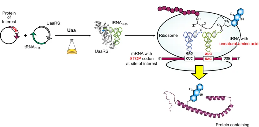
Figure 1.2 Site-specific incorporation of unnatural amino acids.
Protein translation uses transfer RNAs (tRNAs), which are aminoacylated with
their canonical amino acids by aminoacyl-tRNA synthetase enzymes (RSs), to read triplet
codons in messenger RNAs (mRNAs) when paired with the anticodon of the tRNA. The
ribosome facilitates both the decoding of triplet codons on mRNAs by tRNAs, and the
polymerization of the corresponding amino acids in order for the mRNA to be translated
into a polypeptide chain. All proteins are ribosomally synthesized with the limitation of 20
canonical amino acids building blocks. While a wide variety of post-translational
modifications occur on proteins, these are generally restricted in sequence context so that
using them to modify a protein requires the introduction of a tagging sequence which may
6
expand the genetic code, enabling the site-specific incorporation of unnatural amino acids
(Uaas, also known as non-canonical amino acids or ncAAs) into proteins synthesized in
both prokaryotic and eukaryotic cells (Figure 1.2).
The site-specific incorporation of a Uaa into proteins has provided new insights to
study biological processes which are difficult and challenging to address by classical
approaches. Peter Schultz10 developed a genetic code expansion approach that uses an
orthogonal RS/tRNA pair to direct the incorporation of a Uaa into proteins in response to
an unassigned codon (commonly the so-called amber stop codon, UAG) introduced at the
desired site in a gene of interest, which is called amber suppression. Researchers can now
incorporate Uaas into proteins in bacteria (typically Escherichia coli or E. coli),
Saccharomyces cerevisiae, mammalian cells (a few experiments in mice have even been
reported), Caenorhabditis elegans, Drosphila melanogaster.11 To be “orthogonal”, the
RS/tRNA pair must meet many requirements. The specificity of the RS active site needs to
be optimized so that it can transfer the desired Uaa, but not any of the canonical amino
acids to the orthogonal tRNA. The orthogonal tRNA must be recognized only by the URS,
and none of the endogenous RSs for the natural amino acids. Finally, the URS must not
recognize any of the endogenous tRNAs. To meet these complex requirements, Schultz
developed a strategy of importing an RS/tRNA from another organism where the tRNA
structure was known to be substantially different from that of the host organism.12 Initially,
most amber suppression experimenst used orthogonal RS/tRNA pairs based on TyrRS and
LeuRS evolved from Methanocaldococcus jannaschii (e.g. MjTyrRS, MjLeuRS) or E. coli
7
desired Uaa of interest. Recently the pyrrolysyl (Pyl) RS/tRNA pairs (from
Methanosarcina barkeri and Methanosarcina mazei) have grown in popularity as Pyl is
found in archaea, but not in bacterial or mammalian cells.13-16 Thus, PylRS/tRNA pairs are
orthogonal in both cell types and only require evolution of the active site for the desired
Uaa. The methods for directed evolution of the active site have been well-developed and
are usually performed in E. coli or yeast cells.
Figure 1.3 Positive and negative selection strategies for the generation of an orthogonal synthetase/tRNA pair with altered amino acid specificity for an unnatural amino acid (blue star).
Typically, several rounds of positive and negative selection are used to select an
active site variant that allows the incorporation of the Uaa but none of the 20 canonical
amino acids (Figure 1.3)17. The positive selection step relies on the ability of the RS/tRNA
8
resistance gene). This positive selection provides RSs in surviving colonies that can
incorporate the Uaa and/or a natural amino acid. These RSs are then subjected to a negative
selection procedure performed in the absence of the Uaa with the amber codon now
encoded in toxic a gene (such as a toxic ribonuclease or barnase). The RSs that use the
canonical amino acid will read through the amber codon and lead to cell death. After
several rounds of positive and negative selection, only mutant RSs that can selectively
incorporate the Uaa will survive. This method has been used to generate RSs for more than
100 Uaas, which have been widely employed in the production of both recombinant
proteins for in vitro experiments and in live cell experiments.18 However, some limitations
on protein yield exist.
The efficiency of protein expression with Uaa incorporation can be improved by
removing release factor 1 (RF1 for E. coli and eRF1 for eukaryotes).The decoding of the
UAG amber codon normally competes with RF1, which leads to termination of protein
synthesis and decreases protein yield. This issue has been dealt with in several ways. In
one case, the Chin laboratory’s evolution of an orthogonal ribosome that no longer interacts
with RF1 enables the efficient incorporation of a Uaa in response to amber codons at
multiple sites.19-20 In a second example, it has been shown in some bacterial strains (such
as E. coli C321) that the deletion of RF1 can improve the performance of Uaa incorporation
in response to the quadruplet codon UAGA.21 This quadruplet system has been evolved
from Methanocaldococcus jannaschii-derived frame-shift suppressor tRNA/RS pair that
can enhance UAGN (N = any nucleotide) suppression efficiency. In eukaryotes, the release
9
engineered to provide a significant increase in Uaa incorporation in response to the UAG
codon without increasing read-through of other stop codons.22 With the combination of this
engineered eRF1 and an optimized PylRS/tRNA expression system for mammalian cells,
one can increased the yield of protein containing a single site of Uaa substitution 17- to
20-fold and the proteins yield for multiple site substitutions are comparable to a protein yields
from a gene that does not contain a UAG stop codon. With these and other methods to
improve protein yields, Uaa mutagenesis has become a robust technique that is being
applied by an ever-growing number of laboratories.
Analysis of Aminoacyl tRNA Synthetases (RSs)
Since most UaaRSs are achieved through directed evolution as described above,
there are a limited number of cases where detailed analysis of the structure and function of
the resulting UaaRS have been performed. RSs catalyze a two-step reaction that provides
aminoacylated tRNA for protein synthesis. The first step (1) involves the condensation of
amino acid (Aa, or in this case, the Uaa) and ATP to form the enzyme-bound aminoacyl
adenylate intermediate (Aa-AMP or Uaa-AMP) and release pyrophosphate (PPi). The
second step (2) is the reaction between this adenylated intermediate and the 2’- or 3’
-terminal hydroxyl (OH) group on tRNA to form aminoacyl-tRNA (Aa-tRNA or
Uaa-tRNA).
Aa + ATP –> Aa-AMP + PPi (1)
10
The ability of RSs to differentiate an amino acid substrate from other amino acids is almost
solely responsible for the accuracy of protein synthesis.23 Indeed, it is the lack of ribosomal
proof-reading of Uaa-tRNA that permits Uaa mutagenesis to work. Thus, an analysis of
the initial Uaa-AMP formation step can provide significant insight into UaaRS
incorporation. In general, the analysis of RS function has mainly relied on steady state
kinetic pyrophosphate exchange and aminoacylation assays.24 In the pyrophosphate
exchange reaction, the RS is added to the reaction buffer containing amino acid, Mg•ATP,
and [32P]-pyrophosphate in prepared reaction buffer. The [32P]-ATP formed is then
adsorbed on activated charcoal or thin layer chromatography support. The standard
pyrophosphate exchange assay is typically performed at 37 °C in a reaction containing [32P]
Na•PPi, RS, amino acid in concentrations ranging from 0.2 to 10 times the Michaelis
constant (Km), and ATP. The PPi exchange reaction is usually performed in the absence of
tRNA. Therefore, the kcat and Km Michaelis-Menten values derived from this method are
not necessarily identical to the actual rate of amino acid activation in the presence of tRNA.
The steady-state tRNA aminoacylation reaction is traditionally investigated by monitoring
the formation of [3H] or [14C] aa-tRNA over time. This steady-state assay is usually run at
37 °C in buffer containing RS, amino acid, ATP, and tRNA. The limitation of the steady
state aminoacylation assay is that amino acid concentrations are practically limited to
concentrations of about 1 mM, due to the relatively low specific activity of [3H]- or [14
C]-labeled amino acids. Alternatively, the [32P]-labeled tRNA can be used together with
11
Previous kinetic activities of RS charging of a few Uaas have been studied. For
example, the Schultz group has analyzed the kinetics of adenylate formation of O-methyl
tyrosine and tyrosine with ATP catalyzed by mutant TyrRSs using the pyrophosphate
exchange assay. The Km value for tyrosine (5833 ± 902 µM) is approximately 13-fold
higher than that for O-methyl-L-tyrosine (443 ± 93 µM). The kcat for tyrosine (1.8 ± 0.2 x
10-3 s-1) is eight-fold less than that for O-methyl-L-tyrosine (14 ± 1 x 10⁻³ s-1).25
12
To alter the amino acid specificity of the orthogonal TyrRS so that it can charge the
mutRNATyr
CUA with a desired unnatural amino acid, a library of TyrRS mutants was
generated and screened. Based on the crystal structure of the homologous TyrRS (Figure
1.4) from Bacillus stearothermophilus, five residues in the active site of M. jannaschii
TyrRS that are within 6.5 Å of the para position of the aryl ring of bound tyrosine were
mutated. Crystal structures of the corresponding evolved synthetases can be used to
rationalize how specificity was changed.
Kamtekar et al.28 reported the 3.2-Å resolution crystal structure of the
Methanococcus maripaludis phosphoseryl-tRNA synthetases (SepRSs). The kinetic
constants of these M. Maripaludis SepRS mutants for phosphoserine charging activation
has measured by ATP-PPi exchange assay. Umehara et al. reported the directed evolution
of PylRS from Methanosarcina mazei to generate N-acetyl lysyl-tRNA synthetases
(AcKRSs). The kinetic activity of these AcKRSs has also been determined by ATP-PPi
exchange assays.29 Limited structural information is available for PylRSs.30
Genetic Incorporation of Fluorescent Amino Acids
Genetically encoded fluorescent Uaas have been used to label proteins in both
prokaryotes and eukaryotes, but the number of fluorescent Uaas developed is surprisingly
small, given the noteable advantages of Uaa labeling: it provides perfect site-specificity
and minimal perturbation of protein structure and function. Schultz and coworkers
reported the generation of an orthogonal RS/tRNA pair that selectively incorporates
13
including a high fluorescence quantum yield (0.63), a large Stoke’s shift, small size, and
sensitivity to pH and solvent polarity. The incorporation of Hco into proteins was carried
out by using an evolved MjTyrRS/tRNA pair. Protein expression was shown in sperm
whale myoglobin and protein mutants were used as a probe to study urea-induced chemical
denaturation of holomyoglobin. The fluorescence intensity of the hydroxycoumarin moiety
is sensitive to solvent polarity, and it showed correlation between protein unfolding and an
increase in fluorescence of Hco. Hco has also been site-specifically incorporated into signal
transducer and activator of transcription 3 (STAT3) in order to use the labeled protein as a
fluorescent reporter of the phosphorylation status of STAT3.32 Hco was genetically
incorporated into the STAT3b isoform in E. coli at Trp residue 564. A large fluorescence
change was observed when the STAT3 probe was phosphorylated by Src kinase in vitro
and when it was incubated with endogenously activated STAT3. This method enables the
investigation of protein phosphorylation on other STAT protein substrates and other SH2
domain-containing proteins because Trp564 is conserved in all seven mammalian STAT
proteins. It also illustrates a general strategy for using fluorescent Uaas to make sensor
proteins.
Lou et al. reported the site-specific incorporation of three new coumarin lysine
derivatives (Figure 1.5) into proteins in bacterial and mammalian cells using an engineered
PylRS. The genetically encoded coumarin lysines were utilized as optochemical probes for
14
Figure 1.5 Structure of 7-hydroxycoumarin (1, Hco) and three coumarin lysine analogues (2-4)
These three new coumarin lysine derivatives showed unique photochemical
activities. Bromine atom substitution at the 6-position of 3 enables decaging not only with
UV, but also near IR excitation using two photon methods. The extension of the linker
chain in 4 can prohibit photolysis. Therefore, coumarin lysines 2 and 3 can be used as both
fluorescent and light-activated probes for optochemical control of protein function using
UV or near-IR light, while coumarin lysine 4 serves as a stable fluorescent probe that does
not decage under UV excitation. To demonstrate the application of these coumarin lysines
for optical control, photoregulation of firefly luciferase was achieved in live cells by caging
a lysine residue, and excellent turn-on ratios were observed for 2 and 3. As expected, the
stable coumarin amino acid 4 did not undergo photolysis. Furthermore, two-photon and
single-photon excitation of EGFP tagged with 4 was also demonstrated by using different
excitation wavelengths (365, 405, and 760 nm) for the sequential activation of protein
function in live cells. While caged lysine 3 could be activated using two-photon irradiation
at 760 nm, lysine 2 was stable under these conditions but could be decaged with blue light
of 405 nm. This example illustrates how the long lysine linker allows one to attach
O O
HO
H2N COOH
O O HO O X n O N H 1 COOH NH2
15
substrates of varying bulk and yet remain compatible with PylRS charging for protein
incorporation.
Schultz reported the site-specific incorporation of a second fluorophore, L
-3-(2-naphthyl)alanine (5, NAP), shown in Figure 1.6, into proteins in E. coli using M.
jannaschii TyrRS mutants.34 A mutant of mouse dihydrofolate reductase containing NAP
at Tyr163 was generated and characterized to confirm the ability of the NAPRS/tRNA pair
to site-specifically incorporate NAP in response to an amber stop codon.
Lee et al. reported the incorporation of 3-(6-acetylnaphthalen-2-ylamino)-2-
aminopropanoic acid (6, Anap) into proteins using an orthogonal RS/tRNA pair derived
from the E.coli LeuRS and engineered in a two-step process for the bulky Uaa (Figure
1.6). Anap has absorption and emission maxima at 360 and 490 nm, respectively. Its
extinction coefficient is 17,500 cm-1 M-1 and its quantum yield is 0.48 in EtOH (with
excitation at 360 nm). The Anap amino acid was site-specifically incorporated in E. coli
glutamine-binding protein and it was used to probe ligand-induced local conformational
changes in proteins through solvatochromic effects without the need for a FRET pair. The
small size of Anap and ability to introduce it by simple mutagenesis at defined sites should
make it a useful probe of protein structure, molecular interactions, protein folding, and
16
Figure 1.6 Structure of NAP, Anap, danLys, dansylalanine, and terphenyl amino acids. (5-9)
Chatterjee and Schultz also demonstrated the incorporation of Anap in mammalian
cells in response to the TAG codon with high efficiency using AnapRS for expression of
enhanced green fluorescent protein (EGFP) with an Anap mutation at a permissive site
(Tyr40TAG) and a C-terminal histidine tag into Human Embryonic Kidney 293 (HEK293)
or Chinese Hamster Ovary (CHO) cells.36 They further demonstrated that Anap can be used
to image the subcellular localization of proteins in live mammalian cells by using confocal
microscopy. Anap fluorescence was localized in the nucleus of histone H3. In addition to
conventional fluorescence microscopy, Anap fluorescence can also be visualized by
two-photon excitation, which provides deeper tissue penetration, efficient light detection, and
reduced phototoxicity. Two-photon excitation of the Anap-mCherry double-labeled
histone H3 revealed excellent overlap between Anap (excitation, 730 nm, two-photon;
emission, 420−500 nm) and mCherry (excitation, 543 nm; emission, 600−700 nm)
fluorescence in the nucleus.
H2N COOH HN
S O O
N
H2N COOH
5, NAP
H2N COOH
9, Terphenyl H2N COOH
HN O
6, Anap 8, Dansylalanine
H2N COOH HN
S O O
N
17
Anap has also been used as a FRET pair in mammalian cells. Chatterjee and
coworkers developed a genetically encoded FRET that uses Anap as the donor fluorophore
and enhanced green fluorescent protein (EGFP) as the acceptor.37 They co-expressed the
EGFP- Y39TAG mutant in HEK293T human embryonic kidney cells along with an
orthogonal AnapRS/tRNA pair. This expression system can efficiently perform with Anap
concentrations as low as 1 µM in the media. The calculate Förster radius of the Anap-EGFP
FRET pair is 49 Å. They demonstrated the use of this FRET pair to monitor proteolysis
reactions and protein conformational change in the Ca2+-dependent binding of the
calmodulin protein to its substrate peptide M13.
Another application of the Anap fluorophore is that it can be used to incorporate in
the Shaker voltage-gated potassium channel (KV) at key regions that were previously
inaccessible to labeling, and it can be used to study dynamic structural information
collected from the cytosolic side of the channel which was not possible with the other
chemical labeling method because of the large number of unwanted binding sites in the
cytosol.38
The dansyl fluorophore has been shown to be ribosomally incorporated into b
-galactosidase by Chamberlin. e-Dansyllysine 7 (e-DanLys) along with the other two
fluorescent amino acids; 5-hydroxytryptophan, 7-azatryptophan, have been
site-specifically incorporated in vivo using chemically charged tRNAs (note that this method
has not been discussed above due it slack of adoptability by other laboratories in
18
The fluorescent unnatural amino acid
2-amino-3-(5-(dimethylamino)naphthalene-1-sulfonamide)propanoic acid (8, Dansylalanine) was genetically encoded in
Saccharomyces cerevisiae with high fidelity and good yield using an orthogonal RS/tRNA
pair.40 This environmentally sensitive fluorophore was selectively inserted into human
superoxide dismutase and used to monitor unfolding process in the presence of
guanidinium chloride.
Lei Wang also reported the incorporation of dansylalanine 8 into a
voltage-dependent membrane lipid phosphatase, specifically the voltage-sensitive domain (VSD)
of Ciona intestinalis voltage-sensitive phosphatase (CiVSP). They described a
lentiviral-based gene delivery method to stably incorporate Uaas into proteins expressed in
HCN-A94 neural stem cells. This genetically encoded fluorescent Uaa optically reported on the
conformational change of the voltage-sensitive domain in response to membrane
depolarization in differentiated neurons.41
The incorporation of fluorescent unnatural amino acid 4-biphenyl-L-phenyl alanine
(9, Terphenyl) in Figure 1.6 into GFP has been achieved by Douglas Young and
coworkers.42 Terphenyl moieties have relatively high quantum yields (f = 0.49), long
fluorescent lifetimes (t = 4.38 ns) and novel emission spectra with large Stokes shifts (lex/
lem = 280/342 nm). This terphenyl moiety has also been found to be environmentally
sensitive and widely used in various applications including two-photon laser scanning
microscopy, femtosecond fluorescence spectroscopy, and a-helical secondary structure
investigations. Previously, Sidney Hecht’s group demonstrated that various terphenyl
19
studies by using chemically acylated tRNA methods.43-44 The terphenyl amino acid has
been shown by Young to be site-specifically incorporated into GFP at multiple sites and
these GFP mutants has been shown to impact the protonation state of the terphenyl
fluorophore, potentially expanding the utility of the protein as a biosensor.42 Together,
these studies illustrate the wide array of potential applications of fluorescent amino acids.
Fluorescent Labeling Using Unnatural Amino Acids with Bioorthogonal Reactivity
An alternative approach for protein labelling can be performed by site-specific
incorporation of Uaas with “biorthogonal” handles into proteins by genetic code expansion
followed by subsequent labeling of these proteins by specific chemoselective reactions.
(Figure 1.7a) These biorthogonal reactions need to proceed under
biologically/physiologically compatible conditions and yet react only with the desired
partner and not any of the endogenous biomolecule functional groups. Several examples
of these reactions have been shown including reaction of ketones/aldehydes with
hydrazines/hydroxylamines, the classic copper-catalyzed azide-alkyne cycloaddition
reaction (CuAAC), the strain-promoted azide-alkyne cycloaddition reaction (SPAAC),
Staudinger ligations, certain palladium-catalyzed cross-coupling reactions, “photoclick”
reactions, inverse-electron-demand Diels-Alder reactions.17 These chemoselective
reactions usually have rate constant in the order of 10-4 – 104 M-1 s-1 as shown in Figure
20
Figure 1.7 Labeling proteins via incorporation of unnatural amino acids that can be chemoselectively labeled. (a) An unnatural amino acid bearing a unique bioorthogonal functionality is introduced site-specifically into a protein via genetic code expansion and then chemoselectively labeled with an externally added chemical probe. (b) Rate constants of chemoselective reactions for which one of the partners can be genetically encoded in form of an unnatural amino acid. (Adapted figure reproduces from Lang, K., Chin, J.W. Cellular Incorporation of Unnatural Amino Acids and Bioorthogonal Labeling of Proteins, Chem. Rev. 2014, 114, 4764−4806.)
While Uaas containing bioorthogonal amino acids can be used to install a wide
variety of fluorophores, there can be some limitations of the utility of these probes in
studying protein folding. Direct genetic encoding of a fluorescent Uaa allows one to place
a chromophore on the interior of a protein and to label proteins that cannot be reversibly
unfolded and refolded. Having the chromophore attached by a short sidechain rather than
a “click” chemistry linker also reduces the positional uncertainty of the FRET probe. For
21
a-synuclein (Figure 1.8), commonly used linkers would lead to a 30 Å variance in the
distance between the probes. Thus, our work has focused on small fluorophores, relatively
closely tied to the protein backbone (unlike Lys derivatives 2-4 or 7) and with rationally
tunable fluorescent properties (unlike NAP and Anap).
22
Acridone as a Tunable Fluorescent Scaffold for Generating Fluorescent Amino
Acids
The research described in this thesis builds off of previous experiments in which
the amino acid acridonylalanine (Acd, Figure 1.9) was shown to be a genetically
incorporable fluorescent Uaa. This was initially demonstrated by Sisido and coworkers
using in vitro translation of chemically aminoacylated tRNA.45 Subsequently, our
laboratory collaborated with Ryan Mehl to develop an RS-based system for Acd
incorporation.46 However, this AcdRS was prone to misincorporation of a trace byproduct
of the Acd synthesis, and thus Chapter 2 describes its optimization. These optimization
efforts included collaborations with the Saven laboratory to computationally model
different generations of AcdRS resulting from directed evolution experiments, as well as
analysis of purified versions of the RSs, using established methods which are briefly
described above. While Acd is a valuable blue fluorescent amino acid, many aspects can
be improved, including its brightness and modifying its absorption and emission to make
red-shifted variants. Thus, Chapter 3 describes these efforts, resulting in
aminoacridoylalanine (Aad, Figure 1.9). To arrive at Aad, a series of analogs of the
acridone core were prepared and analyzed using photophysical techniques. These data
were also compared to calculations performed by George Petersson which showed
excellent correlation with the experimental data, making us optimistic about predicting Acd
derivatives in the future. Finally, Chapter 4 describes ongoing efforts to improve the
23
Figure 1.9 The structure of acridonylalanine (Acd) and 2-aminoacridonylalanine (Aad) H2N
O OH
NH O
NH2
H2N O
OH NH O
24
CHAPTER 2 : The Optimization of a Permissive Aminoacyl tRNA Synthetase for a Target Unnatural Amino Acid
Adapted with permission from Sungwienwong, I.; Hostetler, Z. M.; Blizzard, R. J.; Porter, J. J.; Driggers, C. M.; Mbengi, L. Z.; Villegas, J. A.; Speight, L. C.; Saven, J. G.; Perona, J. J.; Kohli, R. M.; Mehl, R. A.; Petersson, E. J., Improving target amino acid selectivity in a permissive aminoacyl tRNA synthetase through counter-selection. Org.
25 Background
It is now well-established that protein folding and dynamics play essential roles in
health and disease. For example, the small protein calmodulin (CaM) undergoes a dramatic
conformational rearrangement to carry out its calcium sensor function in eukaryotic
cells.47-48 In a second example, the bacterial repressor-protease LexA uses a complex
sequence of RecA-induced structural change, self-proteolysis, and dissociation of subunits
to sense DNA damage and activate genes that ultimately lead to antibiotic resistance.49-50
Finally, the conformational flexibility of the neuronal protein a-synuclein (aS) is a
liability, as it leads aS to misfold and form amyloid fibrils that contribute to the
pathogenesis of Parkinson’s disease.51-52 Fluorescence spectroscopy is a powerful tool for
studying such processes, as it allows one to observe protein motions in real time under
physiological conditions, including measurements in live cells.53-54 One can even obtain
low resolution structural information using distance-dependent chromophore interactions
such as Förster resonance energy transfer (FRET) and quenching by photo-induced
electron transfer (eT).9 To appropriately model protein motions, one needs a set of probes
that are capable of accurately reporting on distance changes without disrupting the fold and
function of the protein of interest.8
Recent developments in genetic code expansion and biorthogonal chemistry have
made the site-specific incorporation of unnatural amino acids (Uaas) and installation of
fluorophores through post-translational modification straightforward, even in cells or
lysates.17 Unfortunately, the fluorophores used in these approaches are often relatively
26
observed FRET measurements. Additionally, larger fluorophores cannot be introduced
co-translationally to allow packing into the interior of a folded protein. Thus, they will be
restricted to surface-accessible positions, limiting the regions of the protein for which
conformational changes can be studied.
Smaller probes that are more closely tied to the backbone are better able to report
on conformational changes of the protein. The Petersson laboratory has developed small
fluorescent probes and quenchers that should be non-perturbing to proteins and which are
closely tied to the protein backbone. These include thioamide substitutions of the backbone
itself and intrinsically fluorescent Uaas such as acridon-2-ylalanine (Acd or 1).46 These
new approaches are significant because, of the >100 Uaas that have been genetically
encoded in E. coli, only four are fluorescent. 18, 31, 35-36, 40, 46, 55-56
Acd is a blue-wavelength fluorescent amino acid that is a useful fluorophore
because of its small size (222 Å3), near unity quantum yield in water (F = 0.95), unusually
long lifetime (t ~ 15 ns) and high photostability (< 5% degradation after 3 h irradiation).45,
57-58 Previous work in the Petersson laboratory has shown that Acd can be efficiently
quenched by a thioamide through an eT mechanism.59 We have also shown that it can be a
valuable FRET acceptor from Trp or methoxycoumarin, and a donor to more red-shifted
dyes such as nitrobenzoxadiazole (NBD) or fluorescein.46
Prior to our work, Sisido and coworkers had shown that Acd can be ribosomally
incorporated into proteins by in vitro translation using the PURE system. Since protein
yields from this system can be quite low due to its small scale, the Mehl and Petersson
27
by Schultz.10 These methods require the generation of an aminoacyl tRNA synthetase (RS)
that is selective for the Uaa and a tRNA that can be selectively charged by the ncRS to
deliver the tRNA to an unassigned codon, typically the amber stop codon UAG
(tRNACUA).27 An RS was selected from a library of permissive M. janaschii (Mj) tyrosyl
RS mutants that had previously been shown to incorporate bulky aromatic amino acids
such as 4-(2′-bromoisobutyramido) phenylalanine (Brb) and p-benzoyl phenylalanine
(Bzf).60-62 The most active mutant from this library (G2) was used to express
Acd-containing variants of CaM, triose phosphate isomerase, and aS.46 Here, we will refer to
this mutant as AcdRS1.
Previous Acd syntheses reported by Lankiewicz and Sisido used a NaOH
deprotection step and acid-catalyzed Friedel-Crafts cyclization in polyphosphoric acid
(PPA). However, the yields from this route were only 23% from the p-nitrophenylalanine
starting material, and even this material presumably had substantial racemization due to
hydroxide treatment. We improved the synthesis by starting from the natural amino acid
Tyr (2) using a Buchwald-Hartwig coupling to O-methyl anthranilate (4) to form 5.
(Scheme 2.1)46 We also found heating in sulfuric acid with the addition of water to be a
preferable way to induce Friedel-Crafts cyclization, as it resulted in deprotection of the Boc
group and methyl esters as well. We obtained an 86% overall yield from Tyr using the
route shown in Scheme 2.1 with H2SO4 used in the final step (no racemization observed
by high performance liquid chromatography, HPLC). Unfortunately, this route also
produced a decarbonylated by-product, N-phenyl-aminophenylalanine (Npf or 6) in trace
28
than Acd. Thus, although Npf was present at < 1% according to HPLC analysis, roughly
50 % of the expressed protein contained Npf.
We were able to initially address this problem by converting 5 to Acd using
LiOH deprotection followed by PPA cyclization. This eliminated Npf formation,
but limited us to a 44% overall yield from Tyr with complete racemization (a 22%
yield of the requisite L-Acd form). While we were able to express proteins
containing exclusively Acd, this was not a very satisfactory solution, and we sought
to obtain an AcdRS that was sufficiently selective against Npf that we could use the
higher yielding H2SO4 synthetic route. We also recognized that the paradigm of
using a permissive RS library to obtain an initial, working RS and subsequently
optimizing it for a target amino acid would be a strategy that could be employed by
other laboratories if we were successful.
Scheme 2.1 The synthesis of acridon-2-ylalanine (Acd). OH
H2N O OH OTf Boc-HN O OMe 3) PhNTf2, DMAP,
Et3N, CH2Cl2 1) SOCl2, MeOH 2) Boc2O, Na2CO3
NH
H2N O OH O NH2 O OMe Pd(OAc)2 rac-BINAP
Cs2CO3
NH
Boc-HN O
OMe
Tyr, 2 3
(88%)
4
MeO2C
5 (92%)
NH
H2N O
OH
NH
H2N O
OH MeO2C
Acd, 1 Npf, 6 7
29 Synthetase Selection
A typical selection experiment to evolve an RS for a Uaa consists of rounds
of positive selection (performed in the presence of the Uaa and the 20 canonical
amino acids) and negative selection (performed in the presence of only the 20
canonical amino acids). To develop an AcdRS specific for Acd only, we performed
these standard selections as well as negative selections where Npf was included in
the selection media to eliminate those AcdRS mutants charging tRNA with Npf.
This resulted in two additional RSs, AcdRS2a (clone G11, from traditional negative
selection) and AcdRS2b (clone A9, from Npf counter-selection), which both showed
good selectivity in an initial screen in which the Uaas were incorporated into green
fluorescent protein (GFP).
We have performed detailed studies of their in vivo selectivities in
expressions of CaM, aS, and LexA and found that AcdRS2b has superior selectivity
for Acd when compared to AcdRS2a. We have also expressed and purified AcdRS1
and AcdRS2b to measure their Acd and Npf activation kinetics. We are able to
rationalize their selectivities in terms of the X-ray crystal structure of AcdRS1 and
a homology model of AcdRS2b. Our study provides an improved Acd incorporation
method for fluorescent labeling of proteins, and also validates a general strategy for
how one may optimize a permissive RS to eliminate incorporation of an unwanted
30
Figure 2.1 AcdRS selection. Left: Images of E. coli agar plates used in rounds of positive (+ Acd) and negative (+ Npf) selection. Media for both plates also contain sources of the 20 canonical amino acids. Middle: Fluorescence of suspensions of E. coli cells expressing GFP with a TAG codon at position 150 using the indicated RS clone and amino acid mixture. Emission was measured at 528 nm with excitation at 485 nm. Top Right: Image of the AcdRS1 (G2) active site with radical polymerization initiator Brb bound. (PDB ID: 4PBR) A favourable hydrogen bond between the carboxylate of Glu65 and the aniline N-H of Brb can be seen. Bottom Right: Sequences
of Mj RS clones used for incorporation of Acd. Additional sequences of clones from GFP-based screening are given in the experimental method section (Figure 2.6 and Table 2.2).
CaM, LexA and aS constructs with UAG mutations were expressed in E. coli
along with plasmids encoding AcdRS1 and its cognate tRNACUA species. We
analyzed the selectivity of AcdRS1 based on matrix-assisted laser desorption
ionization (MALDI) mass spectrometry (MS) data of both intact and
trypsin-digested, purified proteins, including CaM, LexA, and aS. Data for incorporation
at position 113 in CaM are shown in Figure 2.2, 2.3, and 2.7; additional data for αS
31
proteins are expressed using media containing pure Npf or Acd produced using the
PPA route, a single peak for the intended product is obtained in the MALDI spectra.
When the proteins are expressed in media containing Acd produced using the H2SO4
route, we observe a roughly 45:55 Npf/Acd ratio of the two CaM species, even
though Npf is present only in trace quantities. We also considered the possibility
that Npf could be generated in vivo by several possible routes. For example, a
carboxy-lyase suchas YigC could convert 7 (also a < 1% contaminant in the H2SO4
Acd synthesis) into Npf,63 or some fraction of Acd could be converted to Npf. Our
PPA Acd expression data allow us to exclude metabolic processing of Acd, since
we see no Npf incorporation in this case. When we use media in which we
intentionally include 1% or 10% Npf with Acd, we detect products containing
Npf/Acd ratios of 65:35 and > 95:5, respectively. (Figure 2.8) These data and
similar data for other proteins indicated to us that the selectivity of our nominal
AcdRS—designed to incorporate Acd specifically—in fact favored Npf
incorporation by approximately 100-fold. Despite the incongruity between its
intended use and its actual fidelity, one should keep in mind that AcdRS1 was
selected based on expression yields of GFP in the presence of media containing Acd
from the H2SO4 route (and therefore containing ~1% Npf). Similar contamination,
barely detectable by HPLC analysis, may be present in other stocks of
32 AcdRS Optimization
RS Selection to Remove Npf Activity.
(Note: Synthetases selection experiments were performed by Robert J. Blizzard,
Joseph J. Porter, and Professor Ryan A. Mehl at Department of Biochemistry and
Biophysics, Oregon State University)
To reduce the incorporation of Npf, we screened mutant Mj TyrRS libraries
with positions in the amino acid binding pocket randomized using a GFP expression
screen common to the Mehl laboratory.61, 64 Two rounds of positive and negative
selection were performed according to standard protocols, with the desired Uaa (i.e.,
Acd) present in the media for rounds of positive selection and only the 20 canonical
amino acids present in the media for rounds of negative selection. In parallel, we
also performed a similar two round selection experiment with a novel negative
selection step that included the undesired Npf in the media. Thus, in this second
“counter-selection” protocol, we explicitly selected against Npf incorporation rather
than relying on high activity for Acd to be mutually exclusive of activity for Npf.
The Mj RS libraries were based around the G2 (AcdRS1) and F9 clones, both of
which incorporate Acd, and have very similar sequences. The level of Npf
misincorporation is noticeably higher for F9 than for G2, highlighting the idea that
small sequence changes can have a large impact on selectivity.
From these two selection protocols, eleven RS clones were identified that
showed high levels of Acd incorporation. Of these clones, only A9 derived from
33
selection, the G11 clone demonstrated the highest level of selectivity as measured
by the GFP fluorescence of cell suspensions expressed in media containing either 1
mM Acd or 1 mM Npf. (Figure 2.1 and 2.6) Therefore, the G11 and A9 RSs were
cloned into the pDule2 vector for expression of other proteins, and are referred to as
AcdRS2a and AcdRS2b, respectively.
Comparison of AcdRS Active Sites.
The availability of an X-ray crystal structure of AcdRS1 (G2) allowed us to
examine the sequences of various library members to understand how changes might
confer increased selectivity for Acd relative to Npf. Although we were never able
to obtain suitably diffracting crystals of an Acd complex with AcdRS1, the Mehl
group has previously published a structure of this RS with Brb bound. (Figure 2.1,
Top right) Recall that “AcdRS1” is a permissive RS, as can be seen in an analysis
of its activation of a variety of aromatic Uaas in Cooley et al.64 Brb, Npf, and Acd
share the feature of a nitrogen atom in the para position of the phenylalanine ring,
which seems to be a key recognition determinant as it can hydrogen bond with Glu65.
Both of the RS clones that give the highest selectivity for Acd, AcdRS2a (G11) and
AcdRS2b (A9), have a mutation at this position, but one that maintains the potential
hydrogen bond acceptor functionality. Other library members with Pro, Trp, Ile, or
Val at position 65 had less than ten-fold selectivity for Acd over Npf in the GFP
34
(G2), AcdRS2a (G11), and AcdRS2b (A9) selectivity based on homology modelling
and docking studies is given below.
Analysis of AcdRS Selectivity
In Vivo Characterization of AcdRS Selectivity.
To more rigorously investigate the selectivity of AcdRSs 2a and 2b, we
expressed proteins in E. coli, purified them, and analyzed incorporation by MALDI
MS of whole proteins and trypsin digests. As above, each experiment was carried
out under five media conditions, varying in the amino acid provided and/or the
synthetic route by which it was obtained: with H2SO4 Acd, PPA Acd, Npf, 1%
Npf/Acd, and 10% Npf/Acd. Data for incorporation at position 113 in CaM are
shown in Figure 2.2 and Figure 2.3; data for aS and LexA are shown in the Figure
2.8 and Figure 2.9. As anticipated from the GFP screening data, we found that both
AcdRS2a and 2b have improved selectivity against Npf. However, AcdRS2a still
gives a mixture of Npf- and Acd-containing protein when H2SO4 Acd is used in the
growth media. In contrast, proteins expressed using AcdRS2b contain only Acd,
even when challenged with 10% Npf in the media. This level of selectivity was
observed in CaM, aS, and LexA. (Figure 2.2 and Figures 2.7 – 2.9) It should also
be noted that AcdRS2b selectivity is observed in a variety of media. CaM and aS
are expressed in minimal media or Luria broth (LB) with isopropyl β
35
arabinose auto-induction media. Based on these data, we selected the AcdRS2b
(A9) for further characterization
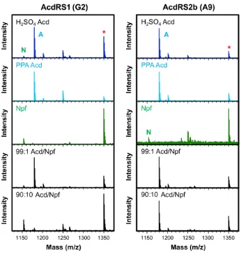
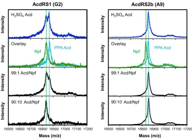
Figure 2.2 In vivo AcdRS selectivity. CaM (UAG codon at 113) was expressed in minimal media containing 1 mM Uaa: Acd, synthesized either using the H2SO4 route or PPA route,
or Npf. Expression was performed with one of the three AcdRSs indicated. Significant incorporation of Npf is seen for AcdRS2a (G11) when using H2SO4 Acd, but only
Acd-containing protein is seen with AcdRS2b (A9).
For a more rigorous, quantitative analysis of selectivity, we analyzed the
CaM trypsin digest data by normalizing the intensity of the peak for the 108-116
fragment, containing Acd or Npf at position 113, to the intensity of the peak for the
117-127 fragment. (Figure 2.3) CaM117-127 should be produced in a 1:1 ratio with
CaM108-116 when the protein is completely digested by trypsin, and this can be
confirmed by varying the digest time and observing that the intensity ratios do not
change (data not shown). Normalization using an internal standard is essential to
interpreting the intensity data correctly. The CaM108-116Acd113 fragment (A in
36
while the CaM108-116Npf113 fragment (N in Figure 2.3) ionizes 6.1-fold worse than
the CaM117-127 fragment. This can be seen by examining the peak ratios for PPA
Acd (i.e., Acd only) and Npf expressions. After peak scaling, one obtains an
MS-based Acd/Npf selectivity ratio (MS Sel) of 2.6 X 10⁻³ for AcdRS1 and 0.20 for
AcdRS2b, calculated as follows:
MS Sel = (Scaled Acd/ Scaled Npf)/(Acd/Npf ratio in media)
The AcdRS1 MS Sel value is in good agreement with our estimate of
100-fold selectivity for Npf based on the less quantitative whole protein MALDI MS
data. While the AcdRS2b MS Sel value may seem surprisingly low given the
absence of any obvious CaM108-116Npf113 peak in the MALDI spectra, it is important
to keep in mind that the RS is only being challenged with at most 10% Npf in the
media and that CaM108-116Npf113 ionizes 30-fold worse than CaM108-116Acd113.
Using these Acd/Npf selectivity ratios, we determined that selection resulted in a
76-fold improvement in selectivity for AcdRS2b relative to AcdRS1. We note that
the effective in vivo Acd selectivities for both AcdRS1 and AcdRS2b are less than
37
Figure 2.3 CaM113 trypsin digest data for AcdRS selectivity analysis. CaM (UAG codon at 113)
was expressed in minimal media containing 1 mM Uaa: Acd, synthesized either using the H2SO4
route or PPA route, Npf, or either a 99:1 or 90:10 mixture of PPA Acd and Npf. Expression was performed with one of the three AcdRSs indicated. The peaks for the (M+H)+ masses of the
CaM108-116Acd113 fragment (A, 1179.8 Da), CaM108-116Npf113 fragment (N, 1153.8 Da), and CaM
117-127 fragment (*, 1349.9 Da) are indicated. The intensities of the CaM108-116Acd113 and CaM
38 In Vitro Characterization of RS Activity.
To better understand AcdRS1 and 2b selectivity, we generated His-tagged
variants of the enzymes, then expressed and purified them for in vitro activity assays.
Charging of tRNA by RSs is a two-step process, where the first step is the
RS-catalyzed reaction of amino acid with ATP to form an aminoacyl-adenylate
intermediate (Aa-AMP), releasing inorganic pyrophosphate (PPi); and the second
step is the reaction of this enzyme-bound adenylate with the 2’ or 3’ hydroxyl group
on A76 at the 3’ end of the tRNA.65
Aa + ATP –> Aa-AMP + PPi (1)
Aa-AMP + tRNA –> Aa-tRNA + AMP (2)
While some prior studies have used assays that measure only the first step of
the aminoacylation reaction to demonstrate that in vitro activities are consistent with
Uaa incorporation,10, 66-67 it has been shown by the Perona laboratory that full tRNA
aminoacylation assays correlate well with in vivo observations of RS activity.68 This
is expected, since amino acid incorporation into protein in vivo can only occur upon
aminoacylation. Thus, aminoacylation of an in vitro transcribed 32P-labeled
tRNACUA was measured under single-turnover conditions at a variety of Acd or Npf
concentrations for AcdRS1 and AcdRS2b. Plots of the first-order rate constants as
a function of amino acid concentration were used to determine kobs and Kd for each