INTERNATIONAL RESEARCH JOURNAL OF PHARMACY
www.irjponline.com
ISSN 2230 – 8407
Research Article
FORMULATION AND EVALUATION OF CURCUMIN LOADED THIOLATED POLYMER COATED
LIPOSOMES FOR APHTHOUS ULCERS
Neha Manish Munot *
1, Kishore N. Gujar
21
PhD Scholar, PAHER’s Pacific University, Debari, Udaipur Rajasthan, Assistant Professor at S.T.E.S’s Smt. Kashibai
Navale College of Pharmacy, Kondhwa , Pune 48 , Maharashtra, India
2
STES’s Sinhgad College of Pharmacy, Vadgaon (Bk), Pune, Maharashtra, India
*Corresponding Author Email: nehamunot@yahoo.comArticle Received on: 08/10/18 Approved for publication: 22/12/18
DOI: 10.7897/2230-8407.100119
ABSTRACT
Aphthous ulcers or Recurrent Aphthous Stomatitis (RAS) are inflammatory lesions of mucous lining of mouth associated with redness, swelling and occasional bleeding from affected area(s). Curcumin, a herbal drug was selected due to its antioxidant, anti-inflammatory, antimicrobial and wound healing ability. Present study deals with formulation, optimization and evaluation of curcumin loaded liposomes for local drug delivery as liposomes have property to get concentrated in inflamed areas. Efficacy of liposomes was enhanced in terms of mucoadhesion, retention in the buccal mucosa and sustained release of drug by coating them with Thiolated Karaya gum. Curcumin loaded liposomes, prepared using thin film hydration method were
optimized by 32 factorial design using Design Expert Software. Batch F8 having entrapment 75.1±0.94 % and controlled the release of curcumin up to
12 h was considered optimized and was coated with thiolated karaya gum to get TKF8. Successful coating was evident from TEM images, increase in particle size from 129.86 ± 9.1 nm to 152.39 ± 1.3 nm, zeta potential from negative to positive -10.5 mV to 9.55mV. TKF8 showed better mucoadhesion
by absorbing 6.98 % more amount of mucin and also had ability to concentrate in the buccal mucosa for 24 h as observed using exvivo studies. It can
be attributed to positively charged thiolated polymer interacting with negatively charged sialic acid residues in the mucus. Drug release from F8 and TKF8 was sustained for 12 h and 24 h respectively and they followed diffusion controlled release (Peppas model). This formulation can be efficient therapeutic strategy for apthous ulcers which would provide relief to the patients.
Keywords: Apthous ulcers, curcumin, liposomes, thiolated karaya gum, mucoadhesion, coated liposomes
INTRODUCTION
Oral cavity is lined with mucosa which may be susceptible to many inflammatory, atrophic and ulcerative conditions like aphthous stomatitis, lichen planus, erythema etc. Aphthous ulcers or Recurrent Aphthous Stomatitis (RAS), commonly referred to as canker sores, are painful, inflammatory lesions of the mucous lining of the oral cavity. The symptoms of RAS include pain, weakness and major alterations in oral functions, such as speech, chewing and swallowing1.There is no specific treatment for RAS.
Treatment is aimed at reducing pain, speeding healing and reducing recurrence. Common treatment modalities of RAS are topical agents. The efficacy of treatments of oral ailments is often challenged by a low residence time of the conventional pharmaceutical formulations in the oral cavity due to salivary secretion, swallowing, food intake and abrasive actions of the soft tissue2. Topical treatment of ulcerative inflammatory diseases is
associated with several general disadvantages, like high permeability of the oral mucosa for drugs which could cause release of drug into the blood circulation and some unwanted side effects. In order to localize the effect of drugs the use of liposomal formulations with encapsulated drug has been investigated 3.
M.T.Ercn et al carried out in-vivo studies on rats for the treatment of oral ulcers with liposomal dexamethasone sodium phosphate and suggested that, in the case of humans, the liposomes should be modified to decrease removal of drug from lesional area.4Also,
liposomes suffer from instabilities such as aggregation, fusion, degradation, hydrolysis and oxidation of phospholipids5. Polymer
coating is a promising way to modify the surface characteristics of liposomes in order to improve their applicability. These
liposomes can be coated with mucoadhesive polymers to increase the residence time at the ulcerated area. Some researchers have reported chitosan-coated liposomes can improve their properties and applicability6. First generation mucoadhesive polymers like
chitosan, karaya gum etc. form weak non-covalent bonds like Van der Waal’s interaction, hydrogen bond or ionic interaction with mucus which cannot prolong the residence time of the dosage form to a great extent. Thiomers or thiolated polymers are a new generation of mucoadhesive polymers capable of forming intra- and interchain disulfide bonds within the polymeric network leading to strongly improved cohesive properties and stability of drug delivery systems7.Due to the formation of strong covalent
(S-S disulphide) bond with mucus glycoproteins, thiomers show the strongest mucoadhesive properties of all so far tested polymeric excipients via thiol disulfide exchange reaction and an oxidation process. Apart from mucoadhesion, thiolated polymers have ability to control the drug delivery8. Hence, in this study,
drug loaded liposomes were coated with thiolated karaya gum for improved efficacy.
Current therapeutic treatments of RAS involve use of immune-modulating steroids that suppress the inflammatory response and reduce symptoms. They also induce unacceptable and sometimes serious systemic side effects. These may render the ulcer surface more susceptible to anaerobic infections or increase the risk of candidiasis and mucosal atrophy9. Therefore, a combination of
from rhizome of Curcuma longa, family zingiberaceae. In 2012, an randomized placebo controlled trial was done using 2% curcumin gel in treatment of minor RAS, which showed that it is a well-tolerated effective antibacterial, anti-tumor agent with potent anti-inflammatory and analgesic properties10. Curcumin
also shows wound healing activities effective in treatment of mouth ulcers by increasing cellular proliferation and collagen synthesis at the wound site as evidenced by increasing in DNA, total protein and type-III collagen content of wound tissue leading to faster rate of epithelilisation, wound contraction11.
Hence, in the present study, curcumin loaded liposomes were coated with thiolated karaya gum for site specific, local delivery of curcumin for oral lesions which would sustain the release of drug, reduce dose and dosing frequency of drug, reduce systemic exposure of drug finally reduce the side effects and improve patient compliance.
MATERIALS AND METHODS
Curcumin (Cu) was kindly gifted by Amser Pvt. Ltd. Goa. Egg Phosphotidylcholine (Egg PC) Lipova E-120 was gifted by Vav Life Sciences Mumbai, Cholesterol (CH), Ethanol, Methanol, t-butanol, acetone were purchased from Research lab India, Thioglycolic acid (80%) were procured from Thomas Baker, Gum karaya and L cysteine were procured from Research Lab Fine Chem Industries, 5,5′-Dithiobis-(2-nitrobenzoic acid) (DTNB) (99%)/Ellman’s reagent and Mucin from bovine submaxillary glands (Type I-S) were procured from Sigma Aldrich. All other materials and solvents were of analytical grade.
Synthesis and evaluation of Thiolated Karaya Gum (TK)
Synthesis of TK is discussed elsewhere and optimized batch of TK was resynthesized as reported by N.M. Munot et al7. Briefly,
TK was synthesized by esterification of Gum Karaya (GK) with TGA (80%) in presence of hydrochloric acid. GK (5 g) was dispersed in 70 of cold water, 2.5 mL of TGA (80%) and 2 mL of 7 N hydrochloric acid were added to it. This reaction mixture was allowed to react at 70 0C for 4 h. It was then poured in 500 mL
methanol. White precipitate of TK so obtained was filtered and washed twice with methanol. Product was then added to ethanol (50 mL) in order to solubilize the impurities, filtered again and dried under IR lamp.
Thiol modification of karaya gum was confirmed using FT-IR spectroscopy, DSC analysis and Ellman's method. FT-IR spectroscopy was performed using FTIR spectrophotometer (Jasco V 630). DSC thermogram of GK and TK was recorded using Differential scanning calorimeter (SIIO 6300 with auto sampler, SIIO, Japan). Degree of thiol group substitution was determined by using DTNB by Ellman’s method12. An accurately
weighed GK (control) and TK (50 mg) were dispersed in 25 mL distilled water separately. DTNB (5mL, 0.03% w/v) was allowed to react with an aliquot of 2.5 mL of polymer solution which was diluted with 2.5 mL of 0.5 M pH 8.0 phosphate buffer for 2 h at room temperature in dark. UV Spectrophotometer (Jasco V-630) was fixed at 412 nm and absorbance of reaction mixture was measured and compared with standard curve prepared using L cysteine reacted with DTNB as mentioned above.
Preparation of curcumin loaded liposomes (CL)
CL were prepared using thin film hydration method. Briefly Egg
PC, CH and curcumin in the weight ratio of 10:5:1 were dissolved in 10 mL of methanol – t-butanol mixture (1:2) to form clear solution in 250mL round bottom flask. Organic solvent was evaporated at 37 0C for 30 min at 100 rpm by using rotary
evaporator (Heidolph, Germany). After complete removal of organic solvent dried thin lipid film formed over the inner surface
of a round-bottom flask was hydrated with 10 mL distilled water to form dispersion. During this process temperature was maintained at 370C and speed at 60 rpm for 30 min. Prepared
dispersion was sonicated for 10 min in bath sonicator (Spectra labs, Navi Mumbai). Then dispersion was kept overnight at 2-8oC
for complete vesiculation. Prepared liposomes were further optimized using factorial design.
Optimization of curcumin loaded liposomes
To study the effect of variables on CL, different batches were prepared using 32 factorial design. Amount of Egg PC and CH
were selected as two independent variables. Entrapment efficiency (EE) and percent drug release were selected as dependent variables. Experimental trials were performed at 3 possible combinations higher, lower and middle. The resulting data were fitted into Design Expert 11 software and analyzed statistically using analysis of variance (ANOVA). The data were also subjected to 3-D response surface methodology to determine the influence of Egg PC and CH on dependent variables as given in Table 1, 2 and 3.
Preparation of thiolated karaya gum coated liposomes loaded with curcumin
Aqueous solution (1%, w/v) of TK was added drop wise into optimized liposomal suspension (F8) at equal volume with magnetic stirring of 500 rpm for 60 min followed by incubation in refrigerator overnight. The final concentration of TK was 0.5% (w/v). Excess TK and curcumin were then removed by ultracentrifugation (Remi, India) at 2000 rpm for 30 min followed by a single washing with distilled water. The resulting TK-coated liposomes (TKF8) were resuspended in phosphate buffer solution13.
Entrapment Efficiency (EE)
The liposomal suspension was centrifuged (Remi, India) at 2000 rpm for 45 minutes to remove the curcumin attached possibly on the surface of liposomes, and then re-dispersed with the PBS. This procedure was repeated several times to make sure that the curcumin in the supernatant was hardly detected. Supernatant was removed and methanol was added to sediment and sonicated for 10 minutes to disrupt the liposomes. The vesicles were broken to release the drug, which was then estimated for the drug content. The absorbance of the drug was noted using Ultraviolet Spectrophotometry (Shimadzu V-630 Jasco, Japan) at the wavelength of 425 nm which is λmax for curcumin. Entrapment
efficiency was then calculated using following equation,
Entrapment Efficiency EE (%) = Entrapped drug X 100 Total drug added
Vesicle size and size distribution
The mean vesicle size and vesicle size distribution of the optimized batches F8 and TKF8 were obtained by particle size analyzer (Sympatec - Nanophox (NX0088)). The instrument measures the vesicle size and its distribution based on the dynamic light scattering theory. The apparatus consists of a He-Ne laser beam of 632.8 nm focused with a minimum power of 10 mW using scattering angle 900 to the sample cell of 10 X 10 mm2
and calculate particle size by 3D cross correlation technique. The average values from at least three measurements were reported, with the polydispersity index (PDI)
Transmission Electron Microscopy (TEM) analysis
Electron Microscopy (TEM). Formulation (F8 and TKF8) 20 µl and 20µl of 1% phosphotangustic acid was taken in Eppendroff tube by micro pipette and mixed gently. A drop of the mixture was placed on to the carbon-coated grid and excess was drained. The grid was allowed to dry, and it was observed under Transmission Electron Microscope (Model CM 200 SUPERTWIN STEM Philips TEM resolution 0.23 nm, voltage 200 kV).
Determination of Zeta potential
Charge on drug loaded vesicles surface (F8 and TKF8) was determined using Zetasizer Version 3.94 (Brookhaven Instrument Corporation). Analysis time was kept for 1 min at pH 6.7 and average zeta potential and charge on the liposomes was determined. Dispersion of liposomes in distilled water was used for analysis. Temperature was kept at 250C. The result obtained
for each batch was the average of three measurements on the same sample aliquot.
Ex vivo drug retention studies for localization of drug in buccal mucosa
Curcumin release from uncoated and coated liposomes (F8 and TKF8) and its retention in the buccal mucosa were determined through Franz diffusion cells (Orchid Scientifics, India) using porcine buccal mucosa (0.5 cm2) received fresh from a local
abattoir. A mixture of 5 mL of simulated saliva fluid (2.38 g Na2HPO4, 0.19 g KH2PO4, and 8.00 g NaCl per liter of distilled
water adjusted with phosphoric acid to pH 6.75) and methanol (60:40) was used as the receptor phase, allowing sink conditions at 37°C ± 0.5. The samples withdrawn directly at appropriate time intervals were analyzed at 425nm by a validated UV Spectroscopy method using double beam spectrophotometer
(Shimadzu V-630 Jasco, Japan). At the end of the experiment (8 h for F8 and 24 h for TKF8), buccal mucosa was extracted with receptor medium and the amount of drug in the mucosa was found.
Mucoadhesion study: Mucin adsorption study
The adsorption of mucin on the surface of the liposomes was measured to evaluate the mucoadhesive properties of the liposomes. First, 1 mL of mucin (1 mg/mL) was stirred with one mL of TK-coated liposomes (1 mg/mL and 2 mg/mL) for 2 hours at 37°C. The same procedure was performed for the noncoated liposomes (F8). The suspension was then centrifuged at 2000rpm for one hour. The amount of free mucin was determined in order to assess the amount of mucin adsorbed onto the liposomes according to the difference between total and free mucin14.
Percent adsorption was determined using the equation below:
Adsorption % = Total amount of mucin − Free mucin ×100 Total amount of mucin
In vitro drug release studies
The release of drug from liposomal formulations was determined using the membrane diffusion technique. Liposomal dispersions (5 mL) sealed in dialysis bag (Himedia) was immersed in 200 mL simulated saliva medium maintained at 37±0.5oC and placed in
orbital incubator shaker (Chromous Biotech # OS-02) rotated at 50 rpm. The samples (2mL aliquots) were withdrawn at scheduled intervals of 1 h up to 10 h and then 24 h. The medium was replaced with equivalent amount of dissolution medium to maintain in vivo sink conditions. The aliquots were analyzed for drug content by UV spectroscopy at 425nm.
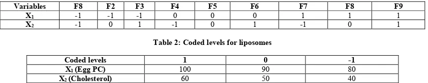
Table 1: Coded formulations for liposomes
Variables F8 F2 F3 F4 F5 F6 F7 F8 F9
X1 -1 -1 -1 0 0 0 1 1 1
X2 -1 0 1 -1 0 1 -1 0 1
Table 2: Coded levels for liposomes
Coded levels 1 0 -1
X1 (Egg PC) 100 90 80
X2 (Cholesterol) 60 50 40
Table 3: Optimization data analysis showing % Entrapment efficiency and % drug release of liposomes
Formulation Coded levels % Entrapment
Efficiency % Drug Release (8h)
X1(Egg PC in mg) X2 (Cholesterol in mg)
F1 80 40 30.52 98.16
F2 80 50 37.38 95.77
F3 80 60 41.14 90.34
F4 90 40 54.59 92.13
F5 90 50 60.69 86.93
F6 90 60 63.19 84.17
F7 100 40 69.28 82.78
F8 100 50 75.1 80.21
F9 100 60 75.19 83.47
Table 4: ANOVA for response surface model for entrapment efficiency of liposomes
Source Sum of Squares df Mean Square F-value p-value
Model 2200.12 5 440.02 1919.77 < 0.0001 significant
A-Egg PC 2036.15 1 2036.15 8883.46 < 0.0001
B-CH 105.25 1 105.25 459.21 0.0002
AB 5.55 1 5.55 24.20 0.0161
A² 44.59 1 44.59 194.53 0.0008
B² 8.58 1 8.58 37.45 0.0088
Residual 0.6876 3 0.2292
Table 5:ANOVA for response surface model for drug release of liposomes
Source Sum of Squares df Mean Square F-value p-value
Model 309.85 7 44.26 364.81 0.0403 Significant
A-Egg PC 121.06 1 121.06 997.70 0.0201
B-CH 31.68 1 31.68 261.10 0.0393
AB 18.11 1 18.11 149.21 0.0520
A²B 6.44 1 6.44 53.06 0.0869
AB² 6.56 1 6.56 54.04 0.0861
Residual 0.1213 1 0.1213
Cor Total 309.97 8
Table 6: Release kinetics summary for liposomes
Model Zero
order T-test 1
st order T-test Matrix T-test Peppas T-test Hix.Crow. T-test
R 0.9369 9.285 0.9525 10.838 0.9574 11.489 0.9736 14.773 0.9674 13.237
K 3.0313 (Passes) -0.0662 (Passes) 14.8532 (Passes) 6.3211 (Passes) -0.0163 (Passes)
Figure 1: Scheme of synthesis of thiolated karaya gum
(a) (b)
Figure 2: FTIR spectrum of Gum Karaya (a) and Thiolated gum karaya (b)
(a) (b)
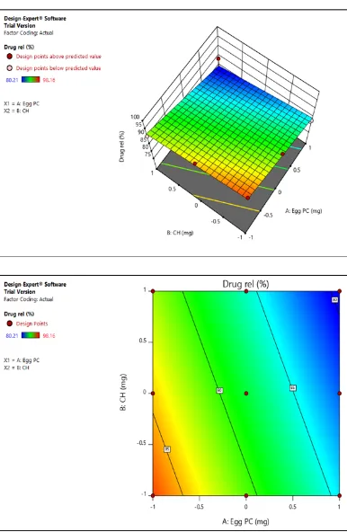
Figure 4: Effect of lipid concentration on entrapment efficiency of liposomes and Contour plot for effect of lipid concentration on entrapment efficiency of liposomes
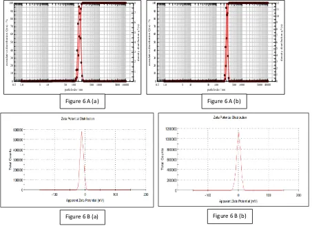
Figure 6: Particle size analysis (6A) and Zeta potential (6B) of Batch F8 [6 A (a), 6 B (a)] and TKF8 - thiolated polymer coated liposomes [6 B (a), 6 B (a)]
(a) (b) Figure 7: Morphology of uncoated liposomes (a) and thiolated polymer coated liposomes (b) using TEM
0 10 20 30 40 50 60 70 80 90 100
c
um
ul
a
ti
ve
di
s
tri
but
ion Q
(x) /
%
0 1 2 3 4 5 6 7 8 9 10 11 12
de
ns
it
y di
s
tri
but
ion q
*(x)
0.5 1.0 5 10 50 100 500 1000 5000 10000 particle size / nm
0 10 20 30 40 50 60 70 80 90 100
c
um
ul
a
ti
ve
di
s
tri
but
ion Q
(x) /
%
0 1 2 3 4 5 6 7 8
de
ns
it
y di
s
tri
but
ion q
*(x)
0.5 1.0 5 10 50 100 500 1000 5000 10000 particle size / nm
Figure 6 A (a) Figure 6 A (b)
Figure 8: In vitro drug release studies
RESULT AND DISCUSSION
Synthesis and characterization of Thiolated polymer
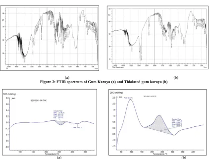
Covalent bonding between GK and TGA carried out by esterification of hydroxyl groups in GK by carboxyl groups in TGA (Fig 1) lead to thiolation of GK. TK was found to contain 5.026 mM of thiol groups per gram of polymer as determined by Ellman’s assay.
Thiolation of GK was confirmed by FTIR spectroscopy that shows all the peaks found in GK but additional peak at 2549 cm -1 due to –SH stretch and more intensity COO stretch at 1749 cm -1 as seen in Fig.2, FTIR spectra of unmodified gum (GK) shows
broad peak of –OH group at 3442 cm-1, CH stretch at 2922 cm-1,
COO asymmetric stretch at 1734 cm-1 and COO symmetric
stretch at 1419 cm-1.
Also, Fig.3, displays DSC thermogram of GK and TK. DSC thermogram of GK showed an endotherm at 254.30C with heat of
fusion -12.39 J/g while DSC of TK showed the broad exotherm at 236.60C with heat of flow 316.1 J/g. The difference in the DSC
pattern indicated modification of gum karaya. This synthesized polymer was used for coating curcumin liposomes which would increase the mucoadhesion and residence time of the formulation ultimately leading to better efficacy.
Formulation and optimization of curcumin loaded liposomes
Liposomes were prepared using thin film hydration technique and method was found to be well suited for the production of liposomes without aggregation. Responses of different batches were obtained using factorial design (Table 3).
Obtained data were subjected to multiple regression analysis using Design Expert 11 software and obtained data were fitted in following equation
Y = b0 + b1X1 + b2X2 + b11X1X1 + b22X2X2 + b12X1X2
Where Y is the dependent variable; b0 is the arithmetic average of
all the quantitative outcomes of nine runs. b1, b2, b12 are the
estimated coefficients computed from the observed experimental response values of Y and X1 and X2 are the coded levels of the
independent variables. The interaction term (X1X2) shows how
the response values change when two factors are simultaneously changed.
Statistical validity of the polynomials was established on the basis of analysis of variance (ANOVA) provision in the Design Expert software. Level of significance was considered at p < 0.05. The best-fitting mathematical model was selected based on the comparison of several statistical parameters, including the coefficient of variation (CV), the multiple correlation coefficient(R2), the adjusted multiple correlation coefficient
(adjusted R2), and the predicted residual sum of squares (PRESS),
provided by the software.
PRESS indicates how well the model fits the data, and for the chosen model, it should be small relative to the other models under consideration. The 3-D response surface graphs and the 2-D contour plots were also generated by the 2-Design Expert® software. These plots are very useful to see interaction effects of the factors on responses.
Response 1- Entrapment Efficiency (EE)
Determination of EE is an important parameter in case of liposomes to avoid drug loss and it may also affect the drug release. In the present study, the observed EE for all batches were in the range of 30.83- 75.35%. To understand the effect of lipid concentration on EE, coefficient observed for EE fitted in equation.
Final equation in terms of coded factors:
Entrapment efficiency(EE)= +60.87+18.42X1+4.19X2 +1.18X1X2 + 4.72
X1²+2.07X2²
i.e. Entrapment efficiency ( EE) = +60.87+18.42(Egg PC) +4.19(CH)+ 1.18 EggPC* CH +4.72(Egg PC)
² +2.07(CH) ²
Final equation in terms of actual factors: =
+60.87111+18.42167Egg PC + 4.18833CH + 1.17750 EggPC* CH + 4.72167 Egg PC² + 2.07167CH²
A positive correlation was observed for both variables X1(Egg
PC and CH, entrapment efficiency found to be increased. EE of the curcumin liposomes were increased with increasing the lipid content. This is may be due to high amount of lipid available to encapsulate the drug. Also, upon increasing the lipid content number of layers coating the drug also increased, this resulted in an increase in the EE. Further increase in the lipid content above optimum concentration, there is no much increase in the entrapment due to the availability of the drug to be incorporated is low.
Drug concentration also affects EE- as we go on increasing drug concentration with constant lipid content it was observed that entrapment also go on increasing but up to a certain limit above that entrapment decreases due to excess of drug which cannot be encapsulated by lipids.
Data were analyzed statistically by one-way analysis of variance (ANOVA) using software.
As seen in the Table 4, The Model F-value of 1919.77 implies the model is significant. There is only a 0.01% chance that a "Model F-Value" this large could occur due to noise. Values of "Probability > F" less than 0.0500 indicate model terms are significant.
The 3D response surface (Fig 4) predicts high entrapment at maximum amount of Egg PC and cholesterol level, hence showing F8 batch as optimized batch.
Response 2- Drug Release
Most important parameter which needs to be evaluated after manufacturing of liposomes, is the drug release from the liposomes as it determines its in vivo performance.
It was observed that the relative amount of Egg PC and CH was found to play important role in vesicle size and entrapment which affects the drug release. To understand the effect of lipid concentration on drug release, coefficient observed for liposome drug release was fitted in Equation.
Final equation in terms of coded factors: Drug Release = +88.22-6.30 X1-2.51 X2
Final equation in terms of actual factors:
Drug Release =+88.21778 - 6.30167Egg PC - 2.51500CH
A negative correlation was observed for both variables X1(Egg PC) and X2 (CH). Thus with increase in the concentration of Egg PC and CH, percentage of drug released was found to be decreased, and thus sustained release of drug could be possible. This can be attributed to increase in the diffusion path length as coating of the lipid on the drug is increased upon increase in lipid content.
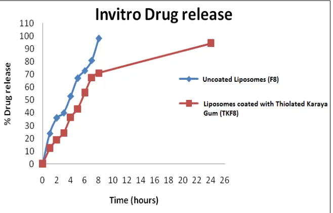
Drug release was found to be almost 100 % within 8 h from formulation batches F1 to F4 (Table 3) whereas formulation F5 to F9 could sustain the release of drug up to 12 hours, which was desirable. Experimental and Predicted values for Optimized Formulation F8were fitted to Design Expert Software version 11, the dependent variables demonstrated that the model was significant for both response variables (EE and drug release). Comparison between the experimental and predicted values for EE was found to be 75.51 and 75.1 respectively; while Drug release was found to be 83.5% and 80.21% respectively.
Most probable optimal formulation F8 show 0.923 desirability, it can be concluded that as predicted values agree with experimental
values, it demonstrates the feasibility of the model in the development of liposomes formulation
Data were analyzed statistically by one-way analysis of variance (ANOVA) using software. From the Table 5 it can be seen that Model F-value of 364.81 implies the model is significant. There is only a 0.04 % chance that a "Model F-Value" this large could occur due to noise. Values of "Prob > F" less than 0.0500 indicate model terms are significant.
3D response surface images show the optimum region for drug release, as F8 batch was showing optimum drug release, it is selected by software.
Drug release kinetics
Data treatment to drug release from liposomes (batch F8) showed that it followed Korsemeyer-Peppas model with n = 0.7896 (Table 6). This indicates the drug release mechanism by diffusion, as proposed by Peppas. On the basis of the Korsmeyer-Peppas model, the best fitting was obtained with n > 1, indicating super case II. When the chain relaxation process is very slow compared with diffusion, the case II transport occurs, which again confirms that the drug release is controlled mainly by diffusion.
Evaluation of thiolated karaya gum coated liposomes loaded with curcumin
Optimized batch of liposomes loaded with curcumin (F8) was coated with thiolated karaya gum for improved mucoadhesion. Phosphatidyl choline is amphiphilic moiety, in which one side with double carbon chains shows a hydrophobic property, and the other side with phosphate (PO4 3−) and choline display a
hydrophilic property and cell affinity. Curcumin and Egg PC could spontaneously form liposomes with solvent removed gradually due to low solubility of curcumin and amphiphilic property of Egg PC15 .The negative surface of the liposomes and
cationic TK forms firm coating by ionic linking, and the free –SH groups are easily oxidized or crosslinked to be stable –S–S– groups, which will be more efficient to stabilize the liposomes than that by plain karaya gum.
Vesicle size and size distribution
The mean vesicle/ particle size of the uncoated liposomes (Batch F8) was found to be 129.86 ± 9.1 nm, polydispersibility index (PDI) of 0.31. After coating (batch TKF8) it increased to 152.39 ± 1.3 nm and PDI of 0.20 (Fig 6 A). This increase in the size of the vesicles can be explained by the interaction between lipid and TK. PDI values indicated that particle size was well controlled with a narrow dispersity.
Determination of Zeta potential
Transmission Electron Microscopy (TEM) analysis
The morphological appearance of uncoated (Fig 7a) and coated liposomes was examined by TEM. Both groups of liposomes present nanosized, spherical shapes, with no rupturing of the membrane wall after coating with TK. The morphology of the coated liposomes TKF8 showed visible layer on the liposomal surface along with increase in the vesicle size, as shown in Fig. 7b.
Ex vivo drug retention studies for localization of drug in buccal mucosa
The amount of Curcumin detected in the receptor medium of Franz diffusion cell was under the detection limits during the ex vivo drug-release studies (the limit of detection value was 0.029). Buccal mucosa was extracted and the amount of drug in the mucosa was found to be 91.28% ± 2.65% and 89.92% ± 1.65% of the initial dose from F8 and TKF8 after 12 h and 24 h respectively. This indicates that prepared both the liposomal formulations did not penetrate through buccal mucosa to reach the receptor compartment of Franz diffusion cell which is representative of systemic circulation in vivo. This shows that, under the study conditions, prepared formulation might potentially be used as a vehicle for local delivery for oral lesions. Although drug was not detected in receptor phase, the fact that curcumin was extracted from the mucosa confirmed that liposomes were localized in the tissue. Results obtained are in accordance with Sinem Yaprak Karavana et at16
Evaluation of mucoadhesion: Mucin adsorption study
Invitro efficacy of uncoated liposomes to sustain the drug release upto 12 h can be marred as they cannot be retained on the affected area due to lack of mucoadhesion. Hence, these liposomes were coated with positively charged thiolated karaya gum which can interact with negatively charged mucin secreted from buccal mucosa. Therefore, the mucoadhesiveness of liposomes was estimated by measuring the amount of mucin adsorbed on the surface of the TK coated liposomes. Compared with the uncoated liposomes (F8), the amount of mucin adsorbed on the surface of the coated liposomes (TKF8) was 6.98 fold greater. This result may be explained by the strong electrostatic interaction between the positively charged TK and the negatively charged acidic sialic moieties of mucin, although hydrogen bonding and hydrophobic interactions may also be involved. In that sense, the charge and the chemical groups on coated liposomes are favorable for mucoadhesion that would increase the residence time of curcumin in the oral lesions, resulting in prolonged drug absorption.
In vitro drug release studies
Drug release from prepared liposomes followed diffusion mechanism is reported earlier (Table 7). It can be seen from the Figure 8 that liposomal vesicles (Batch F8) could sustain the release of curcumin up to 12 h but coating of TK on liposomes (Batch TKF8) further sustained the release of drug up to 24 h. due to inter- and intramolecular crosslinking of thiolated polymer chains which resulted in swelling of the polymer and thus increasing the diffusional path length of drug resulting in gradual and continuous release of drug.
CONCLUSION
Oral lesions like Apthous ulcers or RAS if untreated may lead to trauma and inconvenience. In order to localize the drugs within the oral lesions, the use of liposomal encapsulation has been investigated. Liposomes suffer from the limitation that they cannot be retained on the affected mucosal area for long time.
Hence, in the present study, curcumin loaded liposomes were coated with second generation mucoadhesive polymer, thiolated Karaya gum which would form strong covalent bond with mucin in mucous leading to improved mucoadhesion and retention in the mucous membrane. Coating also led to sustaining the release of drug for 24 h in comparison to 12 h in case of uncoated liposomes.
All these results obtained in the present research work are preliminary evidence of superior efficacy of mucoadhesive liposomes and additional clinical trials should be conducted to confirm these conclusions. Depending on the above mentioned results, it can be said that curcumin loaded thiolated karaya gum coated liposomes may be promising systems as drug carriers in the treatment of inflammatory ulcerative diseases of oral mucosa.
REFERENCES
1. Akintoye S. O., Greenberg M. S. Recurrent Aphthous Stomatitis. Dental Clinics of North America 2005 ; 49 Suppl 1: 31– 47.
2. Tarakji B., Gazal G., Al-Maweri S. A, Azzeghaiby S. N, Alaizari N. Guideline for the Diagnosis and Treatment of Recurrent Aphthous Stomatitis for Dental Practitioners. Journal of International Oral Health 2015; Suppl 7:74–80. 3. Milan Petelin, Marjeta S,Entjurc C, Zorka Stolic, Uros
Skaleric. EPR study of mucoadhesive ointments for delivery of liposomes into the oral mucosa. International Journal of Pharmaceutics 1998;173 :193–02.
4. Farshit F.S., Ozer A.Y., Ercan M.T., Hincal A. A. In-vivo studies in the treatment of oral ulcers with liposomal dexamethasone sodium phosphate. Journal of Microencapsulation 1996; 13 Suppl 3: 293-06
5. Tantisripreecha C., Jaturanpinyo M., Panyarachun B., Sarisuta N. Development of delayed-release proliposomes tablets for oral protein drug delivery. Drug Development and Industrial pharmacy 2012; 38: 718–27
6. Nguyen T.X., Huang L., Liu L., Abdalla A.M.E., Gauthier M., Yang G. Chitosan-coated nano-liposomes for the oral delivery of berberine hydrochloride. Journal of Materials Chemistry B 2014 ; 2:7149–59.
7. N.M.Munot, S.S.Bahulkar, S.S.Surwase. Synthesis, characterization of thiolated karaya gum and evaluation of effect of pH on its mucoadhesive and sustained release properties. Carbohydrate Polymers 2015; 130:183-90. 8. Andreas Bernkop- Schnurch. Thiomers: A new generation of
mucoadhesive polymers. Advanced Drug Delivery Reviews 2005; 57: 1569– 82
9. Holbrook WP, Kristmundsdottir T, Loftsson T. Aqueous hydrocortisone mouthwash solution: clinical evaluation. Acta Odontologica Scandinavica 1998; 56:157 – 60.
10.Manifar S, Obwaller A, Gharehgozloo A, Boorboor Shirazi Kordi HR, Akhondzadeh S. Curcumin gel in treatment of minor aphthous ulcer: A randomized, placebo-controlled trial. Journal of Medicinal Plants 2012; 11:41-49.
11.Thorat Yogesh, Sarvagod Asha, Kulkarni Shital, Hosmani Avinash.Treatment of mouth ulcer by curcumin loaded thermoreversible mucoadhesive gel: a technical note. International Journal of Pharmacy and Pharmaceutical Sciences 2015; 7 Supp 10 :399-402
12.Rahmat, D., Sakloetsakun, D., Shahnaz, G., Perera, G., Kaindl, R., Bernkop-Schnürch, A. Design and synthesis of a novel cationic thiolated polymer. International Journal of Pharmaceutics 2011; 411: 10–17.
13.Martin Werle, Kohei Hironaka, and Hirofumi Takeuchi. Development and In Vitro Characterization of Liposomes Coated with Thiolated Poly(Acrylic Acid) for Oral Drug Delivery. Drug Development and Industrial Pharmacy 2009; 35:209–15
in vitro/in vivo characterization. International Journal of Nanomedicine 2014; 9: 2299–306
15.Li, R., Deng, L., Cai, Z., Zhang, S., Wang, K., Li, L., Zhou, C. Liposomes coated with thiolated chitosan as drug carriers of curcumin. Materials Science and Engineering: C 2017; 80: 156–64.
16.Sinem Yaprak Karavana, Evren Homan Gökçe, Seda Rençber, Seda Özbal, Çetin Pekçetin, Gökhan Ertan. A new approach to the treatment of recurrent aphthous stomatitis with bioadhesive gels containing cyclosporine A solid lipid
nanoparticles: in vivo/in vitro examinations. International Journal of Nanomedicine ; 2012:7:5693–704
Cite this article as:
Neha Manish Munot and Kishore N. Gujar. Formulation and evaluation of curcumin loaded thiolated polymer coated liposomes for aphthous ulcers. Int. Res. J. Pharm. 2019;10(1):103-112
http://dx.doi.org/10.7897/2230-8407.100119
Source of support: Nil, Conflict of interest: None Declared