Chemical, toxicological, anti-inflammatory and
antimicrobial evaluation of Ganoderma lucidum
extracts
Nilsa Sumie Yamashita Wadt1*, Marise K. H. Okamoto1, Edgar Matias Bach Hi2, Erna Elisabeth Bach1 1Department of Healthy, UNINOVE, São Paulo,Brazil. R. Dr. Adolfo Pinto, 109, Barra Funda, CEP 01156-050, São Paulo, SP, Brazil; 2UNILUS, Academic Nucleum in Experimental Biochemistry (NABEX), Santos, São Paulo
INTRODUCTION
Ganoderma lucidum (Fr.) Krast, a basidiomycete belonging to the Ganodermataceae family is one of the most famous traditional Chinese medicinal herbs, used as a healthy food and in medicine (Fang and Zhong, 2002). Fruiting bodies of these fungi produced in nature is not sufficient for commercial use (Berovic et. al., 2003) and by method Jun-Cao it’s possible (Urben, 2004). In Brazil, some people produced also in the same process and is indicated that is a nutriceutical product by ANVISA (Agência Nacional de Vigilância Sanitária) (Anvisa, 2002).
According to Chang and Buswell (1996), the term “mushroom nutriceutical” is used for a new class of new compounds extractable from either the mycelium, or the fruiting body of the mushroom.
Mushroom nutriceuticals may possess both nutritional and medicinal properties. Mushrooms produce several
biologically active compounds that are usually associated with the cell wall. Most notably, a group of polysaccharides comprising high molecular weight sugar polymers that have been reported to contribute to their immune enhancing and tumour retarding effects. It has been reported that the anti-tumour and anti-cancer effects of the polysaccharides are based on the enhancement of the body’s immune systems, including activated macrophages, natural killer cells, cytotoxic T cells, and their secretory products, such as the tumor necrosis factor, reactive nitrogen and oxygen intermediates, and interleukins, rather than direct cytocidal effects (Mizuno et al., 1995; Liu et al., 1996).
Another group of medicinal compounds found in
Ganoderma spp. are triterpenoids, steroid-like compounds, which contribute cytotoxic, hepatoprotective, and hypolipidmic influence on platelet aggregation, inhibition of angiotensine-converting enzyme, and inhibition of histamine release (Paterson, 2006; Sliva, 2006). By the
Ganoderma lucidum (Fr.) Krast, a basidiomycete belonging to the Ganodermataceae family is one of the most famous traditional Chinese medicinal herbs, used as a healthy food and in medicine. In Brazil, some people produced also in the same process and is indicated that is a nutriceutical product. The dry Ganoderma lucidum of three different states of Brazil were milled and extracted with hot water, water and ethanol, and ethanol only. The results obtained demonstrated that extracts with HotWater and Water-ethanolic presented more concentration of beta-glucan and proteins. Extract ethanolic presents higher concentration of free sugar and increased concentration of phenols when compared with others extracts but doesnt have protein. When make a mixture of water extract with alcohol, there is a higher concentration of beta-glucan with protein and phenol. When compared samples from three states, the one from Brasília presented more
beta-glucan, phenols, proteins and free sugar in all extractions. The hydroethanolic extract showed tannins, flavonoids, terpens, steroid nucleus. It showed an inflammatory and antimicrobial activity when tested with specific reaction for each group. It showed an anti-inflammatory activity when tested by cotton pellets implantation in rats and antimicrobial activity in pour plate assay for Staphylococcus aureus and Candida albicans. It did not exhibit acute toxicology in acute model in mouse.
Keywords: Beta-glucan; Ganoderma; Acute toxicology; Anti-inflammatory; Antimicrobial
A B S T R A C T
http://www.ejfa.me/
R E G U L A R A R T I C L E
*Corresponding author:
Nilsa Sumie Yamashita Wadt, Department of Healthy, UNINOVE, São Paulo,Brazil. R. Dr. Adolfo Pinto, 109, Barra Funda, CEP 01156-050, São Paulo, SP, Brazil. Email: [email protected]
authors, these fungi have a medicinal value that can be included treating diseases such as gastric ulcer, chronic hepatitis, hypertension, asthma, arthritis, insomnia, cancer, diabetes and anorexia (He et al., 1992; Lin et al., 1993).
The taxonomy of the genus Ganoderma is considered to be in disarray. This is due to the fact that basidiocarp morphological characters have been used to differentiate the species but is presented equals characteristics and is difficult to distinguish between them. Several studies have used different alternative methods such as biochemical and chromatography analyses. Chemical composition is complex and principle compounds can be said: ergosterol, triterpenes and polysaccharides type beta-glucan linkage 1,3 and 1,6 (Huie and Di, 2004; Zhu et al., 2007).
In fungus produced in Brazil, there are few studies proving the chemical composition and the Ganoderma
pharmacological effect, this way the present work aimed verifying the chemical compounds, anti-inflammatory and anti-microbiological activity, beside the acute toxicity.
MATERIALS AND METHODS
Extraction and analysis of Ganoderma
The fruiting bodies of G. lucidum were cultivated in different places from Brazil (Table 1) and after dried were then finely milled in a hammer mill equipped with a 1mm mesh stainless steel sieve. The sieve powder was stored dry until further use.
Thirty gram of dry mushroom was submitted to four methods for extracting that are: 1) mass homogenizing in 100mL of water and maintained in cool for one hour and
then filtered with filter paper that´s (GCoolW); 2) mass homogenate in 100mL of water, but kept at 60C for one hour (GHotW) and then filtered with paper filter; 3) mass was percolated with 50mL alcohol 70% (GAlc) for one week and kept in bottles; 4) extract 2 mixture with 20% of alcohol extract (GWater-Alc). All extracts were submitted to quantify proteins (Lowry et al., 1951), phenols (Swain and Hillis, 1959) and beta-glucan by Lever method (Lever, 1972).
Reaction for beta-glucan involved 0.1mL of extract diluted in 50mM sodium acetate pH=5 and mixture with 0.1mL of beta-glucanase enzyme (Sigma G4511) then kept for 30min at 37ºC. At the end of the reaction, was added 1.5mL of p-hydroxybenzoic acid hydrazide (Sigma). The samples were heated at 100°C for 10 min, cooled to ambient temperature, and their absorbance read at 410nm. The optical density was converted into mg of reducing sugar using a standard curve prepared with glucose and Laminarin (Sigma). One glucanase unit is defined as the amount of enzyme which will produce 1mg/mL from Laminarin in glucose.
Samples that demonstrated more beta-glucan was used in thin layer chromatography (TLC) and also read in spectrophotometric UVlight. TLC was carried out on Merck silica gel 60 F254 plates (10 cm x 10cm). Aliquots of standards p-coumaric acid, kaempferol, chlorogenic acid, salicylic acid, rutin and extracts were applied as spots at the origin on a plate and developed with Butanol-acetic acid-water (BAW 4:1:5) in a pre-saturated chromatographic chamber. Developed plates were dried in a stream of hot air (hair dryer) and visualized with UVlight (violet ultra-light)
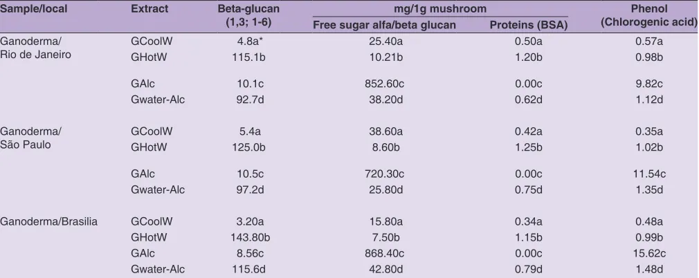
Table 1: Concentration of beta‑glucan, free sugar, proteins and phenols in samples of Ganoderma lucidum submitted to different extracts Sample/local Extract Beta‑glucan
(1,3; 1‑6) Free sugar alfa/beta glucanmg/1g mushroomProteins (BSA) (Chlorogenic acid)Phenol
Ganoderma/
Rio de Janeiro GCoolWGHotW 115.1b 4.8a* 25.40a 10.21b 0.50a1.20b 0.57a0.98b
GAlc 10.1c 852.60c 0.00c 9.82c
Gwater-Alc 92.7d 38.20d 0.62d 1.12d
Ganoderma/
São Paulo GCoolWGHotW 125.0b 5.4a 38.60a 8.60b 0.42a1.25b 0.35a1.02b
GAlc 10.5c 720.30c 0.00c 11.54c
Gwater-Alc 97.2d 25.80d 0.75d 1.35d
Ganoderma/Brasilia GCoolW 3.20a 15.80a 0.34a 0.48a
GHotW 143.80b 7.50b 1.15b 0.99b
GAlc 8.56c 868.40c 0.00c 15.62c
Gwater-Alc 115.6d 42.80d 0.79d 1.48d
and ferric chlorite (1% in alcohol). Biorad software was used to determine the area of each spot on the plate. The extract was also quantified in spectrophotometer UV/light 275-600 nm (Fenton) and comparing readings to standard peak of absorbance.
Two enzymes were also used to determinate the activity, that were peroxidase (POX) and poliphenoloxidase (PPO).
The peroxidase activity was determined measuring the tetraguaiacol absorbance variation formed at the enzymatic reaction in Beckman DU 650 computerized spectrophotometer with wave length of 470 nm, according Moerschbacher et al. (1986). The absorbance was read in 5 minutes and an activity unity was calculated using the tetraguaiacol molar absorptivity of 2,66 x 10 4 L/mol/cm, according Southerton and Deverall (1990).
The poliphenoloxidase activity was determined measuring the orthoquinone absorbance variation in 495 nm formed in the enzymatic reaction. An activity unity (unities/mL) was defined as the enzyme amount that causes the increase of 0,001 absorbance unity per minute, second Srivastav (1987).
Animals testing
The anti-inflammatory and acute toxicity assays were approved by Ethics and Research Committee of Nove de Julho University, with protocol number 34/2010 and 20/2012.
Anti-inflammatory evaluation in rats
In this phase it was used the extract that presented the higher amount of beta-glucan and smaller phenols amount. Method is based in BASILE et al., (1989); Wadt, (2000).
Male Wistar rats weighing 150-180g were obtained from UNINOVE biotery (creation room). The animals were kept in polypropylene cages (three animals per cage) covered with metallic grids in a room maintained at 23ºC, 55+10% humidity, 12h light and 12h dark cycle and feed ad libitum
for one week before the start of the study. Groups of five rats were anesthetized and submitted to cotton pellets implantation method at the dorsal region.
Negative controls were water, ethanol 70% in dosage 1mL/kg and for positive control was used dexamethasone in dosage 0,2 mg/kg (1mL/Kg). Further the extract was given by oral gavage (p.o) in dosage 1mL/kg for seven days. The animals were sacrificed by anesthesia excess (xylazina/ketamine) and the cotton pellets were removed, dried and weighted at analytical balance and the mass difference were statistically evaluated by the ANOVA/ Tukey method.
Acute toxicological evaluation
Method was based on Brito, (1994); Wadt, (2000) and Gomes et al., (2013). Male Swiss mouses (35-40g) were obtained from UNINOVE bioterio. The animals were kept in polypropylene cages (six animals per cage) covered with metallic grids in a room maintained at 23ºC, 55+10% humidity, 12h light and 12h dark cycle and feed ad libitum
for one week before the start of the study.
Each group was composed with six animals and they received, by oral treatment (p.o), the extract, water and ethanol 70% at dose of 1mL/Kg, in single dose. The following signs of toxicity were observed: skin changes, hair (pilo erection), eyes, circulatory and respiratory pattern, abnormal locomotion, tremor and hypnosis. Mortality was observed during the first 24 hours and daily for 14 day. The animals were monitored in relation of their mass, water and feed consumption daily. After this period the animals were sacrificed by lethal dose of anesthetic (xylazin and ketamine), followed by the removal and weighing of the organs, being analyzed the kidneys, lunges, heart, liver and spleen.
Macroscopically analyses were realized in the organs for alteration verification and if histopathological changes occur, studies of organs affected would be performed.
Antimicrobial evaluation
The antimicrobial activity was evaluated by the method of growing in depth (pour plate) casein soy agar against
Staphylococcus aureus (ATCC 6538) and Sabourad dextrose agar for Candida albicans (ATCC 10231). Method was based in Farmacopeia brasileira, (2010); Pinto et al., (2003) and Wadt, (2000).
The microorganisms inoculations were realized in culture medium inclined for the microbial growth activation and these cultures were washed with physiological solution and used at the microbial assay. Decimal dilutions were realized and were utilized 10-4 dilution for plating. A hundred µL of physiological solution, ethanol 70% and extract were added in petri dishes and inoculated 100µL of the microorganism solution, over the sets was added 15 mL of molded culture medium, specific for each microorganism on the dishes and waited for solidification. All samples were made in triplicate.
After the culture sets solidify the dishes were inverted and incubated for 24-48 hours at 36 ºC. The Colony Formative Unities (UFC) was counted and calculated the percentage (%) of microbial growth inhibition.
Phytochemical evaluation
Tannins – precipitation reactions with metals, alkaloids and proteins; Flavonoids – colorimetric reactions with metal, hydroxides, chloride acid and metallic magnesium, and ultra violet light; Anthraquinones – colorimetric reactions in basic mean; Cardioactives – colorimetric reactions for steroidal nucleus identification, lactone ring and sugars; Saponins – persistent foam formation for 15 minutes; Alkaloids – precipitation reactions with reactive in iodine and metals; Methylxanthines – colorimetric reactions of Murexida; Terpenes– odor and chromatography for terpene substances using sulfovanillic developer.
RESULTS AND DISCUSSION
Biochemical analysis
Basidiocarp from all samples (Table 1) have very similar pileus colour (brown) and concentric zones on the surface of the pileus (Kim et al., 1982; Saravanakumar et al., 2010) that can be expressed as G. lucidum.
Analysis in different types of extracts were realized seen that, as the fruiting body is rigid, the polysaccharide is hard to be extracted and the extract presented a brown color due to the presence of oxidized phenols being important evaluate the phenols concentration.
Results obtained in extraction demonstrated that extracts GHotW and GWater-Alc presented more concentration of beta-glucan and proteins. Extract GAlc presents higher concentration of free sugar and increased concentration of phenols when compared with others extracts but doesn’t have protein. When make a mixture of water extract with alcohol, there is a higher concentration of beta-glucan with protein and phenol (Table 1). When compared samples from three states, the one from Brasília presented more beta-glucan, phenols, proteins and free sugar in all extractions.
In polysaccharides it is important studying concentration of beta glucan and also phenols and in respect of this extracts of GWater-Alc, GAlc and GHotW were evaluated in spectrophotometer and TLC. Samples of GCoolW is not study because don’t have high concentration of beta-glucan (Table 1).
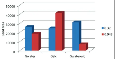
Graphic from spectrophotometer presented a common peak (322nm) in all samples. GWater-Alc presented peaks in 322, 390, 405, 415,425, 445, 475nm; GHotW presented in 322, 350, 375, 385, 445-485, 510nm and GAlc presented in 322, 435-485nm (Fig. 1). Peak in 322nm is correspondent to ferulic acid confirmed by TLC with Rf=0.948 but another band was unknown (Fig. 2).
The presence of ferulic acid in the mushroom demonstrate its importance for the human health, because possesses antioxidant action with activity of protection against the free radicals and related diseases (Nagem et al., 1992; Pratt, 1992; Von Gadow et al., 1997).
However, there are two important enzymes, peroxidase (POX) and polyphenoloxidase (PPO), to be observed regarding its presence in foods or mushrooms. These enzymes act over a variety of substrates, such as: phenols, aromatic amines, naphtols, methoxybenzenes and, in the presence of oxygen, acting over the phenol compounds, oxidizing them and leading to the presence of colored substances, in a process called enzymatic darkening promoting the astringent and bitter flavor (Arslan et al., 1997; Bravo, 1998; Croft, 1998; Durán and Padilla, 1993; Erenel et al., 1993; King and Young, 1999; Mizuno et. al., 1995).
The Ganoderma possesses brown color, such as many mushrooms, and its use depends of heating for the
Fig 1. Spectrophotometer lectures (A240-660) from samples GHotW
(extracted with hot water); GAlc (extracted with alcohol), GWater‑Alc (extracted with hot water and mixture 20% of alcohol extract). Peaks observed: GWater‑Alc 322, 390, 405, 415,425, 445, 475nm; GHotW 322, 350, 375, 385, 445-485, 510nm and GAlc 322, 435-485nm.
Fig 2. Thin layer chromatography from samples GHotW (extracted with
chemical compounds release, being possible the release of also enzymes, because of this fact, it was determined the enzymatic activity in the extracts.
The results (Table 2) indicated that the fungus submitted to extraction with hot water and/or mixed with 20% of ethanolic extract, showed enzymatic activity of the peroxidase and polyphenoloxidase. Yet in the ethanolic extract it was possible to observe high presence of activities from both enzymes when compared with other extracts. These enzymes promote phenolic compounds release when compared with other extracts. The higher the activity of these compounds, more toxic will be the extract for humans’ health.
GHotW (extracted with hot water); GAlc (extracted with alcohol), GWater-Alc (extracted with hot water and mixture 20% of alcohol extract).
Mamatha et al. (2012) said that phenol can be toxic when present at an elevated level in water or food and is known to be carcinogenic. In the case with Pleurotus eryngii
and P. ostreatus that produced peroxidase that oxidized Mn2+ into Mn3+ similar to the action of MnP (manganes-peroxidase), and also high redox potential aromatic compounds (Ruiz- Duenas et al., 2009).
Against that, GAlc can be not used because have higher concentration in phenolic compounds and can be toxic. So was used the hydroethanolic extract than present low concentration of phenols and are also responsible for numerous pharmacological activities (Brunneton, 2004).
Phytochemical analysis
The results showed presence of tannins, flavonoids, terpens, steroidal nucleus, lactone ring and sugars.
The tannins and the flavonoids are phenolic compounds and possesses an elevated antioxidant potential, being a lot used as anti-inflammatory, beyond possessing antimicrobial potential, because both complex with metals inhibiting many metabolically routes from the microorganisms (Simões et al., 2004; Bruneton, 2004).
The terpenes also presented antimicrobial potential inhibiting microbial metabolically routes (Bruneton, 2004).
The presence of steroidal nucleus could indicate a toxically potential from this compound, seen that it can interfere in the hormone formation, cortisol, within others (Simões et al., 2004).
Anti-inflammatory assay
The hydroethanolic solution of G. lucidium (GWater-Alc) presented an anti-inflammatory activity very significant, when evaluated by the statistic method Tukey/Anova, justifying some popular uses that are narrated for this mushroom (Table 3).
The presence of flavonoids and tannins in the G. lucidium
extract (GWater-Alc)indicate antioxidant activity, because these compounds show many hydroxides in its structure. The antioxidant activity protects of cell damage caused by oxygen reactive species (ORS) involved in the inflammation pathology (Fidelis et al., 2014).
The Ganoderma lucidium hydroethanolic (GWater-Alc) solution is easily prepared and can be an alternative for inflammation treatments.
Antimicrobial assay
The depth inoculation (pour plate) method was chosen for being a method that enables the growth of microorganisms without interference in the method, such as, for example, difficulty of diffusion in the agar extract, seen that substances like tannins present in the extract precipitate as proteins, this happens at the technique in disks diffusion, being possible to be a decrease of the disk halo (Pintoet al., 2003).
Table 2: Peroxidase and Poliphenoloxidase activity present in extracts from G.lucidum
Sample/
local Extract Peroxidase U/minPolifenoloxidase
Ganod/RJ GHotW 26.5a* 6.88a
GWater-Alc 27.0a 7.20a
GAlc 58.0b 15.30b
Ganod/SP GHotW 28.5c 7.20a
GWater-Alc 28.2c 7.45a
GAlc 65.4d 18.50c
Ganod/Br GHotW 25.0e 4.12b
GWater-Alc 24.8e 4.80b
GAlc 60.5f 18.20c
*Different letters in columns indicated that value between methods of extraction,
in the same mushroom, are significantly different (p<0.05) Test Tukey (ANOVA). GHotW (extracted with hot water); GAlc (extracted with alcohol), GWater‑Alc (extracted with hot water and mixture 20% of alcohol extract)
Table 3: Average mass (g) from the cotton pellets after oral application, gavage (p.o) of water (GHotW), dexamethasone, extract of Ganoderma lucidium (GWater‑Alc)
Treatment Number of animals
Cotton pellets average mass (g)
Standard
deviation Significance
GHotW 1 mL/Kg 5 0.4254 0.02774
Dexamethasone
1 mL/kg 5 0.2482 0.01711 P<0.001 ***
GWater-Alc
1 mL/Kg 5 0.3122 0.04905 P<0.001 ***
The extractive hydroethanolic solution shown antimicrobial activity against S. aureus (ATCC 6538) inhibiting 100% of microbial growth. The S. aureus is a bacteria Gram positive more sensible to many compounds, being easier its elimination, such in the case of G. lucidium hydroethanolic extract (Subramaniam et al., 2014).
The physiological solution was used as the positive control, because it doesn’t possess inhibitory activity against microorganisms, the ethanol 70% was used as solvent control, the ethanol presents antimicrobial activity, yet that
G. lucidium hydroethanolic extract, even when compared to the ethanol microbial growth presented 100% inhibition, with no growth of any CFU (colony forming unities) in the plate.
The antimicrobial assay was also realized against
Candida albicans, because this yeast is an opportunistic fungus, resistant to numerous anti-fungal, being the causative agent of various diseases, especially in individuals with low immunity (De Barros Machado et al., 2005).
The results demonstrated that the 70% hydroethanolic extract presented an inhibition of 85% of the CFU. These results demonstrated that the hydroethanolic extract show an antimicrobial potential to be used both in drugs, foods and even at agricultural defensives.
Acute toxicological assay
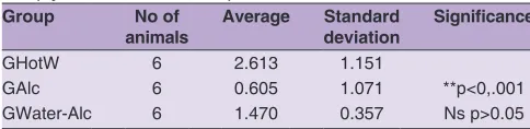
In the animals it wasn’t observed any significant alteration in the corporal mass, neither in the water or feed consumption utilized, for both males and females. These data are shown in Tables 4 to 9.
The only group that presented significant alteration in the corporal mass both male and female was the ethanol 70%, proving that the ethanol, even at single dose, affects the animal metabolism and that it´s according to Oga (1996).
The ethanol 70% was tested, because the same was used in the production of the G. lucidum hydroethanolic extract (GWater-Alc), but this did not interfere in the corporal mass development of the animals, because the ethanol 70% concentration is smaller in the extract, and another possibility, is that the G. lucidum (GHotW) possesses
substances that protect the organism from damage caused by the ethanol.
The water and feed consumption, both male and female, did not show significant alterations for every group, indicating that the animals did not have their appetite or thirsty amended. There weren’t the death of animals in any group. The organs macroscopically did not show pathological alterations. So, the hydroethanolic extract (GWater-Alc) was not toxic in the essayed dose.
Table 4: Analysis of corporal mass development from females comparing the groups: Gwater, GAlc (Ethanol 70%) and GWater‑ Alc (hydroethanolic extract)
Group No of
animals Average Standard deviation Significance
GHotW 6 2.613 1.151
GAlc 6 0.605 1.071 **p<0,.001
GWater-Alc 6 1.470 0.357 Ns p>0.05
**: Very significant, Ns: Non significant
Table 5: Analysis of corporal mass development from males comparing the groups: Gwater, GAlc (Ethanol 70%) and GWater‑ Alc (hydroethanolic extract)
Group No of
animals Average Standard deviation Significance
GHotW 6 5.432 0.3842
GAlc 6 1.647 1.006 ***p<0.001
GWater-Alc 6 4.633 2.277 Ns p>0.05
***: Extremely significant, Ns: Non significant
Table 6: Analysis of feed consumption mass in 14 days from males comparing the groups: Gwater, GAlc (Ethanol 70%) and GWater‑Alc (hydroethanolic extract)
Group No of
days Average Standard deviation Significance
GHotW 14 4.640 0.2546
GAlc 14 4.780 0.6223 Ns p>0.05
GWater-Alc 14 4.590 0.5515 Ns p>0.05
Ns: Non significant
Table 7: Analysis of the consumption water volume in 14 days from males comparing the groups Gwater, GAlc (Ethanol 70%) and GWater‑Alc (hydroethanolic extract)
Group No of
days Average deviationStandard Significance
GHotW 14 3.160 0.3394
GAlc 14 2.540 0.6505 Ns p>0.05
GWater-Alc 14 3.035 0.7566 Ns p>0.05
Ns: Non significant
Table 8: Analysis of feed mass consumption in 14 days by females comparing the groups Gwater, GAlc (Ethanol 70%) and GWater‑Alc (hydroethanolic extract)
Group No of
days Average Standard deviation Significance
GHotW 14 4.670 0.5374
GAlc 14 4.685 0.7566 Ns p>0.05
GWater-Alc 14 4.610 0.2687 Ns p>0.05
Ns: Non significant
Table 9: Analysis of water volume consumption in 14 days by females comparing the groups Gwater, GAlc (Ethanol 70%) and GWater‑Alc (hydroethanolic extract)
Group No of
days Average Standard deviation Significance
GHotW 14 3.090 0.8202
GAlc 14 3.480 0.2121 Ns p>0.05
GWater-Alc 14 3.290 0.0565 Ns p>0.05
CONCLUSION
Ganoderma lucidum possess its basidiocarp too rigid, needing to be crushed to produce some effect and depending on the extract type, presents beta-glucan release. It possess protein, phenol, peroxidase and polyphenoloxidase enzymes, plus tannins, flavonoids, terpenes, steroidal nucleus, lactone ring, and sugars. The hydroethanolic extract presents significant anti-inflammatory activity, and antimicrobial activity against
Staphylococcus aureus and Candida albicans. The hydroethanolic extract from Ganoderma lucidum did not show acute toxicity for both males and females.
ACKNOWLEDGEMENTS
We wish to thank for providing fruit bodies from G. lucidum
producers, mainly to Naturalmed (Paulo Americo), Juncao Brazil (Maria Camargo) and also Arailde Urben from Embrapa-Brasilia. This work was supported by CNPq (Grant #474681/2013).
Author contributions
All authors contributed equally in this article.
REFERENCES
Brazil. 1959. Pharmacopoeia of United States of Brazil. 2ªed. Siqueira Printing Industry. Sao Paulo, p.1265.
Brazil. 2010. Pharmacopoeia of Brazil. 5ªed. Atheneu Publishing House. Sao Paulo.
National Agency for Sanitary Vigilance (ANVISA). 2002. Technical regulation of bioactive substances and probiotics insulated with claim functional or health. Resolution RDC nº2, of January, 7 of 2002. Republished in Union Official Daily in 07/17/2002. Bach, E. E. 2010. Biotecnologia aplicada a cogumelos. In: Cogumelos
Medicinais: Aspectos de Cultivo e Aplicações/Marli Gerenutti, Organização. Sorocaba: EDUNISO; p. 19.
Bach, E. E. 2008. Extração do princípio ativo de Agaricus blazei. Anais IV SICOG IV Simpósio Internacional sobre cogumelos no Brasil e, III Simpósio Nacional sobre cogumelos comestíveis (Revista documentos Embrapa). Rio Grande do Sul, Brasil. 4: 91-98. Basile, A. C., J. A. A. Sertié, T. Oshiro and K. D. V. Caly. 1989. Topical
anti-inflamatory activity and toxicity of Cordiaverbenacea.
Fitoterapia. 60: 260-263.
Berovic, M., J. Habijanic, I. Zore, B. Wraber, D. Hodzar, B. Boh and F. Pohleven. 2003. Submerged cultivation of Ganoderma lucidum biomass and immunostimulatory effects of fungal polysaccharides. J. Biotechnol. 103: 77-86.
Bravo, L. 1998. Polyphenols: Chemistry, dietary sources, metabolism and nutrition significance. Nutr. Rev. 56: 317-333.
Brito, A. S. 1994. Manual de ensaios toxicológicos in vivo. Campinas: Editora da UNICAMP; p. 122.
Bruneton, J. 2004. Farmacognosia - Fitoquímica e Plantas Medicinales. Zaragoza: Acribia; p. 120.
Chang, S. T. and J. A. Buswell. 1999. Ganoderma lucidum (Curt: Fr.) P. Karst. (Aphyllophoromycetidease): A mushrooming medicinal mushroom. Int. J. Med. Mush. 1: 139-146.
Costa, A. F. 1978. Farmacognosia. 2nd.ed., Vol. 3. Fundação Calouste
Gulbekian, Lisboa. p. 190.
De Barros Machado, T., I. C. Leal, R. M. Kuster, A. C. Amaral, V. Kokis, M. G. De Silva and K. R. Dos Santos. 2005. Brazilian phytopharmaceuticals evaluation against hospital bacteria. Phytother. Res. 19: 519-525.
Durán, R. M. and B. Padilla. 1993. Actividad antioxidante de los compuestos fenólicos. Grasas y Aceites. 44: 101-106.
Erenel, G. and D. Erbas. 1993. Aricioglu, a. Free radicals and antioxidant systems. Mater. Med. Polona. 85: 37-43.
Fang Q. H. and J. J. Zhong. 2002. Effect of initial pH on production of ganoderic acid and polysaccharide by submerged fermentation of Ganoderma lucidum. Process. Biochem. 37: 769-774. Fidelis, Q. C., T. A. N. Ribeiro, M. F. Araújo and M. G. Carvalho. 2014.
Ouratea genus: Chemical and pharmacological aspects. Rev. Bras. Farmacogn. 24: 1-19.
Gomes, M. R. F., R. S. Schuh, A. L. B. Jacques, G. G. Dorneles, J. Montanha, P. M. Roehe, S. Bordignon, E. Dallegrave, M. B. Leal and R. P. Limberger. 2013. Biological assessment (antiviral and antioxidant) and acute toxicity of essential oils from
Drimysangustifolia and D. brasiliens. Rev. Bras. Farmacogn. 23: 284-290.
He, Y., R. Li, Q. Chen, Z. Lin, D. Xia and L. Ma. 1992. Chemical studies on immunologically active polysaccharides of Ganodermalucidum (Leyss. ex Fr.) Karst. Chung Kuo Chung Yao Tsa Chih. 17: 226-256.
Huie, C.W. and X. Di. 2004. Chromatographic and electrophoretic methods for Lingzhi pharmacologically active components. J. Chromatogr. B. 812: 241-257.
Kim, B. K., E. C. Choi, K. S. Chung, C. Y. Kang, S. H. Kim, J. S. Kim, Y. J. Kim, K. L. Lee and J. K. Lee. 1982. The constituents of higher fungi of Korea: Antitumor polysaccharides from the carpophores of some Basidiomycetes. Arch. Pharmacol. Res. (Seoul). 5: 21-24.
Lin, J. M., C. C. Lin, H. F. Chiu, J. J. Yang and S. G. Lee. 1993. Evaluation of the anti-inflammatory and liver-protective effects of anoectochilusformosanus, ganodermalucidum and gynostemmapentaphyllum in rats. Am. J. Chin. Med. 21: 59-69.
Liu, F., V. E. C. Ooi, W. K. Liu and S. T. Chang. 1996. Immunomodulatory and antitumouractivities of polysaccharide-protein complex from the culture filtrates of a local ediblemushroom, Tricholoma lobayense. Gen. Pharm. 27: 621-624.
Lowry, O. H., N. J. Rosenbrough, A. L. Farr and R. J. Randall. 1951. Protein measurement with the Folin phenol reagent. J. Biol. Chem. 193: 265-275.
Mamatha, J., A. B. Vedamurthy and S. D. Shruthi. 2012. Degradation of phenol by turnip root enzyme extract. J. Microbiol. Biotechnol. Res. 2: 426-430.
Mizuno, T., H. Saito, T. Nishitoba and H. Kawagishi. 1995. Antitumour active substances from mushrooms. Food Rev. Int. 11: 23-61. Moerschbacher, B. M., B. Heck, K. H. Kogel, O. Obst and H. J. Reisner.
1986. An elicitor of the hypersensitive lignification response in wheat leaves isolated from the rust fungus Pucciniagraminis f. sp. Tritici. Z. Naturfosch. 41: 32-37.
Nagen, T. J., T. T. O. Albuquerque and L. C. G. Miranda. 1992. Ácidos fenólicos em cultivares de soja: Ação antioxidante. Arq. Biol. Tecnol. 35: 129-138.
Oga, S. 1996. Fundamentos de Toxicologia. 2ª ed. Atheneu, São Paulo. p. 474.
Pinto, T. J. A., M. T. Ohara and T. M. Kaneko. 2003. Controle biológico de qualidade de produtos farmacêuticos, correlatos e cosméticos. Atheneu, São Paulo. 2nd ed. p. 158.
Pratt, D. E. 1992. Natural antioxidants from plant material. In:Ho, C. T., Lee, C. Y., Huang, M. T. Phenolic Compounds in Food and Their Effects on Health. American Chemical Society, Washington, D. C. p. 54-71. (ACS Symposium Series, n.507).
Ruiz-Duenas, F. J. M., Morales, E. Garcia, Y. Miki, M. J. Martinez and A. T. Martinez. 2009. Substrate oxidation site in versatile peroxidase and other basidiomycete peroxidases. J. Exp. Bot. 60: 441-452.
Saravanakumar, P., V. Karthikeyan, S. Patharajan, T. Kannan, K. Sathya and P. T. Kalaichelvan. 2010. Effect of 1,3-β-d glucan of
Ganoderma lucidum of fibro sarcoma induced mice. Pharmacol. 1: 958-970.
Simões, C. M., E. P. Schenkel, G. Gosmann, J. C. P. Mello, L. A. Mentz and P. R. Petrovick. 2004. Farmacognosia. Editora da Universidade UFRGS, Porto Alegre. 5ª ed. p. 1102.
Sliva, D. 2006. Ganoderma lucidum in cancer research. Leuk. Res. 30: 767-768.
Southerton, S. G. and B. J. Deverall. 1990. Changes in phenylalanine ammonia-lyase and peroxidase activities in wheat cultivars expressing resistance to the leaf-rust fungus. Plant Pathol. 39: 223-230.
Srivastava, S. K. 1987. Peroxidase and poly-phenol oxidase in Brassica
juncea plants infected with Macrophomina phaseolina(Tassai) Goid, and their implication in disease resistence. J. Phytopathol. 120: 249-254.
Subramaniam, S., M. Keerthiraja, A. Sivasubramaniam. 2014. Synergistic antibacterial action of β-sitosterol – D-glucopyranoside isolated from Desmostachy abipinnata
leaves with antibiotics against commom humam pathogens. Rev. Bras. Farmacogn. 24: 44-50.
Swain, R. and W. E. Hillis. 1959. The phenolic constituents of Prunus domestica. I. The quantitative analysis of phenolic constituents. J Sci Food Agric. 10: 63-68.
Urben, A. F. 2004. Produção de Cogumelos Por Meio de Tecnologia Chinesa Modificada. 2nd ed. Embrapa, Brasília. p. 50.
Von Gadow, A., E. Joubert and C. F. Hansmann. 1997. Comparison of the antioxidant activity of aspalathin with that of other plant phenols of rooibos tea (Aspalathus linearis), a-tocopherol, BHT and BHA. J. Agric. Food Chem. 45: 632-638.
Wadt, N.S.Y. 2000. Study of onto genetic variation of the active ingredients of Leonurus sibiricus L. and their pharmacological actions. Doctoral dissertation (Doctorate in Sciences -phD). Pharmaceutical Sciences College (FCF) – Sao Paulo University (USP). Sao Paulo.
Zhu, X. L., A. F. Chen and Z. B. Lin. 2007. Ganoderma lucidum