Comparative Study of VDR and the Electrical Activity of the
Anterior Temporal and Masseter Muscles using Physiologic
rest Position, Phonetics and Swallowing methods
SHIRIN LAVAF* and ARASH AZIZI MEYSAM PAKRAVAN
School of Dentistry, Ahvaz Jundishapur University of medical sciences, Ahvaz, Iran. DOI: http://dx.doi.org/10.13005/bpj/878
(Received: October 20, 2015; accepted: November 21, 2015) ABSTRACT
Vertical dimension of rest (VDR) or physiologic rest position (VDR) is defined as a stage of prosthetic treatment for edentulous patients. Mandibular rest position is used to record VDR which is fulfilled via common methods such as Physiologic rest position, Phonetics and Swallowing methods. This study aims to compare the rate of VDR and electrical activity of the anterior temporal and masseter muscles using physiologic rest position, phonetics and swallowing methods. This is a clinical trial conducted in the Rehabilitation faculty of Ahvaz Jundishapur University of Medical Sciences during 2009-2010. The statistical population consists of 30 individuals (15 men and15 women; age range 18-30 years old). The conditions for inclusion to this study is having 28 natural teeth with the Class I (Class I molar relationship), normal range of overjet and overbite, without anterior and posterior crossbites. Mean of VDR was recorded after swallowing (7/48+0.012), phonetics (8.14 0.19) and physiologic rest position (8.22 0.19). Mean of voltage of anterior temporal muscles was recorded after swallowing (7/360.216), phonetics (6.19 0.095) and physiologic rest position (6.19 0.119). it can use all the mentioned methods to acquire precise distance for VDR in clinic and it can use mean of acquired sizes under any difference.
Key words: VDR, Electromyography, phonetic voltage.
INTRODUCTION
The person is exposed to impairment with the loss of tooth, under which a change occurs in structure and functions of facial muscles; in addition, edentulous is a threat to health of masticatory system. Different components in masticatory system include teeth, neuromuscular system and cranial and facial structures [1]. In edentulous state due to needing to perfect teeth to retrieve masticatory system, knowing functional anatomy of the masticatory system and how the components of this system associate to each other is required [2]. The lost function and beauty can be returned to the patient by means of dental prosthesis, helping for health of remaining oral tissue. Recording jaw relations in these patients is made for alignment
much problem as these changes are gradual and just faces tangible changes in his face and reduction in his masticatory system by the passage of years [6]. Access to vertical height at the state of vertical dimension of occlusion (VDO) has been mentioned as one of the important principles to heal the patients who need the methods for mouth reconstruction [7]. When the person’s natural teeth exist, there will be Vertical dimension of occlusion by these teeth [8]. In all the patients without teeth and patients who have lost their posterior teeth or have lost their VDO due to teeth wear, acquisition of methods to record the best result for VDO seems difficult [7]. Vertical height of face in the status of Vertical dimension of rest position is used to record VDO, which this is under influence of different factors such as loss of teeth, age of person and bone loss after edentulous; this method is used to determine the patient’s position[9, 10, 11]. Formula VDO= VDR – FWS is used to record VDO. FWS refers to a distance between upper or lower teeth when mandible is in physiological rest position, mentioned that the distance at the premolar region is between 2-4 mm [12]. To set mandible at rest position and record VDR, there are a variety of methods such as phonetics and swallowing methods [7]. Further, it has been suggested to repeat measurement for several times regardless of the used methods and use mean of obtained sizes in case of difference [13]. This is because some studies have shown that this organ cannot be considered as an authentic reference point in determining VDO due to no constant mandible [14]. Fur ther, cephalometric examination indicates that rest position of toothless jaw changes even in short time [15]. It seems that there is no certain rule to determine vertical dimension of occlusion from scientific perspective [14]. In general, a variety of methods to determine VDO have been suggested including mechanical methods such as Anthropometric measurements (16), cephalometric (17), records before pulling teeth ( 18 ) , the patient profile ( 19 ) , and face and physiological measurement ( 21,20 ) such as beauty , swallowing and speaking. In some cases, Computer programs based on the person’s personal judgment is used to determine VDO used (22). Results of several studies indicated that use of denture alters the electrical activity of muscles by increasing VDO, which it can determine denture replacement time by recording EMG of
masseter and temporal muscles [23, 24, 25]. Unsuitable VDO causes several orofacial disorders such as bruxism, masticatory muscle pain and temporomandibular joint problems (TMJ Disorders)[26]. On the other hand, increase or decrease of VDO in the area around the mouth and the face has been followed by substantial effects including beauty and masticatory force [9, 27]. Ambramicheltali(1998) compared size of vertical distance in rest position in clinic and vertical distance in rest position by means of Electromyographi and deduced that there is a millimeter difference in the distance between two vertical resting position at the clinic (phonetics and swallowing ) and EMG[28]. Further, Frenso(2007) evaluated activity of temporal muscles using EMG, Phonetics and Swallowing methods and deduced that there is no significant difference between measured VDR via different methods[29].
With regard to difference in the results of the mentioned studies, this study aims to examine amount of VDR by means of three Physiologic rest position, Phonetics and Swallowing methods and examine electrical activity of the anterior temporal and masseter muscles under mentioned conditions.
MATERIALS AND METHODS
For anterior temporal muscle, one of the electrodes is placed in 1 cm above the zygomatic arch and 1.5 cm behind the border of the orbit. The other electrode is placed 1.5 cm below the first electrode. Masseter muscle electrode is placed in the middle of the right muscle. Number of muscle channel includes channel by channel (2) and (4): temporal muscle masseter muscle, respectively. After ensuring the safety and soundness of device, the person was asked to practice phonetics which contains pronunciation of the letter (M) and the electrical activity of muscles was recorded in 15 seconds(3 seconds of rest, 5 seconds of muscle contraction, 7 seconds of rest after function). After pronunciation of the word and relaxation of muscles by EMG during record time, VDR distance between two points on the nose and chin that was already marked, measured and recorded. Then saliva was swallowed at 15 seconds and again VDR distance was measured and recorded at rest after the function. In the third function, the person was asked to press his/her teeth for 2 seconds and then he/ she stopped pressing and the technician placed mandibular muscles in the position of rest using touch, ensured on total rest of anterior temporal and masseter muscles via EMG, during which VDR distance was measured and recorded. After recording data of all the individuals, the information was analyzed via Myo-Dat program.
RESULTS
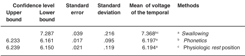
Analysis of variance with repeated measures displayed that there is a significant difference between phonetics, swallowing and rest methods in the average voltage of the temporal muscle (p <.001). Fur thermore, Tukey’s HSD test indicated a significant difference between ‘swallowing’ and ‘phonetics and rest methods’ in the average voltage of the temporal muscle (p <.001), but there is no significant difference between phonetics and rest methods in the average voltage of the temporal muscle (p >.0.05)(Table 1).
DISCUSSION AND CONCLUSION
The present study was conducted to examine the levels of VDR measured via p h y s i o l o g i c r e s t p o s i t i o n , p h o n e t i c s and swallowing methods; in this study, electrical activity of the anterior temporal and masseter muscles in the right side of subjects was recorded via device EMG. A number of previous studies mentioned lack of electrical activity of muscles during rest [30, 31], but most of studies believed in little electrical activity of muscles during rest of muscles [32, 33, 34], that the present research confirms these investigations. In this study, mean of voltage of anterior temporal muscles was recorded
Table 1: Comparison of the temporal muscle voltage in physiologic rest position, phonetics, and swallowing methods. The letters a, b, and c indicate statistically significant difference
(p<0.05).
Confidence level Standard Standard Mean of voltage Methods
Upper Lower error deviation of the temporal
bound bound
7.287 .039 .216 7.368bc a Swallowing
6.233 6.161 .017 .095 6.197a b Phonetics
6.239 6.150 .021 .119 6.194a c Physiologic rest position
Table 2: Comparison of voltage of the masseter muscles in physiologic rest position, phonetics and swallowing methods. The letters a, b, and c indicate statistically significant
difference (p<0.05).
Confidence level Standard Standard Mean of voltage Methods
Upper Lower error deviation of the temporal
bound bound
.230 6.53 6.36 .042 6.450bc a Swallowing
.062 5.55 5.50 .011 5.528a b Phonetics
.072 5.52 5.47 .013 5.501a c Physiologic rest position
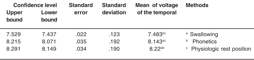
Analysis of variance with repeated measures displayed that there is a significant difference between phonetics, swallowing and rest methods in mean of VDR (p <.001). Further, Tukey's HSD test indicated that there is a significant difference between 'swallowing' and 'phonetics and rest methods' in t mean of VDR (p <.001)(Table 3).
Table 3: Comparison of mean of VDR in Physiologic rest position, Phonetics and Swallowing method. The letters a, b, and c indicate statistically significant difference (p<0.05)
Confidence level Standard Standard Mean of voltage Methods
Upper Lower error deviation of the temporal
bound bound
7.529 7.437 .022 .123 7.483bc a Swallowing
8.215 8.071 .035 .192 8.143ac b Phonetics
8.291 8.149 .034 .190 8.22ab c Physiologic rest position
after swallowing (7/36 0.216), phonetics (6.19 0.095) and physiologic rest position (6.19 0.119).
Mean of voltage of anterior masseter muscles was recorded after swallowing (6/45 0.23), phonetics (5.52 0.062) and physiologic rest position (5.50 0.072). Difference in gender of participants (male and female) does not indicated a significant effect on electrical activity of anterior temporal and masseter muscles in physiologic rest position, phonetics and swallowing methods, which these results are consistent with the studies by Rugh & Michelotti. Results of this research indicated that there is a significant difference between mean of electrical activity of anterior temporal and masseter muscles after swallowing and mean of voltage of these muscles after physiologic rest position and phonetics(p>0.05), but there is no significant difference between mean of voltage of these muscles in physiologic rest position and phonetics.
of this research indicate this point, that is, preparedness of muscle is expected more for movement facilitation in swallowing regarding high level of muscle activity. Mean of VDR was recorded after swallowing (7/48 0.012), phonetics (8.14 0.19) and physiologic rest position (8.22 0.19). A significant difference on VDR was seen in three methods (p>0.05) that the highest associates to physiologic rest position and the least vertical distance in physiologic rest position associates to physiologic rest position after swallowing. This result is consistent with the results of study by Michelotti & Frenso, indicating that VDR in rest position of muscle is greater than VDR after clinical tests of swallowing and phonetics, because after clinical tests, masticatory muscles must be placed in the least contraction for jaw maintenance status that the contraction causes reduction of VDR. On the other hand, these results can be justified with neuropsychological theory of post contraction facilitation [36], that is, since level of muscle activity is greater in closing the mouth in swallowing activity, preparedness of muscle will be greater in facilitation of next movement, causing more contraction and
electrical activity of involved muscles for movement facilitation. Thus, it is expected that VDR followed by swallowing has the least rate and measured VDR in rest position has the highest rate. Each of three methods under study is used by the dentists to determine VDR, because each of three methods is simple. But results of this research indicate a significant difference in rate of VDR recorded by these three methods. Indeed, three VDR measurement methods can be justified, but none of them is more authentic than others [7]. Therefore, it is suggested that VDR has been measured via three methods and it is recorded as vertical height of face in rest position under any difference of mean. Further, it is suggested to examine effect of the factors including head, neck and body position, sleep, gender, age, excitement, pain, stress on VDR.
ACKNOWLEDGMENT
The present study was financially supported by Ahvaz Jundishapur University of Medical Sciences (Grant No.: U-88307).
REFERENCES
1. Ravanmehr, H. Gerami, A , Atlantic cephalometric, first print , Tehran University Jihad, Tehran University of Medical Sciences , 1994 , 43 .
2. Ash M, Ramfjord S. Occlusion, 4th ed, Philadelphia: WB Saunders; Ch. 12: 1994; 197-219.
3. Dawson P. Evaluation. Diagnosis and treatment of occlusal problems. 2nd ed. St. Louis: Mosby; 1989; P.56.
4. Mc Gee JF.Use of facial measurement in determining vertical dimension. J Prosthet Dent.1965;15:241-247
5. Landbadakis J. Karkasis HC. Changes in the mandibular rest position after removal of remaining teeth and insertion of complete dentures. J P D. 1992; 68: 74-77.
6. Hellsing G. Ekstrand K. Abiligy of edentulous human beings to establishing the facial vertical dimension. J Prosthet Dent. 1968; 20(1): 37-42.
7. Frenso MJ. Electromyographic evaluation of
anterior temporal and suprahyiod muscle using habitual methods to determine clinical rest position. J craniomondibular practice 2007 ; 25 (4) : 257-263
8. Boucher carlo , Bolender Charles l. Prosthodontic treatment for edentulous patients 12 thed.2004
9. Mohindra NK, Bulman JS. The effect of increasing vertical dimension of occlusion on facial aesthetice. Br Dent J 2002; 192(3): 164-68.
10. Tallgren A, Lang BR, Walker JF, Ash MM Jr. Roentgen cephalometric analysis of ridge resorbtion and changes in jaw and occlusal relationships in immediate complete denture wearers. J Oral Rehbil 1980; 7: 77-94. 11. Anderson JD. Biological and clinical
Implant-Supported prostheses, 12th ed. St. Louis: Mosby; 2004.268.
12. Jalali . M. treatment of Patients without teeth, : the SID ; 2004.s. 327-326
13. Wikler S. Essential of complete dentures prosthodonties. J Dent Res 1979;55-60 14. Sheppared IM, Sheppard SM. Ver tical
dimension Measurments. J Prosth Dent 1975; 34: 269-77.
15. carossa S, et al. The unreliability of facial measurement in the determination of the vertical dimension of occlusion in the edentulous patients. J Oral Rehabil 1990 ; 17(3): 287-290.
16. Or thlieb JD, Laurent M, Laplanche O.Cephalometric estimation of vertical dimension of occlusion. J Oral Rehab 2000; 27:802-807.
17. Bassi F, et al. Evaluation of the utility of cephalometric parameters in constructing complete denture, part I: placement of posterior teeth. J Oral Rehab 2001; 28: 234-38.
18. Heartwell CH, Rahn AO. Syllabus of complete dentures. 4th ed. Philadelphia : Lea & Febiger; 1986; P. 277.
19. Koller MM, et al. A comparative study of two methods for the orientation of the occlusal plane and the determination of the vertical dimension of the occlusion in edentulous patients. J Oral Rehab 1992; 19:413-25. 20. Fayz F, Eslami A. Determination of occlusal
vertical dimension, A literature review. J Prosth Dent 1988; 59(3): 321-23.
21. pound S. Let/S/ be your guide. J Prosth Dent 1977; 38: 482-89.
22. Vanwiligen JD, Rashbass C, Melchior HJ. Byte- Ryte, an aparatus for the determination of the prefered vertical dimension of occlusion required for the construction of the complete denture prosthesis. J Oral Rehabil 1985; 12:23-25.
23. Nakamura M. The Effect of vertical Dimension change on mandibular Movement and muscle Activity. Int. J Prosthet Dent 1998 ; 1 : 279-301.
24. Verkindere M. Electromyographic study of mastication as a function of the denture in humans. J Biol Buccale 1988; 16(3) : 169-178
25. Nuno licona A. Electromyographic activity (EMG) of masseter and temporal muscles in edentulous patients before and after complete denture. Pract odontol 1990; 11(8) : 54-56.
26. Yagi T, et al. Adjustment of the occlusal vertical dimension in bite- raised guinea pig. J Dent Res 2003; 82(2): 127-130.
27. Rugh JD, Johnson RW. Vertical dimension discrepancies and masticatory pain/ dysfunction, In: Solberg WK, Clark G (eds). Abnormal Jaw Mechanics, Chicago: Quintessence; 1984:117.
28. Ambra michelotti. Mandibular rest position and electrical activity of the masticatory muscle. J prosthet Dent 1997; 78(1) : 48-83 29. Beker M. Mandibular elevator muscles.J
Dent Res 1993;101:314-33
30. Yemm R. Passive control in mandibular rest position. J Prosther Dent 1969;22:30-6 31. Hickey JC. Stability of mandibular rest
position. J Prosther Dent 1961;11:566-72 32. Rugh JD.Vertical dimension:a studi of clinical
rest position and jaw muscle activity. J Prosthet Dent 1981;45:670-5
33. Wessberg GA.Evaluation of mandibbular rest position in subject with diverse dentofacial morphology. J Prosthet Dent 1982;48:451-60
34. feldman S.Rest vartical dimention determined by electromyograph with biofeedback as compared to conventional methods. J Prosthet Dent 1978;40:216-9
35. John P.Muscles testing and