Exercise Training, Immobilisation and Castration Effects on
Skeletal Muscle Na
+,K
+-ATPase
Thesis submitted in fulfilment of the requirements for the degree of
Doctor of Philosophy
Submitted by Muath Altarawneh
Principal supervisor: Professor Michael J. McKenna Co-supervisor: Dr Aaron C. Petersen
Institute for Health and Sport (iHeS) Victoria University
II Abstract
The Na+,K+-ATPase (NKA) is a key protein involved in the maintenance of skeletal muscle excitability during contractions, and comprises two subunits (α and β), each of which are expressed as multiple isoforms in skeletal muscle (α1 - α3 and β1 - β3). Therefore
any modulation of NKA content or of individual NKA isoforms has the potential to affect muscle function. This thesis comprises four studies that investigated the effects of conditions intended to induce downregulation or upregulation of the NKA in skeletal muscle, utilising hindlimb immobilisation, testosterone suppression and increased physical activity through various training regimes. A theme of the thesis was specific effects of changes in physical activity and on muscle NKA.
Study one. This study investigated the effects of hindlimb immobilisation and testosterone suppression via castration surgery on soleus muscle [3H]ouabain binding site content and NKA isoform abundances. Eight week old male Fischer rats underwent sham or castration surgery, and then after 7 days were subjected to 10 days of immobilisation of one hindlimb. For both sham and castration groups, soleus muscles were obtained 7 days after surgery from non-immobilised controls, following 10 days immobilisation and after 14 days of recovery, from both the cast and non-cast leg. Within the sham group, after immobilisation, the [3H]ouabain binding site content in the cast leg was 26% lower than in the non-cast leg (p = 0.023) and 34% lower (p = 0.001) than in the non-immobilised control group (P = 0.012), but did not differ at 14 days recovery compared to either the non-cast leg or non-immobilised control group. There were no differences in the NKA α1, α2, α3, β1 or β2 isoform abundances in the cast leg compared to either the
III group and remained depressed by 34% (p = 0.001) at 14 days recovery after immobilisation. The α2 isoform in the cast leg was 60% lower than in both the non-cast
leg (p = 0.004) and non-immobilised control group (p = 0.004) and remained 42% lower than the non-immobilised control group at 14 days recovery (p = 0.039). The β1 isoform
in the cast leg after immobilisation was 26 % lower than in the non-cast leg (p = 0.018), but did not differ at 14 days recovery compared to either the cast leg or non-immobilised control group. The β2 isoform in the cast leg after immobilisation was 71%
lower than the non-cast leg (p = 0.004) and 65% lower than non-immobilised control group (p = 0.012), but did not differ at 14 days recovery, compared to either the non-cast leg or non-immobilised control group. There were no differences in the abundances of the α1 and α3 isoforms between legs or groups. Thus the [3H]ouabain binding site content
and α2 were decreased with immobilisation, and remained depressed at 14 days recovery
in the castration group, with the NKA α2, β1 and β2 isoform abundances also decreased
with immobilisation compared to sham group. The β3 isoform abundance could not be
detected in either sham or castration groups. Hence testosterone suppression was associated with impaired restoration of immobilisation-induced lowered NKA α2 isoform
and of the number of functional NKA in rat soleus muscle.
Study two. This study investigated which NKA isoforms (α1 - α3 and β1 - β3)
accompanied an anticipated upregulation of skeletal muscle NKA content following sprint exercise training. Fifteen healthy young adults (11 males, 4 females) underwent either 7 weeks sprint training comprising repeated 30-s maximal sprint cycling efforts, three days/week (n = 8, ST) or a 7 wk control period (n = 7, CON). A vastus lateralis biopsy was taken at rest, prior to and following ST or CON and analysed for NKA content and NKA α1 - α2 and β1 - β2 isoform abundances. The muscle NKA content tended to
IV participants responded to training with an increased NKA content. However, there were no significant changes with ST for any of NKA α1, α2, β1, or β2 isoform abundances. The
α3 and β3 isoforms could not be detected. No sprint performance improvements occurred
after ST (appendix 8). The unchanged [3H]ouabain binding site content of the whole group was surprising but was consistent with unchanged abundances in the NKA isoforms. This likely reflects a Type II error, but might also reflect an inadequate training stimulus as consistent with the lack of performance improvement. Further research with a larger sample size is required to ascertain which isoforms are upregulated with increased NKA content with ST.
Study Three. This study investigated which NKA isoforms (α1 - α3 and β1 - β3)
accompanied an anticipated increase in skeletal muscle NKA content after resistance exercise training. Twenty-one healthy young males underwent 7 weeks of resistance training (RT, n=16) or control period (CON, n=5). Participants underwent a vastus lateralis muscle biopsy at rest, prior to and following RT or CON, for measurement of NKA content and NKA α1 - α3, and β1 - β3 isoform abundances. After RT, the muscle
NKA content increased by 12% (p = 0.012), NKA α1 isoform increased by 32% (p =
0.013) and the α2 isoform increased by 10% (p = 0.001), with no significant changes in
CON. There were no differences in the β1 or β 2 isoform abundances following RT. The
α3 and β3 isoforms could not be detected. Thus the resistance training-induced increase in
muscle NKA content was accompanied by increases in both the α1 and α2 NKA isoform
abundances. It is speculated that specific adaptations accrued that function to both resist fatigue during intense resistance training sessions, as well as facilitate recovery after sets within training, in these healthy young adults.
V and NKA α1 - α3 and β1 - β3 isoform abundances in patients with chronic kidney disease
(CKD) and also explored possible differences in rest muscle in CKD compared against healthy participants. Fifteen patients (six females, nine males) underwent 12 weeks of either MICT (n = 5), or HIIT (n = 8) on a motorised treadmill, whilst 2 patients acted as controls. Fifteen healthy, age- and sex-matched participants acted as controls. The HIIT comprised four intervals of 4 min duration at 85 - 95% HRmax, with an intervening 3 min
of active recovery at 65% HRmax, whilst MICT comprised 30 min training at 65% HRmax.
A vastus lateralis muscle biopsy was taken at rest, prior to and post-training, for measurement of NKA content and NKA α1 - α3 and β1 - β3 isoform abundances. There
were no significant differences between CKD and CON in rest muscle (n = 15) for NKA content (p = 0.459), α1 (p = 0.984), α2 (p = 0.235), β1 (p = 0.247) or β2 (p = 0.138)
isoforms. No significant differences in muscle NKA content were found following MICT (p = 0.165) or HIIT (p = 0.278), and pooled data from both groups (n = 15) also revealed no increase after training (p = 0.110). No significant differences in NKA α1, α2, β1 or β2
isoform abundances were found following either MICT or HIIT, but there was a significant increase in α2 in the pooled data after training (p = 0.035). The α3 and β3
isoforms could not be detected. Hence training protocols that might be anticipated to enhance muscle NKA content and NKA isoform abundances in healthy participants failed to do so in patients with CKD. The lack of increase in pooled data argues against a possible type II error. Further study is required to verify whether processes underlying upregulation in muscle NKA are impaired in CKD and whether upregulation is possible in CKD with different training protocols or training types such as resistance training. In conclusion, this thesis demonstrated that hindlimb immobilisation reduced [3H]ouabain binding site content in both sham and castration groups. The muscle NKA α2, β1 and β2
VI reduced in the castration group. Importantly, this thesis demonstrated that castration impaired the recovery of [3H]ouabain binding site content and muscle NKA β2 isoform
abundance after reductions with hindlimb immobilisation. This thesis revealed that in humans, resistance training was effective in increasing muscle NKA content and NKA α1
and α2 isoforms, whilst sprint training only tended to increase NKA content but without
VII Declaration
“I, Muath Altarawneh, declare that the Doctor of Philosophy dissertation entitled ‘Exercise Training, Immobilisation and Castration Effects on Skeletal Muscle Na+,K+
-ATPase’ is no more than 100,000 words in length including quotes and exclusive of tables, figures, appendices and bibliography references and footnotes. This thesis contains no material that has been submitted previously, in whole or in part, for any other academic degree or diploma. Except where otherwise indicated this dissertation is my own work”.
Signature Date
VIII Acknowledgements
I would like to thank my supervisor, Prof. Michel McKenna for his fundamental role in my PhD work. This thesis would not have been possible without his great support and guidance every step of the way. Thank you for your encouragement, vast expertise and patience.
This thesis involved multiple projects contributions with a number of different health researchers and professionals. I independently performed all of the muscle tissue analyses using the [3H]ouabain content binding site and Western Blotting techniques, statistical analysis and wrote up thesis chapters. I was also heavily involved in the conduct of the sprint training study contributing to the experimental trials and blood analyses.
To my co-supervisor Dr Aaron Petersen, thank you for all the help and continual support. Thank you for teaching me the [3H]ouabain content binding site and Western Blotting techniques and for your specific contributions to the resistance training study and supply the muscle tissue.
I would like thank Dr. Erik Hanson for his collaboration and supply of the rat muscle tissue for the immobilisation - castration study.
Special thanks to Trevor Farr for collaboration and supply of the muscle tissue for the sprint training study.
IX Abbreviations
Muscle unit
Na+,K+-ATPase sodium-potassium adenosine
5’triphosphatase
NKA Na+,K+-ATPase
ATP adenosine 5’ triphosphate
t-tubules tranverse tubules
SOL soleus muscle
EDL extensor digitorum longus muscle
α Alpha subunit
β Beta subunit
[3H]ouabain binding tritiated ouabain binding (pmol.(g wet wt.)-1 3-O-MFPase 3-O-methylfluorescein phosphatase (nmol.(g wet wt.)-1 pmol.g-1
picomoles per gram
kDa kilodalton
µg microgram
Electrolytes
K+ potassium ion
[K+] potassium ion concentration mM
[K+]i intracellular potassium ion
concentration
[K+]e extracellular potassium ion
concentration
Na+ sodium ion
[Na+] sodium ion concentration mM
[Na+]i intracellular sodium ion concentration
[Na+]e extracellular sodium ion concentration
Ca2+ Calcium ion
Aerobic power
VO2 oxygen consumption
VO2peak peak oxygen consumption L.min-1
VO2max maximal oxygen consumption
X Publications and Presentations
The following manuscripts that have been prepared for submission.
Muath M. Altarawneh, Erik D. Hanson, Andrew C. Betik, Aaron C. Petersen, Alan Hayes, Michael J. McKenna. Effects of testosterone suppression, hind limb immobilization and recovery on [3H]ouabain binding site content and Na+, K+-ATPase isoforms in rat soleus muscle. Submitted to Journal of Applied Physiology
Muath M. Altarawneh, Aaron Petersen, Trevor Farr, Andrew Garnham, James Broatch, Shona Halson, David Bishop1 and Michael J. McKenna Resistance training upregulates skeletal muscle Na+,K+-ATPase content, with elevations in α1 and α2 but not
β isoforms. To be submitted to European Journal of Applied Physiology.
Conference Presentations:
Altarawneh M, A Petersen1, T Farr, A Garnham, J Broatch, CK Argus , S Halson, D Bishop and MJ McKenna. Resistance training upregulates skeletal muscle Na+,K+ -ATPase content, with elevations in both α1 and α2 but not β isoforms. Europhysiology
XI Table of Contents
Abstract ... II Declaration... VII Acknowledgements ... VIII Abbreviations ... IX Publications and Presentations ... X Table of Contents ... XI Table of Figures ... XVIII List of Tables ... XXII
Chapter 1. General introduction ...1
Chapter 2. Literature Review...5
2.1 Muscle contraction, sodium and potassium ion concentrations ...5
2.1.1 The relationship between membrane potential and [K+] ...5
2.1.2 Effects of elevated of muscle [K+] e and reduced [K+]i on muscle fatigue ...7
2.1.3 [Na+] and muscle fatigue ...8
2.2 Sodium-Potassium Adenosine Triphosphatase (Na+,K+-ATPase, NKA) in skeletal muscle ...9
2.2.1 Definition and function of NKA ...9
2.2.2 Structure of NKA ...9
2.2.2.1 The α subunit isoforms ...9
2.2.2.2 The β subunit isoforms ...11
2.2.2.3 The phospholemman subunit ...12
2.2.2.4 [3H]ouabain binding site content ...14
2.2.2.5 Comparisons of muscle NKA variables in human and rat skeletal muscle ...15
XII
2.3 Effects of Age on skeletal muscle NKA ...18
2.4 Effects of selected hormones and sex differences on skeletal muscle NKA ...23
2.4.1 Hormonal regulation ...23
2.4.1.1 Thyroid hormones ...23
2.4.1.2 Glucocorticoids ...23
2.4.1.3 Insulin ...24
2.5 Effects of sex differences and sex hormones (testosterone and estradiol) on skeletal muscle NKA ...24
2.5.1 Estradiol effects ...25
2.5.2 Testosterone effects ...25
2.6 Intracellular [Na+] regulation and NKA in skeletal muscle ...26
2.7 Inactivity, immobilisation and exercise training effects on skeletal muscle NKA ...27
2.7.1 Inactivity and immobilisation effects on skeletal muscle NKA ...27
2.7.2 Effects of exercise training on skeletal muscle NKA content and isoform abundances...30
2.7.2.1 Effects of sprint training ...30
2.7.2.2 Effects of resistance training ...31
2.7.2.3 Effects of submaximal intensity training ...32
2.7.2.4 Effects of combined different type of exercise training ...33
2.8 Chronic kidney disease (CKD) and skeletal muscle NKA ...43
2.8.1 Effects of CKD on muscle function ...43
2.8.2 Effects of CKD on muscle NKA and isoform abundances ...43
2.8.3 Effects of exercise training on skeletal muscle NKA and isoform abundances in CKD. ...45
2. 9 Aims and hypotheses ...47
2. 9.1 Study One ...47
2.9.2 Study Two ...47
XIII
2.9.4 Study Four ...48
Chapter 3: Effects of hind limb immobilisation and castration on [3H]ouabain binding site content and NKA isoform abundance in rat soleus muscle ...49
3.1 Introduction. ...49
3.2 Methods and procedures ...52
3.2.1 Study Design ...52
3.2.2 Castration and Sham Surgery Procedures ...52
3.2.3 Immobilisation Procedure...53
3.2.4 Animal sacrifice and muscle sampling ...53
3.2.5 [3H]ouabain binding site content ...55
3.2.6 Western Blotting ...55
3.2.7 Statistical Analysis ...60
3.3 Results ...61
3.3.1 [3H]ouabain binding site content ...61
3.3.1.1 Comparison of non-immobilised controls in castration vs the sham groups ...61
3.3.1.2 Within-sham group comparisons ...61
3.3.1.3 Within-castration comparisons ...61
3.3.2 NKA isoform abundances ...63
3.3.2.1 NKA α1 isoform abundance ...64
3.3.2.1.1 Comparison of non-immobilised controls in castration vs sham groups ...64
3.3.2.1.2 Within-sham group comparisons ...64
3.3.2.1.3 Within-castration comparisons ...64
3.3.2.2 NKA α2 isoform abundance ...66
3.3.2.2.1 Comparison of non-immobilised controls in the castration vs sham group ...66
3.3.2.2.2 Within-sham group comparisons ...66
3.3.2.2.3 Within-castration comparisons ...66
XIV 3.3.2.3.1 Comparison of non-immobilised controls in castration vs non-immobilised
sham groups ...68
3.3.2.3.2 Within-sham group comparisons ...68
3.3.2.3.3 Within-castration group comparisons ...68
3.3.2.4 NKA β1 isoform abundance ...70
3.3.2.4.1 Comparison of non-immobilised controls in castration vs sham groups ...70
3.3.2.4.2 Within-sham group comparisons ...70
3.3.2.4.3 Within-castration group comparisons ...70
3.3.2.5 NKA β2 isoform abundance ...72
3.3.2.5.1 non-immobilised controls in castration vs sham groups ...72
3.3.2.5.2 Within-sham group comparisons ...72
3.3.2.5.3 Within-castration group comparisons ...72
3.4 Discussion ...74
3.4.1 Effects of hindlimb immobilisation and testosterone reduction on [3H]ouabain binding site content ...74
3.4.2 Effects of hindlimb immobilisation and of testosterone reduction on NKA isoform abundances...76
3.4.3 Conclusions ...81
Chapter 4: Effects of sprint training on skeletal muscle NKA content and isoform abundance in humans...83
4.1 Introduction ...83
4.2 Methods ...85
4.2.1 Participants and overview ...85
4.2.2 Sprint Training Program ...85
4.2.3 Muscle samples. ...86
4.2.4 [3H]-ouabain binding site content ...86
4.2.5 Western blotting ...87
XV
4.3 Results ...88
4.3.1 [3H]ouabain binding site content ...88
4.3.2 NKA isoform abundance ...90
4.3.2.1 Muscle NKA α isoform abundances...91
4.3.2.2 Muscle NKA β isoform abundances ...92
4.4 Discussion ...93
4.4.1 The effects of sprint training on skeletal muscle NKA content ...93
4.4.2 The effects of sprint training on skeletal muscle NKA isoform abundances ...94
4.4.3 Conclusions ...96
Chapter 5: Effects of resistance training on skeletal muscle NKA content and isoform abundance ...97
5.1 Introduction ...97
5.2 Methods ...99
5.2.1 Participants and overview ...99
5.2.3 Resistance Training Program...100
5.2.4 Muscle sampling. ...100
5.2.5 [3H]-ouabain binding site content ...100
5.2.6 Western blotting ...101
5.2.7 Statistics ...101
5.3 Results ...102
5.3.1 Skeletal muscle NKA content ...102
5.3.2 Skeletal Muscle NKA isoform abundances ...103
5.3.2.1 Skeletal Muscle NKA α isoform abundances ...104
5.3.2.2 Skeletal muscle NKA β isoform abundances ...105
5.4 Discussion ...106
5.4.1 The effects of resistance exercise training on skeletal muscle NKA content ...106
XVI Chapter 6. Effects of moderate and high intensity training on skeletal muscle NKA
content and isoform abundances in patients with chronic kidney disease ...110
6.1 Introduction ...110
6.2 Methods ...112
6.2.1 Participants and overview ...112
6.2.2 Training Program ...113
6.2.3 Muscle samples. ...114
6.2.4 [3H]-ouabain binding site content ...114
6.2.5 Western blotting ...114
6.2.6 Statistics ...114
6.3 Results ...116
6.3.1 Comparisons between patients with CKD and healthy controls ...116
6.3.1.1 Muscle NKA content ...116
6.3.1.2 Muscle NKA isoform abundances ...117
6.3.1.2.1 Muscle NKA α isoform abundance ...118
6.3.1.2.2 Muscle NKA β isoform abundance ...119
6.3.2 Effects of moderate and high intensity training on muscle NKA content and NKA isoform abundances ...120
6.3.2.1 Muscle NKA content ...120
6.3.2.2.1 Muscle NKA α isoform abundances ...121
6.3.2.2.2 Muscle NKA β isoform abundances ...123
6.4 Discussion ...125
6.4.1 NKA content and NKA isoform abundances in patients with CKD vs healthy people ...125
6.4.2 Effects of moderate intensity continuous and high intensity interval exercise training on muscle NKA content and isoforms ...126
XVII
7.1 General discussion ...130
7.1.1 Immobilisation, testosterone and recovery in rat skeletal muscle ...130
7.1.2 Training adaptation in human skeletal muscle ...131
7.2 Conclusions ...138
7.3 Recommendations for future research ...140
References ...143
Table of Figures
Chapter 2
Figure 2.1. Resting membrane potential (Em)-[K+] relationship for soleus (●) and EDL (□) mucles. ... 6 Figure 2.2. Molecular structure of Na+ , K+ -ATPase. ... 13 Figure 2. 3. Effects of aging on muscle [3H]ouabain binding in human skeletal
muscle. ... 20 Chapter 3
Figure 3.1. Experimental design overview showing A) time line of surgery, immobilisation and recovery, and B) groups subgroups and sample sizes. ... 54 Figure 3.2. Validation of antibodies used to quantify NKA isoforms, showing the name of each antibody and the dilution factor. ... 58 Figure 3.3. Effects of 10 d hindlimb immobilisation and castration in rats on soleus muscle on [3H]ouabain binding site content from (A) sham and (B) castration groups. ... 62 Figure 3.4. Representative immunoblots of NKA α1, α2, α3, β1 and β2 isoforms in
homogenates of rat soleus muscle. ... 63 Figure 3.5. Effects of 10 d hindlimb immobilisation and castration in rats on soleus muscle NKA α1 isoform protein abundance from (A) sham group, (B) castration
group. ... 65 Figure 3.6. Effects of 10 d hindlimb immobilisation and castration in rats on soleus muscle NKA α2 isoform protein abundance from (A) sham group, (B) castration
XIX Figure 3.7. Effects of 10 d hindlimb immobilisation and castration in rats on soleus muscle NKA α3 isoform protein abundances from (A) sham group, (B) castration
group. ... 69 Figure 3.8. Effects of 10 hindlimb immobilisation and castration in rats on soleus muscle NKA β1 isoform protein abundance from (A) sham group, (B) castration group. ... 71
Figure 3.9. Effects of 10 d hindlimb immobilisation and castration in rats on soleus muscle NKA β2 isoform protein abundances from (A) sham group, (B) from
castration group. ... 73 Chapter 4
Figure 4.1. Skeletal muscle [3H]ouabain binding site content in healthy untreaind young adults A) before and after 7 wk of sprint training and B) individual responses in the ST training to training. ... 89 Figure 4.2. Representative immunoblots of NKA α1, α2, β1 and β2 isoforms in
homogenates of human vastus lateralis muscle. ... 90 Figure 4.3. Skeletal muscle NKA isoform protein relative abundance for (A) α1 and (B)
α2 before and after 7 wk of sprint training in healthy, untrained young adults. ... 91
Figure 4.4. Skeletal muscle NKA isoform protein relative abundance for (A) β1 and (B)
β2 before and after 7 wk of sprint training in healthy, untrained, young adults ... 92 Chapter 5
Figure 5.1. Skeletal muscle NKA [3H]ouabain binding site content before and following 7 wk of resistance training in healthy young adults. ... 102 Figure 5.2. Representative immunoblots of NKA α1, α2, β1, and β2 isoforms in
homogenates of the human vastus lateralis muscle. ... 103 Figure 5.3. Skeletal muscle NKA (A) α1 and (B) α2 isoform abundances before and
XX Figure 5.4. Skeletal muscle NKA (A) β1 and (B) β2 isoform abundances before and
following 7 wk of resistance training in health young adults. ... 105 Chapter 6
Figure 6.1. Comparison of skeletal muscle NKA content between between patients with CKD and healthy controls. ... 116 Figure 6.2. Representative immunoblots of NKA α1, α2, β1 and β2 isoforms in
homogenates of human vastus lateralis muscle. ... 117 Figure 6.3. Comparison of skeletal muscle NKA (A) α1 and (B) α2 isoform aboundances
between CKD patients and healthy controls. ... 118 Figure 6.4. Comparison of skeletal muscle NKA (A) β1 and (B) β2 isoform aboundances
between CKD patients and healthy controls. ... 119 Figure 6.5. Skeletal muscle NKA content before and following 12 wk of moderate intensity continuous (MICT), or high intensity interval training (HIIT). ... 120 Figure 6.6. Skeletal muscle NKA (A) α1 and (B) α2 isoform abundances before and
following 12 wk of moderate intensity continuous, (MICT) or hgh intensity interval training (HIIT). ... 122 Figure 6.7. Skeletal muscle NKA (A) β1 and (B) β2 isoform abundances before and
following 12 wk of moderate intensity continuous, or high intensity interval training ... 124 Chapter 7
Figure 7.1. Percentage changes in skeletal muscle [3H]ouabain binding site content (A)
and α2 isoform (B) α1 isoform (C) in rats after hindlimb immobilisation in sham and
XXI high intensity interval training (HIIT). Results from control groups are not included. ... Error! Bookmark not defined. Figure 7.2. Percentage change in skeletal muscle NKA β1 isoform (A) and β2, isoform (B)
in rats after hindlimb immobilisation in sham and castration groups, in healthy human adults and after resistance training (RT), sprint training (ST) and in CKD patients after moderate intensity continuous (MICT) and high intensity interval training (HIIT). α3 isoform only detected in rat muscle and not presented here, Resulsts from
List of Tables
1 Chapter 1. General introduction
The primary functions of skeletal muscle are to generate mechanical force and power, maintain posture, and control body movement via converting chemical energy into mechanical energy and thus contributing to body generation (Sarvazyan et al. 2014; Frontera and Ochala 2015). The ability of muscle to undergo contraction depends initially on membrane excitability due to large chemical gradients for sodium ([Na+]) and
potassium ([K+]) ion concentrations across the sarcolemma (Nielsen and Clausen 2000). Reduced trans-sarcolemmal gradients of [Na+] and [K+] can result in a loss of cellular excitability and contribute to muscle fatigue (Sejersted and Sjøgaard 2000). In skeletal muscle, the sodium-potassium adenosine triphosphatase (Na+,K+-ATPase, NKA), also called the Na+,K+-pump, has a major regulatory function in maintaining the
electrochemical gradients of [Na+] and [K+] across the cell at rest, as well as during and following muscle contractions (Clausen 2008; Kaplan 2002). Thus regulation of NKA is critical for cellular excitability and muscle function (Juel et al. 2013; Green 2004; Clausen 2013b). In skeletal muscle, the NKA comprises an α and β subunit each with multiple isoforms (α1 - α3) and (β1 - β3) (Murphy et al. 2004; Thomassen et al). The NKA in muscle
can be downregulated and upregulated by a number of factors including inactivity, various hormones and physical activity (Therien and Blostein 2000; Ewart and Klip 1995; Clausen 2013b; Nielsen and Harrison 1998).
2 common theme across all four chapters was the influence of a change in physical activity through different interventions on muscle NKA. Hindlimb immobilisation for 1 - 4 weeks decreased NKA, as measured by [3H]ouabain binding site content, in rat soleus muscle
by 20 - 30% (Kjeldsen 1986; Zemkova et al. 1990), by 23 - 25% in guinea pig gastrocnemius muscle (Leivseth et al. 1992) and by 39% in sheep vastus lateralis muscle (Jebens et al. 1995) but without measures of specific isoforms. Hindlimb immobilisation in rat soleus muscle for 6 hours to 3 days reduced the NKA α2 isoform “electrogenic
activity” by 72% to 89%, with no change in α1 isoform “electrogenic activity” (Kravtsova
et al. 2015; Kravtsova et al. 2016). However, the effects of longer immobilisation on NKA isoforms and also on α3, β1, β2 and β3 are unknown. Importantly, study has investigated
the effects of immobilisation using the combined measures of the [3H]ouabain binding site content and NKA isoform abundances after hindlimb immobilisation.
Testosterone is a male sex hormone and plays a key role in muscle mass and strength (Leichtnam et al. 2006; Kvorning et al. 2006). A decline in testosterone is associated with a loss of muscle mass and strength (Sinha et al. 2014), but no study has yet investigated the possible effects of testosterone suppression on skeletal muscle NKA. Whether testosterone suppression via castration would reduce the skeletal muscle [3H]ouabain binding site content and NKA isoform abundances particularly after immobilisation is unknown. Study 1 (Chapter 3) therfore investigated the effects of hindlimb immobilisation and testosterone suppression via castration on the [3H]ouabain binding site content and NKA isoform protein abundances in rats soleus muscle.
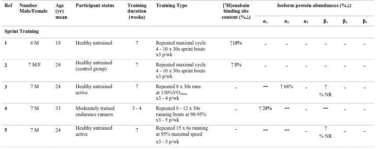
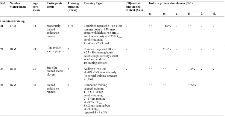
Sprint training in humans comprising repeated 30 s maximal bouts, increased skeletal muscle NKA content by 8 - 16% (McKenna et al. 1993; Harmer et al. 2006). Sprint training comprising repeated 30 s bouts at 130%VO2max elevated the NKA α2 isoform by
3 et al. 2007). In contrast, sprint training comprising repeated 30 s running bouts at 90 - 95% max running speed, increased the NKA α1 isoform by 29%, but with no changes in
α2 and β1 isoforms (Iaia et al. 2008). Neither study measured NKA α3, β2 and β3 isoform
abundances or NKA content. Hence measuring NKA α3, β2 and β3 isoform abundances
with NKA content is important to further understand the NKA adaptability in muscle after sprint training.
Resistance training in human increased muscle NKA content by 16% (Green et al. 1999b). In contrast, resistance training did not increase muscle NKA content in another study, although the NKA content was increased in data pooled from three separate trained groups by 15% (Medbø et al. 2001). A further study found that resistance training increased each of NKA α1, α2 and β1 isoforms, by 37%, 21% and 33%, respectively (Dela
et al. 2004), although the NKA α3, β2 and β3 isoforms and NKA content were not
investigated.
Thus it is unclear from both repeated 30 s sprint or resistance training studies which isoforms are associated with the adaptation of NKA content following training, since no study has combined measures of muscle NKA content and NKA isoform α1 - α3 and β1 -
β3 abundances following training. Therefore, this thesis investigated the effects of each
of sprint (Study 2, Chapter 3) and resistance exercise training (study 3, Chapter 4) on skeletal muscle NKA content combined with α and β isoform abundances in healthy young adults, to clarify which NKA isoforms were upregulated with an increase in skeletal muscle NKA content.
5 Chapter 2. Literature Review
2.1 Muscle contraction, sodium and potassium ion concentrations
Muscle contractions result from action potential (AP) propagation along the plasma membrane and into transverse tubules (t-tubules), where the AP is detected by voltage sensors, known as dihydropyridine receptors. The interaction between the dihydropyridine receptors and the sarcoplasmic reticulum ryanodine receptor causes Ca2+
release from sarcoplasmic reticulum into the cell cytoplasm and consequent cross bridge interaction (Allen et al. 2008; Lamb 2000). Each AP comprises an influx of Na+ ions into, and efflux of K+ ions from the muscle cells, creating Na+ and K+ ion currents across the cell membrane. The intracellular [K+] ([K+]i) is maintained at around 140 - 165 mM by
active K+ pumping into the cell from the extracellular environment, where the [K+] ([K+] e)
is around 5 mM. Through cellular extrusion of Na+, the NKA constrains the rise in intracellular [Na+] ([Na+]i) that would otherwise occur with repeated AP (Clausen 2010; Sejersted and Sjøgaard 2000). Hence, the NKA maintains a low intracellular [Na+] to [K+] ratio in the face of an inward concentration gradient for Na+ and an outward gradient for K+ and fluxes in both ions due to each AP (Mobasheri et al. 2000. Thus precise regulation
of the NKA with muscle contractions to maintain cellular [Na+] and [K+] is needed. 2.1.1 The relationship between membrane potential and [K+]
The resting membrane potential (Em) ranges between 75 - 89 mV, and allows muscle fibres to generate and propagate AP (McKenna et al. 2008; Sejersted and Sjøgaard 2000). Any decline in Em (depolarisation) resulting in membrane depolarisation induced inactivation of voltage Na+ channels, impairs AP propagation and results in a loss of muscle excitability (Allen et al. 2008). In skeletal muscle K+ plays a vital role in maintaining Em, thus any change in [K+] across the sarcolemma could potentially impact
6 Several studies have shown a correlation between the rise in [K+]
e and the decline of Em
in muscle. In rats soleus muscle, high frequency stimulation at 100 Hz increased [K+]e up
to 14 mM, which induced a decrease in Em from -74 to -53 mV (Cairns et al. 1995). In mouse muscle, high frequency stimulation at 120 Hz in soleus and at 200 Hz in extensor digitorum longus (EDL) muscles, elevated [K+]e from 7 to 14 mM and reduced Em
by~35% in both soleus and EDL muscles (Fig 2.1) (Cairns et al. 1997). In rat EDL muscle, AP firing frequency at 6, 15 and 30 Hz increased [K+] in the t-tubular system from 5.3 mM to 6.8, 8.1 and 11.2 mM which was associated with a 0.97, 2.08 and 4.4 mV decrease in Em, respectively (Fraser et al. 2011). In human vastus lateralis muscle, as a results of changes in the [K+]i, [K+]e, [Na+]i and [Na+]e, the Em was calculated to fall from 83 to
-75 mV following intense knee extensor exercise (Sjogaard et al. 1985). These calculations however, underestimated the rise in [K+]e in muscle.
Figure 2.1. Resting membrane potential (Em)-[K+] relationship for soleus (●) and EDL (□) mucles.
7 2.1.2 Effects of elevated of muscle [K+]
e and reduced [K+]i on muscle fatigue
During muscle contractions, muscle [K+]e increased up to 10 - 12 mM, which was
proposed to lead to fatigue development (Nordsborg et al. 2003; Nielsen et al. 2004a). Several in-vitro studies have demonstrated that elevated [K+]e caused a reduction in
tetanic force. In isolated rat soleus muscle, increased extracellular [K+]e from 4 to 10 mM
resulted in a 40% reduction in twitch and tetanic force, whilst increased [K+]e to 12.5 mM
reduced tetanic force by 95% (Clausen et al. 1993). Similarly, in isolated rat soleus muscle, an increase in [K+]e from 4 to 11 mM caused an 85% reduction in tetanic force
(Pedersen et al. 2005). In isolated mouse soleus muscle, an increase in [K+]e from 5 to 10
mM induced a 40% reduction in tetanic force (Juel 1988). In frog sartorius muscle, elevated [K+]e from 3 to 7 mMdecreased tetanic force by 41% (Bouclin et al. 1995).
In human vastus lateralis muscle, [K]i declined from 161 to 141 mM (Sjogaard 1983) and
from 165 to 129 mM during repeated muscle contractions (Sjogaard et al. 1985), which was suggested to contribute to muscle fatigue (Sjogaard 1983). In skinned fibres from rat EDL muscle, [K]i decreased from 113 to 60 mM and resulted in a 30% decline in twitch
force (Nielsen et al. 2004b). During 40 Hz stimulation, [K]i decreased by 32 mM in rat
soleus muscle and by 48 mM in EDL muscle, which was associated with 29 and 10 % reduction, respectively, in muscle force (Juel 1986). These findings have demonstrated that increased [K+]e during muscle contraction may play an important role in muscle
fatigue (Juel et al. 2000; Fitts 1994). However, changes in chloride (Cl-) ion conductance
8 of muscle tetanic force as a result of increased [K+]
e in rat soleus muscle was recovered
via reducing Cl- conductance (Pedersen et al. 2005). This relationship between [K+], Cl -conductance and Em is still unknown in human skeletal muscle.
2.1.3 [Na+] and muscle fatigue
In rat isolated soleus and EDL muscles, a reduction in [Na+]e from 147 to 30 mM and 25
mM decreased both twitch and tetanic force by ~55% (Cairns et al. 2003). Similarly, decreased [Na+]e from 147 to 25 mM reduced tetanic force by ~66% (Overgaard et al.
1997). In skinned fibres from rat EDL muscle, elevated [Na+]
i from 20 mM to 50 mM
reduced the ability of the t-tubules to support AP (Nielsen et al. 2004b). In rat isolated soleus muscle, there was a significant negative correlation between increased [Na+]i and
reduced muscle force during 60 Hz stimulation (Nielsen and Clausen 1996). In frog semitendinosus muscle, increased [Na+]i from 16 to 49 mM was associated with decrease
in Em from -38 to -74 mV (Balog and Fitts 1996).
9 2.2 Sodium-Potassium Adenosine Triphosphatase (Na+,K+-ATPase, NKA) in
skeletal muscle
2.2.1 Definition and function of NKA
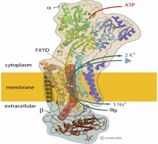
The NKA (Fig 2.2) is a membrane-associated protein complex that is ubiquitously expressed in most eukaryotic cells (Vague et al. 2004; Clausen 2013b; Clausen et al. 2017). The NKA was first identified in 1957 by Skou (Skou 1957) for which he was awarded the Nobel prize for chemistry (Clausen 1998). In skeletal muscle, the NKA are thought to be primarily localised in the sarcolemma and the t-tubules (Clausen 2003a). The NKA produces and maintains steep transmembrane [Na+] and [K+] gradients via
cellular uptake of two K+ and extrusion of three Na+ ions, whilst hydrolyzing ATP generated from cellular glycolysis (Nyblom et al. 2013; Reinhard et al. 2013). The NKA mediated Na+/K+ exchange is electrogenic and thus NKA activity contributes to membrane hyperpolarisation, which is essential for muscle excitability (Clausen 1996b). In addition to this ion-pumping function, the NKA is also involved in complex intracellular signalling. This may occur via direct protein-protein interactions between NKA and its neighbouring proteins, triggering a signalling cascade, culminating in gene transcription (Aperia et al. 2016; Reinhard et al. 2013).
2.2.2 Structure of NKA
The NKA comprises four α subunit isoforms (α1 - α4) and three β subunit isoforms (β1 -
β3), with both α and β subunits required for NKA function (Fig 2.2). A third subunit is a
member of the FXYD family (Kaplan 2002; Bibert et al. 2008). 2.2.2.1 The α subunit isoforms
10 and is responsible for the catalytic processes of the enzyme comprising the binding of Na+ and K+ ions and their transport across the membrane, fuelled by the hydrolysis of ATP (Scheiner‐Bobis 2002; Clausen 2003a).
In human skeletal muscle, all NKA isoforms except α4 are found (Murphy et al. 2004). In
human soleus muscle, the α1 isoform was found to be located in the plasma membrane,
with only around 4% located in an intracellular membrane fraction (Hundal et al. 1994). The α2 isoform was mainly detected in the plasma membrane, and around 75% in
intracellular membrane fractions, while the α3 isoform was also mainly located in the
plasma membrane (Hundal et al. 1994). The α1 isoform was reported to be equally
distributed in type I and type II single muscle fibres, whereas the α2 isoform had a 37%
higher abundance in type II than type I fibres in human muscle biopsy samples (Thomassen et al. 2013). A subsequent study in human single muscle fibres found both the α1 andα3 isoforms to be similarly expressed in type I and II fibres (Wyckelsma et al.
2015), and in contrast to Thomassen et al (2013) also found no α2 isoform differences
between fibre types (Wyckelsma et al. 2015).
In rat skeletal muscle, the α1 isoform was detected in both soleus (SOL) and extensor
digitorum longus (EDL) muscles and was present in sarcolemma and in t-tubules (Kristensen and Juel 2010). The α1 had a higher abundance in oxidative than in glycolytic
muscles in the rat (Kristensen and Juel 2010; Fowles et al. 2004; Thompson and McDonough 1996). In contrast, the α2 was equally distributed in oxidative and glycolytic
muscles (Fowles et al. 2004; Juel et al. 2001; Thompson and McDonough 1996). In murine skeletal muscle, both the α1 and α2 isoforms were present in the sarcolemma
(Williams et al. 2001).
The α1 isoform appears to play a “cellular housekeeping role” regulating [Na+] and [K+]
11 ions and considering the higher abundance of α1 in the sarcolemma (Crambert et al. 2000).
The α2 plays a major role in Na+ and K+ transport in working muscle. The α2 has a lower
affinity for K+ than α
1 in resting muscle (DiFranco et al. 2015), and the α2 activity
increased rapidly in contracting muscle in response to increased [K+]e (DiFranco et al.
2015). Research using gene knockout techniques in muscle show the importance of α1
and α2 isoforms. In mice, partial global knockout of the α1 isoform reduced contractile
force in EDL muscle (Lingrel et al. 2003). The complete global knockout in mice of the α2 isoform caused the animal to either be borne dead or to die shortly after birth (Lingrel
et al. 2003). In mice, skeletal muscle specific α2 knockout reduced each of muscle
strength, endurance and exercise tolerance, suggesting that the α2 plays a significant role
in maintaining contractions and resisting fatigue in skeletal muscle (Radzyukevich et al. 2013). This was also despite an upregulation of the α1 isoform (Radzyukevich et al. 2013).
The α3 isoform is predominantly expressed in neurones (Matchkov and Krivoi 2016;
Clausen et al. 2017; Bøttger et al. 2011). It was earlier suggested that the α3 isoform could
be activated and work as a spare isoform to help restore membrane potential, when the Na+ and K+ fluxes were higher and with NKA α1 and α2 working at maximal rates during
depolarization and repeated action potentials (Blanco & Mercer, 1998). However, the exact role of the α3 isoform in skeletal muscle, is unclear, and further research is required
to determine the functional of NKA α3 in skeletal muscle. 2.2.2.2 The β subunit isoforms
The β subunit spans the membrane once, with the N-terminus localised on the intracellular side of the membrane (Reinhard et al. 2013). The β subunit is glycosylated, and is composed of about 370 amino acids (Kaplan 2002) exhibiting a molecular mass of about 40 to 60 kDa (Mobasheri et al. 2000; Kaplan 2002), which varies for the β1, β2 or β3
12 for regulating NKA activity and also transporting and stabilising of movement of the α subunit from the endoplasmic reticulum to the plasma membrane (Hundal et al. 1994). Each of the β1, β2, and β3 isoforms have been detected in rat (Arystarkhova and Sweadner
1997; Ng et al. 2003) and in human skeletal muscle. In humans, the β1 and β3 isoforms
were found to be similarly expressed in both type I and II fibres (Wyckelsma et al. 2015), whereas the β2 was 27% more abundant in type II fibres (Wyckelsma et al. 2015;
Thomassen et al. 2013). The β1 was detected mainly in the plasma membrane with a small
amount being found in the intracellular fraction (Hundal et al. 1994). In the rat, the β1
isoform has a higher abundance in muscles comprised predominantly of slow twitch fibres, while the β2 has a higher abundance in muscles comprised predominantly of fast
twitch fibres (Fowles et al. 2004; Zhang et al. 2006). The β3 isoform was found to be
similarly abundant in rat red and white gastrocnemius muscles (Ng et al. 2003). The β1
and β2 isoformswere detected in the sarcolemma and t-tubules in rat skeletal muscle
(Hundal et al. 1994).
Whilst it is known that the β-subunit is essential for the transport of the α-subunit to the plasma membrane (Geering 1990; Mcdonough et al. 1990; Chow and Forte 1995), the function of each of the NKA β isoforms is still unclear. However, in basal conditions, the β1 isoform shows a greater affinity to Na+ and lower affinity to K+ compared to β2
(Crambert et al. 2000). Unlike with the α isoforms, no research on β isoforms knockout animal models has yet been conducted.
2.2.2.3 The phospholemman subunit
13 has a relative mass of only 7 - 11 kDa, and is primarily expressed in skeletal muscle (Li et al. 2004; Mishra et al. 2011; Geering 2008).
14 2.2.2.4 [3H]ouabain binding site content
The [3H]ouabain binding site content technique is the traditional method used to measure NKA content in skeletal muscle, being based on the high binding affinity of cardiac glycosides to the NKA α subunitwith a stoichiometry of 1:1 (Hansen 1984). The NKA α1 isoform has a low affinity to cardiac glycosides in rat muscle, thus the standard
[3H]ouabain binding site content technique is unable to detect the α
1 isoform (Clausen
2003a; Hansen 2001). In rat EDL and soleus muscles, the α1 isoform represents around
15 - 25% of the total pool of NKA (Hansen 2001). Another study in rat EDL muscle showed that, the NKA α1 isoform only accounts for 13% whilst the α2 isoform comprised
87% of the total α isoforms; however, the NKA α3 isoform was not detected (He et al.
2001). Thus, in rat skeletal muscle, the [3H]ouabain site content assay does not detect all αsubunits. In contrast, in human skeletal muscle, the NKA α1, α2, and α3 isoforms each
have a similar ouabain affinity and thus all are detected using the [3H]ouabain binding
site content method (Wang et al. 2001). Therefore, in human skeletal muscle, the [3H]ouabain binding site content is a quantitative measure of and can also be referred to as the NKA content, whereas in rodent skeletal muscle this is better expressed just as [3H]ouabain binding site content (Nørgaard 1986; Nørgaard et al. 1984). Therefore, in this thesis these measures will be described as [3H]ouabain binding site content in study
one which utilised rat skeletal muscle and thereafter as NKA content in Studies two, three and four, which all utilised human skeletal muscle. The typical NKA content, measured as [3H]ouabain binding site content, reported in human skeletal muscle is shown in Table
15 2.2.2.5 Comparisons of muscle NKA variables in human and rat skeletal muscle
The key comparisons in skeletal muscle NKA between human and rat, as detailed in previous sections are as follows:
1. In human skeletal muscle, the NKA α1, α2, and α3 isoforms each have a similar
ouabain affinity, thus the [3H]ouabain site content reflects the whole NKA content. In rat muscle, the [3H]ouabain site content reflects the α
2 content due to
lower of α1 isoform affinity for [3H]ouabain.
2. In both human and rat muscle, the NKA α1 isoform is expressed in the sarcolemma
while the α2 isoform is present predominantly in the intracellular membrane
fraction.
3. In human, the α1 α2 andα3 are all ~equally abundant in both type I and type II
fibres. In rat muscle, the α1 is more abundant in oxidative than in glycolytic
muscle, and the α2 is similarly abundant in oxidative and glycolytic muscles.
4. In human muscle, the β1 and β3 isoforms are similarly expressed in both type I and
II fibres, whereas the β2 more abundant in type II fibres. In rat, the β1 isoform has
a higher abundance in type I fibre, and the β1 isoform has a higher abundance in
16 Table 2.1. Vastus lateralis muscle [3H]ouabain binding site content (NKA content)
measured in muscle biopsy samples in healthy human males and females aged between 18 - 69 years.
Study Age
(Mean yr)
Number (sex M/F)
[3H]-ouabain site content (pmol.g wet wt-1)
Klitgaard et al., 1989 68 6 M 237
Klitgaard et al., 1989 28 6 M 278
Green et al., 1993 19 9 M 339
McKenna et al., 1993 19 6 M 333
Madsen et al., 1994 30 39 M 307
Evertsen et al., 1997 18 11 M 343
Green et al., 1999a 21 16 M 284
Medbø et al., 2001 19 8 M 356
Fraser et al., 2002 26 8 M 311
Leppik et al., 2004 27 7 M
1 F
333
Petersen et al., 2005 24 8 M 7 F
291
Aughey et al., 2007 31 11 M 355
McKenna et al., 2012 24 8 M 8 F
350
McKenna et al., 2012 67 9 M 8 F
352
Perry et al., 2013 69 9 M
10 F
357
Wyckelsma et a l., 2017 69 9 M 6 F
17 2.2.2.6 NKA activity
The activity of NKA in skeletal muscle at rest is low, but increases rapidly during muscle contractions (Nielsen and Harrison 1998; Clausen 2008). At rest, the NKA activity is only at 2 - 6% of its maximal theoretical capacity, but when the isolated muscle was loaded with Na+, reached up to 90% of its maximal activity (Clausen et al., 1987). The maximal activity of NKA has been measured in-vitro in human skeletal muscle by the 3-O-methylfeuorescein phosphatase (3-O-MFP) activity assay (Fraser and McKenna 1998) and impairments linked with fatigue (McKenna et al 2008). Submaximal cycling at ~75% VO2peak until fatigue reduced maximal in-vitro 3-O-MFP by ~11 - 13 % (Leppik et al.
2004) and incremental cycling to fatigue reduced 3-O-MFP by ~30% (Sandiford et al. 2005). Isometric exercise on an isokinetic dynamometer caused a reduction in NKA activity by ~35% (Fowles et al. 2002) and isokinetic contractions decreased 3-O-MFP by ~11 - 14% (Fraser et al. 2002; Petersen et al. 2005). Submaximal cycle ergometer exercise caused a 12% decrease in 3-O-MFP (Aughey et al. 2005). The 3-O-MFP assay has several disadvantages in measuring NKA activity after exercise, since it does not involve the hydrolysis of ATP, and relies on the K+ stimulation without the Na+ sensitivity (Juel et al. 2013). Given the role of phospholemman (PLM) phosphorylation in increasing the NKA activity via Na+ activation, this assay thus may not reflect the activity of NKA
caused by phospholemman with exercise.
A more recent technique used to quantify the NKA activity, measures phosphate (Pi) production from hydrolysis of ATP and Na+-dependent activation (Juel et al. 2013).
18 detecting changes in NKA activity with exercise. However, Hostrup et al (2014) reported a reduction in Na+-dependent activity after exercise (Hostrup et al. 2014). Due to methodological concerns regarding both the 3-O-MFP and the ATP hydrolysis method utilised by Juel et al (2013), the activity of NKA in skeletal muscle was not measured in this thesis.
2.3 Effects of Age on skeletal muscle NKA
Few studies have investigated the effects of aging on skeletal muscle NKA in humans, with findings inconclusive. No significant difference was found in vastus lateralis muscle NKA content between older (mean age 68 years) and young, untrained (mean age 28 years) participants (Klitgaard and Clausen 1989). No differences were also detected in NKA content between young versus older (mean age 23.9 vs 66.8 years) participants (McKenna et al. 2012), or in 20 participants ranging from 25 - 80 years old (Nørgaard et al. 1984). In contrast, the NKA content in older adults aged between 69 - 81 years was 25.5% lower than those aged 55 - 68 years (Perry et al. 2013), however, this might be due to their chronic physical activity levels rather than their age (Wyckelsma and McKenna 2016). There was no difference in muscle NKA content between aged and young adults (mean age 25.5 vs 69.4 years) (Wyckelsma et al. 2016). Subsequent pooled analyses found that no age effect on NKA between younger adults aged 18 - 30 years compared to those aged from 55 to76 years (Wyckelsma and McKenna 2016) (Fig 2.4). Thus the evidences points no age effect on NKA.
The NKA isoform abundances were first reported lower by 24% for α2 and 23% for β3
isoform in older (mean age 66 years) than young adults (mean age 29 years), with no differences in other NKA isoforms (α1, α2, β1 and β2) (McKenna et al. 2012). However,
19 protein is highly expressed in Type II than Type I fibers, no differences were found inany of NKA isoform abundances between young and old adults (Wyckelsma et al. 2016). When NKA isoforms were measured in muscle single fibres, the NKA α1 had a ~71%
greater abundance in aged muscle in Type I fibre compared to young. The NKA α3 and
β2 isoforms in aged muscle were both lower by ~47% and ~85%, respectively, in Type II
fibers compared to young (Wyckelsma et al. 2016). The NKA β3 was greater in aged
muscle in both Type I and II fibers by ~96% and ~285%, respectively compared to young (Wyckelsma et al. 2016).
In animal models, clearer age effects have been reported. The muscle [3H]ouabain binding site content from 85 day old rats was 58% lower in soleus (SOL) and extensor digitorum longus (EDL) muscles, when compared to infant rats (Clausen et al. 1982). In female guinea pigs, the [3H]ouabain binding site content was 60% lower in EDL muscle from birth compared to maturity (Kjeldsen et al. 1984b). In rat soleus muscle, the [3H]ouabain
binding site content increased from 120 to 580 pmol.g wet wt-1 from 2 to 28 days of life, and then decreased to150 - 200 pmol.g wet wt-1 from 4 to 21 weeks of life (Kjeldsen et al. 1982). No difference was found in the [3H]ouabain binding site content in either SOL or EDL muscles between 3 and 14 week old rats (Abdel-Azia et al. 1985). The α2 isoform
was lower by 30 - 40% in red and white gastrocnemius muscle in rats aged 18 and 30 months compared to 6 months (Sun et al. 1999). However, in SOL muscle, the α1 isoform
abundances increased by ~100% in one month old rats compared to those aged 6 months (Sun et al. 1999). In white and red gastrocnemius muscle, the α1, β1, and β3 isoforms were
more abundant, while the α2 and β2 isoform abundances decreased in 30-month compared
20 In summary, whereas studies in rat muscle showed that aging is associated with a decline in [3H]ouabain binding site content and NKA isoform abundances, no clear age effects are seen in human skeletal muscle.
Figure 2. 3. Effects of aging on muscle [3H]ouabain binding in human skeletal muscle.
Muscle [3H]ouabain binding site content was collated from data collected on healthy young and healthy older adults from the McKenna research group between 2012 and 2016. (A) Shows the data combined into two discrete age groups, with no signifecant difference analysed by unpaired t-test (p = 0.53). (B) Shows all data plotted into relevant decades of life, analysed with no signifecant difference by one-way ANOVA (p = 0.30); the mean of each group is also shown as a horizontal line (From Wyckelsma and McKenna 2016).
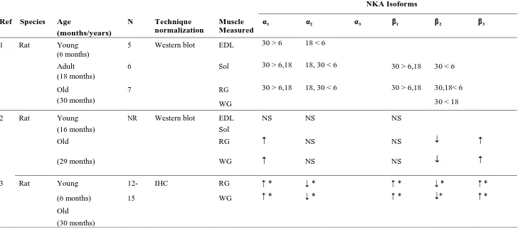
21 Table 2.2 Effects of age on NKA isoform abundances in rat and human skeletal muscle
NKA Isoforms
Ref Species Age
(months/years)
N Technique
normalization
Muscle Measured
α1 α2 α3 β1 β2 β3
1 Rat Young
(6 months)
5 Western blot EDL 30 > 6 18 < 6
Adult (18 months)
6 Sol 30 > 6,18 18, 30 < 6 30 > 6,18 30 < 6
Old 7 RG 30 > 6,18 18, 30 < 6 30 > 6,18 30,18< 6
(30 months) WG 30 < 18
2 Rat Young NR Western blot EDL NS NS NS
(16 months) Sol
Old RG ↑ NS NS ↓ ↑
(29 months) WG ↑ NS NS ↓ ↑
3 Rat Young 12- IHC RG ↑ * ↓ * ↑ * ↓ * ↑ *
(6 months) 15 WG ↑ * ↓ * ↑ * ↓* ↑ *
Old
22
Reference 1, Sun et al., 1999; 2, Ng et al., 2003; 3, Zhang et al., 2006; 4, McKenna et al., 2012; 5, Wyckelsma et al., 2016. Age, mean ± SD. Symbols: ↓,
denotes decrease; –, no change; ↑, increase, data in parentheses denotes % difference between groups. ↑ * not quantitative but increased compared to
young ↓ * not quantitative but decreased compared to young. NR not reported, NS not significant. Muscles: EDL, Extensor Digitorum Longus; Sol,
Soleus; RG, Red Gastrocnemius; WG, White Gastrocnemius; VL, Vastus Lateralis. (From Wyckelsma and McKenna 2016).
Table 2.2 continued NKA Isoforms
Ref Species Age
(months/years)
N Technique
normalization
Muscle Measured
α1 α2 α3 β1 β2 β3
4 Human Old
66.8 ± 6.4
17 Western blot (GAPDH)
VL – ↓ 24% – – – –
Young 23.9 ± 2.2
16
5 Human Old
69.4 ± 3.5
17 Western blot
(Calibration Curve)
VL – – – – – ↑ 250%
Young 25.5 ± 2.8
14 VL Type
I fibres
↑ 71% – – – – ↑ 96%
VL Type II fibres
23 2.4 Effects of selected hormones and sex differences on skeletal muscle NKA
2.4.1 Hormonal regulation
Numerous hormones regulate skeletal muscle NKA and further detail can be found in earlier major reviews (Clausen 2003a; Ewart and Klip 1995). Thyroid hormones, glucocorticoids and insulin all have a marked effect on NKA content and thus are briefly reviewed here.
2.4.1.1 Thyroid hormones
Thyroid hormones exert a major influence on NKA regulation. In humans, the NKA muscle content was 50% lower in patients with hypothyroidism and 68% greater in patients with hyperthyroidism, compared to euthyroid controls (Kjeldsen et al. 1984a) . The muscle NKA content was also reported to be 89% higher in hyperthyroid patients compared to euthyroid control (Riis et al. 2005) . In hyperthyroid rats, injection of a single dose of triiodothyronine (T3, 5-20 ug/100g body wt) increased the [3H]ouabain site binding site content in SOL, gastrocnemius and EDL muscles by 9.8-, 5.1-, and 2.6-fold, respectively, compared to the hyporthyroid rat (Kjeldsen et al. 1986a). In isolated rat SOL muscle, injection of a single dose of triiodothyronine (T3, 20 ug/100g body wt) for 8 d increased the [3H]ouabain binding site content by 103% (Everts and Clausen 1988). 2.4.1.2 Glucocorticoids
24 respectively, compared to placebo (Nordsborg et al. 2008). In rat skeletal muscle, treatment with dexamethasone increased the [3H]-ouabain site binding site content by 23– 52% in EDL, soleus, gastrocnemius and diaphragm muscles (Dørup 1996).
2.4.1.3 Insulin
Insulin regulates the NKA in skeletal muscle acutely via increasing the affinity for intracellular Na+ (Sweeney and Klip 1998; Pirkmajer and Chibalin 2016; Kitasato et al.
1980; Lytton 1985) Insulin also increased NKA content. In human patients with diabetes, the vastus lateralis muscle NKA content was 17 and 22% greater in patients with treated non-insulin-dependent diabetes mellitus and insulin-dependent diabetes mellitus than in control participants (Schmidt et al. 1994). Insulin increased the [3H] ouabain binding site content by ~90% after 60 min of insulin incubation in plasma membrane fraction of frog skeletal muscle (Omatsu-Kanbe and Kitasato 1990), although the methodology was later criticised (Clausen 2003a). In untreated diabetic rats, lower insulin was associated with an 18% reduction in the [3H] ouabain binding site content, conversely, after treatment with insulin, the level of [3H]ouabain binding site content increased by ~23% (Schmidt et al. 1994).
2.5 Effects of sex differences and sex hormones (testosterone and estradiol) on skeletal muscle NKA
25 recreationally active males and females (Murphy et al. 2007). In contrast, in elite skiers, the NKA content was found to be 18% higher in young adult males than females (Evertsen et al. 1997). These differences seen in elite level athletes may be result of training, whilst the participants in the other studies were untrained. Whilst findings from these studies suggest that sex difference is not associated with differences in [3H]ouabain binding site content, further research is required to verify this and also to determine the possibility of sex differences on NKA isoform abundances.
2.5.1 Estradiol effects
Estradiol is the major estrogen hormone in women and produced by the ovaries, and has a vital role in reproductive cycle (Cohen et al. 2003), brain development and function (Vermeulen et al. 2002), lean mass (Brown 2008) and stimulating muscle damage repair via activation and proliferation of satellite cells (Enns and Tiidus 2010). No studies appear to have investigated estradiol effects on NKA in skeletal muscle. However, several studies have investigated the effects of estradiol therapy on NKA in cardiac muscle in rats. Injection of 40 mg.kg-1 estradiol for 24 hours was found to stimulate NKA activity in heart sarcolemma by 360% (Džurba et al. 1997; Obradovic et al. 2015). A dose of 0.5 mg.kg-1 of estradiol delivered via injection for 3 days increased the NKA activity in heart sarcolemma by 85% (Obradovic et al. 2015). However, an acute 40μg.kg-1 dose of estradiol for only 20, 30 and 40 min before analysis had no effect on the cardiac plasma membrane NKA α2 isoform abundance (Koricanac et al. 2011).
2.5.2 Testosterone effects
26 concentration, which can also occur as a result of different clinical conditions such as trauma and obesity; this decrease is associated with a decline in muscle mass and strength (Leichtnam et al. 2006; Brown 2008). Testosterone reduction can also occur as result of androgen deprivation therapy inprostate cancer patients (Mostaghel et al. 2007; Sharifi et al. 2005). Few studies have investigated the effects of testosterone on NKA. In rats, testosterone reduction via castration resulted in a 47 -73% decrease in NKA activity in rat erythrocyte membranes compared to a sham group (Sheng-qi et al. 1994). Injection of 50 µg testosterone for 2 days in castrated rats increased NKA activity 4-fold in the cerebral cortex compared to a sham group (Guerra et al. 1987). No studies have investigated the impacts of testosterone reduction on skeletal muscle NKA content, or on NKA isoform abundances. Given the importance of NKA for muscle function, it is beneficial to understand the impact of testosterone on NKA content and isoform abundances, with important implications particularly for people who suffer from lower testosterone. Therefore, the first study in this thesis investigated the effects of suppression in testosterone due to castration on skeletal muscle [3H]ouabain binding site content and NKA isoform abundance in rat soleus muscle.
2.6 Intracellular [Na+] regulation and NKA in skeletal muscle
Elevated [Na+]i occurs with acute contractions (exercise), is one of the vital regulators for
NKA in skeletal muscle, including possible mediator of NKA upregulation (Nielsen and Clausen 1997; Clausen 1996a; Sejersted and Hallén 1987). The maximal activity of NKA in an isolated soleus muscle was increased up to 90% after being loaded with Na+ (Clausen
and Flatman 1987). Following30 s stimulation in isolated rat soleus muscle, the [Na+]i
27 [Na+]
i by 58%, which was associated with an increase in NKA activity by ~15-fold
(Everts and Clausen 1994).
2.7 Inactivity, immobilisation and exercise training effects on skeletal muscle NKA
2.7.1 Inactivity and immobilisation effects on skeletal muscle NKA
Numerous studies have indirectly investigated the effects of inactivity on muscle NKA content in humans, by measuring the impacts of injury. In participants with shoulder impingement syndrome, the NKA content in deltoid muscle decreased by 26% compared to the non-injured shoulder (Leivseth and Reikerås 1994). In patients after complete spinal cord injury, the NKA content in vastus lateralis muscle was decreased by ~34% compared to the deltoid muscle (Ditor et al. 2004). In chronic spinal cord injury patients, 75%, 52% and 38% decreases in NKA α1, α2 and β1 isoform abundances were reported
compared to the control group, respectively (Boon et al. 2012). In healthy young adults with a torn anterior cruciate ligament, the vastus lateralis muscle NKA content was 20 % less and the α2 isoform abundance was 63% lower in the knee-injured leg than the
non-injured leg (Perry et al. 2015). The above findings from human injury studies, strongly suggest a reduction in NKA content occurs with muscle inactivity, but might also be due to the injury or iatrogenic effects (Madsen et al. 1993).
The vastus lateralis muscle [3H]ouabain-binding site content and the NKA isoform abundances in mixed muscle fibre homogenates were unchanged after 23 day of unilateral lower limb suspension (ULLS); however, in single muscle fibres, the NKA α3 isoform
abundance decreased by 66% in type I, and NKA β1 isoform abundance decreased by 40%
28 content in rat soleus muscle by 20 - 22% (Kjeldsen et al. 1986b) and by 25% (Zemkova et al. 1990). In guinea pigs, hindlimb immobilisation for 3 and 4 weeks decreased the [3H]ouabain binding site content in the gastrocnemius muscles by 25% and 23%,
respectively (Leivseth et al. 1992). In female sheep, nine weeks of hindlimb immobilisation reduced the [3H]ouabain binding site content in the vastus lateralis muscle by 39% (Jebens et al. 1995). Following immobilisation, the [3H]ouabain binding site
content returned to baseline levels after only 7 days (Zemkova et al. 1990) and after 4 - 5 weeks of recovery (Leivseth et al. 1992). Table 2.3 presents the effects of hindlimb immobilisation on [3H]ouabain binding site content in animal skeletal muscle.
Recent studies have found that short-term hindlimb suspension in the rat for only 6 - 12 hours and for 1 - 3 days, decreased the electrogenic transport activity of the NKA α2 by
~96% and ~71%, respectively, but not the NKA α1 isoform, as measured by the
ouabain-sensitive changes in resting membrane potential (Kravtsova et al. 2016; Kravtsova et al. 2015). Neither study investigated the α3 or β1 - β3 isoforms. Nothing is known about the
effects of hindlimb immobilisation on NKA α3 or on β1 - β3 isoform abundances, and the
recovery of NKA isoform abundance following hindlimb immobilisation has also not previously been investigated. Therefore, the first study in this thesis investigated the effects of hindlimb immobilisation in rats on the soleus muscle [3H]ouabain binding site
content and the abundance of each of the NKA α1- α3 and β1 - β3 isoforms, as well as the
29 Table 2.3 Effects of hindlimb immobilisation on [3H]ouabain binding site content in animal skeletal muscle.
Reference: 1, Kjeldsen et al. 1986; 2, Zemkova et al. 1990; 3, Jebens et al. 1995; 4, Leivseth et al. 1992. Symbols: (↓) decrease; (↔) no change; (-) not measured; (∆) changes and (NR) not reported. Muscles: SOL, Soleus; WG, White Gastrocnemius; VL, Vastus Lateralis.
Ref Species Age
(weeks)
Number Duration (weeks)
Muscle Measured
[3H]ouabain binding site content
(% ∆ changes )
Time to Recover
1 Rat 12 5 1 Sol ↓ 20 - 22%
2 Rat 8-10 NR 1 Sol ↓ 25% 3 days
3 Sheep NR NR 9 VL ↓ 39%
![Figure 2.1. Resting membrane potential (Em)-[K+] relationship for soleus (●) and](https://thumb-us.123doks.com/thumbv2/123dok_us/8018377.1333421/28.596.173.444.406.635/figure-resting-membrane-potential-em-k-relationship-soleus.webp)
![Table 2.1. Vastus lateralis muscle [3H]ouabain binding site content (NKA content)](https://thumb-us.123doks.com/thumbv2/123dok_us/8018377.1333421/38.596.88.509.168.761/table-vastus-lateralis-muscle-ouabain-binding-content-content.webp)