Snakebite envenoming
José María Gutiérrez1, Juan J. Calvete2, Abdulrazaq G. Habib3, Robert A. Harrison4, David J. Williams5,6 and David A. Warrell7
1
Instituto Clodomiro Picado, Facultad de Microbiología, Universidad de Costa Rica, PO Box 11501-2060, San José, Costa Rica
2Instituto de Biomedicina de Valencia, Consejo Superior de Investigaciones Científicas (CSIC), Valencia, Spain
3College of Health Sciences, Bayero University, Kano, Nigeria
4Alistair Reid Venom Research Unit, Liverpool School of Tropical Medicine, Liverpool, UK 5Charles Campbell Toxinology Centre, School of Medicine & Health Sciences, University of Papua New Guinea, Boroko, National Capital District, Papua New Guinea
6Australian Venom Research Unit, Department of Pharmacology and Therapeutics, University of Melbourne, Parkville, Victoria, Australia
7
Nuffield Department of Clinical Medicine, John Radcliffe Hospital, University of Oxford, Oxford, UK
Correspondence to:
J.M.G.
Abstract | Snakebite envenoming is a neglected tropical disease that kills >100,000 and
maims >400,000 people every year. Impoverished populations living in the rural tropics
are particularly vulnerable; snakebite envenoming perpetuates the cycle of poverty. Snake
venoms are complex mixtures of proteins that exert a wide spectrum of toxic actions. The
high variability in snake venom composition is responsible for a plethora of clinical
manifestations in envenomings, ranging from local tissue damage to potentially
life-threatening systemic effects. Intravenous administration of antivenom is the only specific
treatment to counteract envenoming. In addition, analgesics, ventilator support, fluid
therapy, haemodialysis and antibiotic therapy are used. Novel therapeutic alternatives
based on recombinant antibody technologies and new toxin inhibitors are being explored.
Confronting snakebite envenoming at a global level demands the implementation of an
integrated intervention strategy involving the WHO, the research community, antivenom
manufacturers, regulatory agencies, national and regional health authorities, professional
health organizations, international funding agencies, advocacy groups and civil society
insititutions.
[H1] Introduction
manifestations. Some toxins in venoms provoke local tissue damage, often resulting in permanent sequelae, whereas others induce systemic effects, including neurotoxic manifestations (leading to, for example, respiratory paralysis), bleeding, acute kidney injury, rhabdomyolysis (generalized breakdown of muscle fibres), cardiotoxicity, autonomic hyperactivity or thrombosis. Venoms from snakes of the family Viperidae (viperids) cause local effects and systemic manifestations associated with bleeding, coagulopathies and hypovolaemic shock2. Venoms from snakes of the family Elapidae (elapids) predominantly induce neurotoxic manifestations, such as neuromuscular paralysis2.
The superfamily Colubroidea — or advanced snakes — consists of >2,500 species with a wide geographical distribution and an extended evolutionary history. The superfamily includes all venomous snakes classified in the taxon Caenophidia, order Squamata, suborder Serpentes. The most dangerous species are classified within the families Viperidae (true vipers and pit vipers) and Elapidae (elapids, for example, cobras, kraits, mambas and sea snakes) (Fig. 1b–i)3,4. Additionally, some species of the families Lamprophiidae (lamprophiids; subfamily Atractaspidinae, for example burrowing asps or stiletto snakes) and several subfamilies of non-front-fanged colubroid snakes are also capable of inflicting envenomings2.
posed by snakebites elsewhere. Consequently, snakebite envenoming have historically received little attention from funding bodies, public health authorities, the pharmaceutical industry and health advocacy groups, thereby impairing the development of effective interventions to reduce the social impact of snakebites9,10.
This Primer summarizes the main issues of snakebite envenoming, including the epidemiology, the composition of snake venoms, and the pathophysiology, clinical manifestations, prevention and clinical management of snakebite envenomings. The global efforts being carried out for reducing the impact of this pathology are described, together with future trends to better understand and confront this neglected tropical disease.
[H1] Epidemiology
Snakebite envenoming is a major public health problem in the developing world (Fig. 2). It is an important cause of morbidity and mortality especially in the impoverished areas of the warmer tropics and subtropics, such as sub-Saharan Africa, South to South-East Asia, Papua New Guinea and Latin America6,7,11. Snakebite envenoming occurs in at least 1.8–2.7 million people worldwide per year, with combined upper estimates of mortality ranging from 81,410 to 137,880 (Refs5,12). At least 46,000 of these deaths occur in India alone6. In sub-Saharan Africa, where data are fragmented, mortality estimates range from 7,000 to 32,000 deaths per year5,7,12, but are likely be underestimated given that, in West Africa alone, annual mortality has been estimated at 3,557–5,450 (Ref.13).
5–14 years6. Children are exposed while helping in agricultural duties, playing or placing their hands in rodents’ burrows.
Snakebite envenoming is an occupational and environmental disease of the young and agricultural workers. The specific populations at risk differ between countries. For example, tea pickers are at risk in southern India and Sri Lanka, rubber tappers in Liberia, Thailand, Malaysia, other South East Asian countries and Brazil, and sugar cane workers in South Africa, Saint Lucia and Martinique16–19. In Myanmar, snakebite envenoming is the 5th leading cause of death, particularly affecting rice paddy farmers20. Fishermen using hand nets and lines in warmer tropical seas are also at risk, as are families of agricultural workers. Pregnant women are a highly vulnerable group, and snakebite has been recognized in Nigeria and Sri Lanka as an important cause of abortion and antepartum haemorrhage, as well as maternal and fetal loss21.
Snakebite is also an environmental hazard of indigenous nomadic peoples, hunter-gatherers, tribes, firewood collectors and indigents (extremely poor individuals). This risk has been documented in South America19, Africa (for example, the Hadza hunter-gatherers of Tanzania, the African Bushmen of the Kalahari desert or Southern Africa, the nomadic Fulani and Turkana pastoralists (sheep or cattle farmers) of the savanna in West Africa and Kenya)22, India and Sri Lanka. Envenoming has been an important cause of death among the indigenous communities of Australia, of the coastal lowlands of New Guinea23 and of the Amazon (Yanomami and Waorani ethnic groups)18,19.
estimate suggested for contributions grouped under ‘venomous animal contact’ in the Global Burden of Disease study27, underscoring a notorious underreporting of snakebites in official records. When both premature deaths and disability from snakebites are factored for 16 countries in West Africa, the combined burden surpasses the worldwide burdens for other neglected tropical diseases such as buruli ulcer, echinococcosis, leprosy, trachoma, yaws, yellow fever and podoconiosis. The burden of snakebites in sub-Saharan Africa is also higher than the burden of trypanosomiasis, leishmaniasis and onchocerciasis13.
[H1] Mechanisms/pathophysiology
[H2] Snake venom
duplication event) as venom toxins in different taxa37. Gene duplication, evolutionary divergence and post-translational quaternary associations in homomer or and heteromer multiprotein complexes38 add to the complexity of snake venoms, by generating mixtures of proteins belonging to a handful of multigenic protein families; some of these protein families exhibit remarkable intrafamily variability39.
Unveiling the spatial and temporal distribution of variations in venom composition within and between species is essential to understand the evolutionary processes and the ecological constraints that molded snake venoms to their present-day variability and to define the phylogeographic boundaries of species. Insights into the selective pressures that resulted in local adaptation and species-level divergence in venoms can shed light on the mutual relationship between evolutionary and clinical toxinology: toxins bearing the highest prey incapacitation activity are often also the most medically important molecules in the context of a human envenoming. Thus, identifying the molecular basis of venomous snake adaptation to their natural ecosystems may assist in the identification of those toxins that must be neutralized to reverse the effects of venom, thereby guiding the rational development of next-generation snakebite therapeutics40.
[H3] Analysis of snake venom composition. The growing interest in different aspects of venom biology has catalysed the development of ‘omics’ methodologies aimed at the qualitative and quantitative characterization of venom toxins, including the proteomics of venoms (that is, venomics; Box 1). In particular, the combination of next-generation transcriptomics41,42 and proteomics workflows has demonstrated unparalleled capabilities for venom characterization in unprecedented detail. Figure 3 summarizes the relative distribution of the main types of toxic components in viperid and elapid venoms.
heterologous venoms43. The combination of antivenomics and in vivo neutralization tests constitute a powerful toolbox for evaluating the preclinical efficacy of an antivenom44.
[H2] Mechanism of action
Snakes inject their venoms through a specialized delivery system, which includes a set of fangs located in the frontal region of the maxillary bones in viperids (Fig. 1a), elapids and lamprophiids, whereas fangs have a posterior location in non-front-fanged colubroids. Depending on the size of the fangs, venom is injected either subcutaneously or intramuscularly. Once delivered, some venom toxins exert local pathological effects in neighboring tissues, whereas others are distributed systemically through the lymphatic system and blood vessels, enabling toxins to act at various organs2. Activities of main toxin families are shown in Box 2, whereas the potential consequences of snakebite envenoming are shown in Figure 5.
and innervation to be successful. In viperid venoms, which affect muscle fibres and damage the vasculature and nerves, skeletal muscle regeneration is impaired, often resulting in permanent sequelae48,49.
In addition to myonecrosis, blood vessel integrity is also affected. Snake venom metalloproteinases (SVMPs) in viperid venoms hydrolyse key components of the basement membrane of capillaries, particularly type IV collagen, causing the weakening of the mechanical stability of microvessels. As a consequence, the haemodynamic biophysical forces operating in the circulation cause distension and, eventually, disruption of the capillary wall, resulting in extravasation50. SVMP-induced microvascular damage can also be a consequence of the disruption of endothelial cell-cell adhesions51. SVMPs and hyaluronidases hydrolyse extracellular matrix components, including various types of collagens, hyaluronic acid and proteoglycans, affecting the structure and function of not only microvessels but also other tissue components, thereby playing a part in venom-induced local tissue damage52.
SVMPs also induce skin damage by degrading the dermal-epidermal interphase, with the consequent formation of blisters53. Some cobra (Naja sp., family Elapidae) venoms induce extensive cutaneous necrosis owing to the action of cytotoxins of the three-finger toxin family54, so named for having a common structure of three loops extending from a central core, which destabilize plasma membranes of various cell types in different tissues through a non-enzymatic mechanism55. Locally-acting toxins (that is, PLA2s and SVMPs) also affect intramuscular nerves49 and other vascular components such as lymphatic vessels, arterioles and venules56.
damage-associated molecular patterns (DAMPs), which are likely to potentiate the inflammation and, possibly, contribute to further tissue damage58.
Some mediators also induce pain57. Catalytically-inactive PLA2 homologues excite pain-related sensory neurons via ATP release and activation of purinergic receptors59.
[H3] Neurotoxicity. Snake venoms of most elapid species, and of some viperid species, contain neurotoxins that induce a descending flaccid neuromuscular paralysis, which can involve the life-threatening blockade of bulbar (muscles of the mouth and throat responsible for speech and swallowing) and respiratory muscles. Two main types of neurotoxins are found in snake venoms: α-neurotoxins and β-neurotoxins.
α-Neurotoxins belong to the three-finger toxin family and exert their action post-synaptically at neuromuscular junctions60. They bind with high affinity to the cholinergic receptor at the motor end plate in muscle fibres, thereby inhibiting the binding of acetylcholine and provoking flaccid paralysis60.
PLA2s can also act intracellurlarly after entering the cytosol by endocytosis or through the damaged plasma membrane62. Within the nerve terminus, PLA2s cause further degenerative events in mitochondria66.
Other neurotoxins are dendrotoxins and fasciculins, present in the venoms of the African elapids mambas (Dendroaspis sp.; family Elapidae). Dendrotoxins block voltage-gated potassium channels at the presynaptic nerve terminal67. Fasciculins, which also belong to the three-finger toxin family, are inhibitors of acetylcholinesterase68. Their combined action results in excitatory effects and fasciculations (involuntary muscle contraction). Some cysteine-rich secretory proteins (CRISPs) in venoms induce paralysis of smooth muscle2.
[H3] Cardiovascular and haemostatic disturbances. Systemic haemorrhage occurs in envenomings by viperids and by some species of non-front-fanged colubroids, and can also develop in envenomings by Australian elapids. In viperid venoms, the main toxins responsible for systemic haemorrhage are SVMPs, especially those of the PIII class. These toxins have a multi-domain structure containing exosites (molecular sites distinct from the active catalytic site that serve as secondary binding sites) that enable them to target the microvasculature50,69. Bleeding can occur in different organs with several pathophysiological consequences. For example, intracranial haemorrhage has been described in envenomings, causing ischaemia and stroke, and leading to neurological sequelae70,71. The mechanism of action of systemically-acting haemorrhagic SVMPs is likely to be similar to that described for local haemorrhage, that is, cleavage of key substrates at the basement membrane of capillaries and at cell–cell junctions, resulting in the mechanical weakening of the microvessel wall and extravasation50,51.
fibrin73. SVMPs also release tissue factor74 and affect endothelial function in various ways. Although these procoagulant components can cause intravascular coagulation, in the majority of cases they induce a consumption coagulopathy, resulting in defibrinogenation and incoagulability that are reflected in the alteration of blood clotting tests70. This condition may contribute to systemic bleeding, especially in venoms containing haemorrhagic toxins that disrupt the integrity of blood vessels2,70. Some Australian elapid venoms, which lack haemorrhagic SVMPs but cause coagulopathy secondary to the action of serine proteinase prothrombin activators, often induce systemic bleeding70.
Many snake venoms affect platelets. SVMP-mediated microvascular damage and C-type lectin-like proteins contribute to the drop in platelet numbers75. Moreover, disintegrins, C-type lectin-like proteins, snake venom serine proteinases and some SVMPs impair platelet aggregation by blocking platelet receptors or by interacting with von Willebrand factor73,76,77. Thrombocytopenia has been associated with an increased risk of systemic bleeding in envenomings by haemorrhagic venoms. By contrast, the venoms of two endemic Caribbean viperid species induce severe thrombosis leading to infarcts in lungs, brain and heart, despite not being directly procoagulant78. Thrombosis is likely dependent on SVMP-induced systemic endothelial dysfunction. Acute pituitary insufficiency secondary to thrombi formation and focal haemorrhage in the anterior pituitary glands occurs in bites by some viperids79.
Hyponatremia, possibly caused by venom natriuretic factor, may play a part in cardiovascular disturbances in some envenomings81. A direct cardiotoxic effect might also add to the multifactorial setting of haemodynamic disturbances; sarafotoxins are responsible for cardiotoxicity in atractaspid venoms2. Sepsis has been described in snakebite victims as a consequence of infection, further contributing to cardiovascular dysfunction and shock.
[H3] Acute kidney injury. Some viperid and some elapid snakebite envenomings can lead to acute kidney injury82. Depending on the type of venom, the following mechanisms have been associated with the pathogenesis of renal damage: ischaemia secondary to decreased renal blood flow resulting from the haemodynamic alterations caused by systemic bleeding and vascular leakage; proteolytic degradation of the glomerular basement membrane by SVMPs; deposition of microthrombi in the renal microvasculature (that is, thrombotic microangiopathy), which might also cause haemolysis; direct cytotoxic action of venom components, such as cytotoxic PLA2s, in renal tubular cells; in the cases of venoms inducing systemic myotoxicity, accumulation of large amounts of myoglobin in renal tubules, with consequent toxicity82,83.
[H3] Rhabdomyolysis. Envenomings by sea snakes, some Australian terrestrial elapids, and some viperid species are associated with systemic myotoxicity, that is, rhabdomyolysis2. This effect is due to the action of myotoxic PLA2s at the systemic level owing to the binding of these toxins to receptors in muscle fibers. Myotoxins disrupt the integrity of the plasma membrane of muscle cells, as described for locally-acting myotoxins, causing calcium influx and cellular degeneration47. Thus, large amounts of muscle cytosolic proteins, such as creatine kinase and myoglobin, are released. Deposition of myoglobin in the renal tubules may contribute to acute kidney injury82.
[H1] Diagnosis, screening and prevention
its geographical range, complicate decision making84. Most snakebites are managed by nurses or health assistants in district and rural hospitals, clinics, dispensaries and health posts. In some cases, referral to a provincial tertiary hospital with specialists, intensive care units and laboratories might be possible.
[H2] Clinical presentation
Victims of snakebite envenoming present with local and systemic symptoms of envenoming, as well as anxiety and symptoms associated with the treatment they received before arriving in hospital or health care post. Fear can cause misleading symptoms such as vomiting, sweating, tachycardia, acroparaesthesiae (abnormal sensation in extremities), carpopedal spasm (tetany causing painful cramps of hands, wrists and feet), tachypnoea and hyperventilation leading to syncope, and functional neurological disorders. Widely practiced traditional first-aid treatments for snakebites include tight bands or tourniquets85, local incisions, ingestion of emetic herbs or topical application of herbs, or application of ice, suction, fire or electric shocks at the site of bite. Not only is the effectiveness of these traditional treatments not proven, but they can result in treatment-associated comorbidities even in the absence of envenoming such as a painful, swollen, ischaemic or even gangrenous limb, bleeding or infections.
The specific clinical manifestations associated with bites of viperids, elapids or non-front-fanged colubroid snakes are described below.
or abscess) (Fig. 6a,b)19,86,87. Systemic effects include early syncope and collapse with transient loss of vision and consciousness; hypotension and shock; cardiac tachyarrhythmia or bradyarrhythmia; severe bleeding diathesis, spontaneous systemic bleeding from nose, gums (Fig. 6c), respiratory, gastrointestinal and genitourinary tracts and sites of recent trauma or healing wounds, and subarachnoid, cerebral, and antepartum or postpartum haemorrhages; and abortion and fetal death19,86,87.
Variant syndromes consist of symptoms in addition to those described above and are associated with envenoming by particular species. These symptoms include early ‘anaphylactic’ (autonomic) symptoms (for example, urticaria, angioedema, shock, sweating, vomiting and diarrhoea), acute kidney injury, generalized increase in capillary permeability, for example chemosis (Fig. 6d), acute or chronic pituitary failure, neuromyotoxicity and in situ arterial thrombosis (causing ischaemic infarcts in brain, kidney, lungs, heart or elsewhere).Envenoming by some rattlesnakes (Crotalus sp.) causes neuromyotoxic symptoms, characterized by fasciculations in North America and elapid-like descending paralysis with rhabdomyolysis and acute kidney injury in South America19. Envenoming by bushmasters (Lachesis sp.) can cause dramatic early autonomic symptoms, sometimes with vaso-vagal features, and severe local envenoming19. A review of the species causing these symptoms is beyond the scope of this Primer, but additional information is provided in Supplementary file S1 and reviewed elsewhere19,86,87.
bulbar and respiratory paralysis. Paralysis is sometimes reversible following treatment with acethylcholinesterase inhibitors or specific antivenoms, and recovers over time in all cases, provided that respiration is adequately supported19,23,86–89. Local symptoms include absent-to-moderate pain, paraesthesiae and local swelling, without blistering or necrosis.
Variant syndromes consist of severe local envenoming with immediate radiating pain and rapidly extending tender swelling, blistering, superficial, patchy (presence of ‘skip lesions’) soft tissue necrosis (Fig. 6g) and secondary infection, tender regional lymph node enlargement, autonomic overactivity and fasciculations, severe abdominal pain resembling renal or biliary colic (which gradually increases in intensity), excruciating pain radiating up the bitten limb and acute kidney injury associated with rhabdomyolysis, hyponatraemia, spontaneous bleeding and coagulopathy, and microangiopathic haemolysis. Venom spat into the eyes by spitting elapids can lead to ophtalmia, resulting in intensely painful chemical conjunctivitis with lacrimation and swelling of eyelids, risk of corneal ulceration, anterior uveitis, and secondary infection leading to permanent blindness (Supplementary file S1) 19,86,87.
[H3] Non-front-fanged colubroids. Bites from non-front-fanged colubroid snakes (including the African boomslang (Dispholidus typus), vine snakes (Thelotornis sp.), Asian keel-backs (Rhabdophis sp.) and South American racers (Philodryas sp.)) are associated with slowly or late evolving ecchymoses, systemic bleeding, coagulopathy and acute kidney injury with minimal local envenoming. There have been fatalities from envenoming by the African and Asian species, but South American non-front-fanged colubroid snakes seem to be less dangerous. Mild local envenoming can be caused by bites of many colubroid species, some of which are kept as pets in the West (for example, hognose snakes Heterodon sp.) (Supplementary file S1)90,91.
secondary infections including osteomyelitis (infection of underlying bone) develop during subsequent weeks or month. Complete resolution of swelling and restoration of normal function in the bitten limb may take weeks. Systemic envenoming may be heralded by vomiting or syncope within minutes of the bite. Coagulopathy and bleeding develop within a few hours and can persist for ≥2 weeks in untreated patients. Neurotoxic signs can progress to generalized flaccid paralysis and respiratory arrest within 30 minutes to a few hours. Patients with neurotoxic envenoming usually recover within a few days with assisted ventilation, but some may need respiratory support for as long as 10 weeks19,86,87. [H2] Diagnosis
To deduce the nature and severity of envenoming, a sequential clinical history of symptoms must be obtained and signs of envenoming rapidly elicited (Box 3), so that appropriate, urgent, life-saving treatment can be given. Rapid clinical assessment must include vital signs, measurement of postural blood pressure to exclude hypovolaemia, formal testing for ptosis and signs of more advanced paralysis especially causing respiratory failure, and examination for spontaneous systemic bleeding.
Patients usually know that they have been bitten, except those who suffer painless nocturnal bites by kraits (Bungarus, FamilyElapidae) while asleep92,93. Differential diagnoses of snakebites include bites by arthropods (for example, spiders), lizards, rodents or fish, stings (for example, by hymenopteran, scorpions or centipedes), or punctures by plant spines or thorns, nails, splinters or other sharp objects. Definite snakebites that result in negligible or no symptoms may have been ‘dry bites’ (transcutaneous bites without envenoming94)inflicted by venomous snakes or bites by non-venomous species. If the dead snake, or a photograph of it, is available, an expert herpetologist can identify the species. Otherwise, descriptions by victims or bystanders can be helpful, supported by recognition of evolving characteristic patterns (syndromes) of symptoms95 and aided by biochemical measurements and imaging.
leucocytosis, indicating a general inflammatory response, confirms systemic envenoming. Low haematocrit (volume percentage of red blood cells in blood) reflects severe haemorrhage, whereas high haematocrit reflects haemoconcentration from leakage of plasma into the tissues as a result of increased capillary permeability. Severe thrombocytopenia is associated with severe bleeding diathesis and sometimes with microangiopathic haemolysis, diagnosed by the presence of schistocytes in a blood film, causing acute kidney injury. Incoagulable blood is a cardinal sign of systemic envenoming by viperids, Australian and Oceanic elapids and non-front-fanged colubroid snakes. The simple 20 minute whole blood clotting test (20WBCT) involves placing a few millilitres of venous blood in a new, clean, dry, glass vessel, leaving it undisturbed at room temperature for 20 minutes, and then tipping it once to see if it has clotted96,97. Lack of clotting indicates severe consumption coagulopathy or an anticoagulant venom87. Laboratory tests such as prothrombin and activated partial thromboplastin times, fibrin degradation products and D-dimer are more sensitive indices of disseminated intravascular coagulation and fibrinolysis. Levels of creatine kinase of >10,000 units per litre indicate severe rhabdomyolysis. Blood urea or serum creatinine and potassium concentrations should be measured in patients at risk of acute kidney injury. Urine should be tested on admission for the presence of haemoglobin, myoglobin, blood and protein.
Electrocardiographic abnormalities include sinus bradycardia, ST–T changes, and varying degrees of atrioventricular block or evidence of myocardial ischaemia. Myocardial infarction can occur secondary to shock in patients with pre-existing coronary artery disease. Echocardiography will detect pericardial effusion and myocardial dysfunction and bleeding into the pleural and peritoneal cavities. Wound ultrasonography has also been advocated to detect local bleeding98. CT and MRI is increasingly available for assessing intracranial haemorrhages and infarcts.
serum) treatment. High concentrations of venom antigen (that is, from wound swabs or wound aspirates) can be detected within 15–30 minutes, but commercial venom detection kits are available only in Australia (produced by Seqirus, Parkville. Australia)103. Venom detection kits are highly sensitive but insufficiently specific to distinguish between venoms of closely related species. Detection of venom in a wound swab does not prove that the patient has been envenomed and is not, on its own, an indication for antivenom treatment. For retrospective species diagnosis, including forensic cases, tissue around the fang punctures, wound and blister aspirate, serum and urine should be stored for enzyme immunoassays. Detection of venom gland mRNA by reverse-transcription PCR104 or snake-derived DNA in bite wound swabs105 are being developed as highly specific methods for determining the identity of the biting species.
[H2] Prevention
The most effective method of preventing snakebites (Box 4) is through education directed at high-risk communities and designed and driven from within those communities86,87. A full range of media should be employed, including radio, TV, mobile phone apps, social media, posters, puppet and drama performances and village-based public meetings. Awareness of snakebite envenoming must be increased together with advice on safer walking, working106 and sleeping107.Transport of bite victims to clinics where they can receive medical care can be improved, even in areas that are inaccessible to conventional ambulances, for example, using boats or volunteer village-based motorcyclists108. Wasting time by visiting traditional therapists should be tactfully but firmly discouraged.
[H1] Management
[H2] First-aid
bitten limb (to reduce dissemination of venom through veins and lymphatic system), removal of rings and tight objects around the bitten limb and application of a pressure-pad or pressure-bandage over the bite wound109,110. The patient must be transported rapidly and preferably passively to the nearest place affording medical care. Pain should be controlled using paracetamol (acetaminophen) or opioids, but not aspirin or NSAIDs because of the risk of exaggerating bleeding problems. Minimizing the risk of fatal shock and upper respiratory obstruction (through bulbar paralysis or fluid aspiration) during transit is achieved by placing the victim in the recovery position and inserting an oropharyngeal airway (a tube to maintain the airway). Ineffective and damaging traditional treatments such as incisions, suction and tight tourniquets must be discouraged. In suspected cases of neurotoxic envenoming by cobras, death adders (Acanthophis sp.), some Latin American coral snakes (Micrurus sp.), and other elapids whose venoms act mainly on postsynaptic receptors of the neuromuscular junction, administration of atropine and neostigmine (an acetylcholinesterase inhibitor) to improve neuromuscular transmission has been suggested as first-aid111.
[H2] Hospital management
Patients who claim to have been bitten by a snake should be admitted for a minimum of 24 hours. They should be clinically assessed as described above. An intravenous line and necessary resources for immediate resuscitation should be in place before an in situ compression bandage or tourniquet is removed as this may precipitate dramatic deterioration112. In patients who are breathless and centrally cyanosed (lips, tongue and mucosae are blue because the blood is poorly oxygenated), the airway should be restored and oxygen given by any possible means. If the patient is in shock, the foot of the bed should be raised immediately, and intravenous fluid infused. Pain is variable but might be very severe and should be treated appropriately, as described above.
on arbitrary criteria are, therefore, inherently unreliable or even potentially dangerous. However, several studies have shown that admission levels of venom antigenaemia (concentration of venom antigens in serum or plasma detected by enzyme immunoassays) were of prognostic value113,114. Snakebite victims should be carefully observed and their blood pressure, pulse rate, level of consciousness, presence or absence of ptosis and spontaneous bleeding, extent and magnitude of local swelling and urine output should be measured. If clinical compartment syndrome (marked swelling of muscles contained in a tight fascial compartment that might jeopardise blood supply) is suspected, intra-compartmental pressure should be monitored. Assessing the level of consciousness of patients with neurotoxic envenoming can be difficult because their generalized flaccid paralysis makes the commonly used Glasgow Coma Scale misleading. For example, a patient may not be able to open their eyes, speak or obey commands, but if cardiorespiratory support is adequate and their paralysed upper eyelids are raised, they may be found to be fully conscious and able to signal ‘yes’ or ‘no’ in response to simple questions by flexing a finger or toe. After resuscitation and attempted species diagnosis, the most critical management decision is whether the patient requires antivenom.
Polyvalent (polyspecific) antivenoms are raised against the venoms of the most medically important snake species in a particular geographical area. Examples are the Indian antivenoms effective against the ‘big four’ national species, that is, Naja naja (family Elapidae), Bungarus caeruleus (family Elapidae), Daboia russelii (family Viperidae) and Echis carinatus (family Viperidae). By contrast, monovalent (monospecific) antivenoms are raised against the venom of a single species, for example, European ViperaTab effective for Vipera berus (family Viperidae).
Antivenoms have proved effective against many of the lethal and damaging effects of venoms for more than a century. Antivenom administration can reverse anti-haemostasis, hypotension and post-synaptic neurotoxicity and, if given early, prevent or limit pre-synaptic neurotoxicity, rhabdomyolysis and local tissue necrosis19,86,87,115. In the management of snakebites, the most important clinical decision is whether or not to give antivenom. Antivenoms are highly specific and, therefore, will neutralize only the venoms used in their production, together with those of a few related species, and so, in a particular case of snakebite, an appropriate antivenom must be selected, based on identification of the snake responsible for the envenoming. In addition, antivenoms are costly, often scarce, poorly distributed in areas where they are most needed and may require cold-chain for transport and storage117. Over the past few decades, several major antivenom manufacturers (such as Syntex, Behringwerke and Sanofi-Pasteur) have stopped production, mainly for commercial reasons, creating serious shortages in the countries that they previously supplied, especially in Africa118. Only a minority of patients bitten by a snake fulfil the criteria for antivenom use (Table 1).
pyrogenic reactions especially during the first 2 hours after starting antivenom treatment. The incidence and severity of these dose-related early adverse antivenom reactions can be reduced by prophylactic adrenaline administration124. If reactions do occur, they should be treated at the earliest sign (often itching and appearance of urticarial plaques, restlessness, nausea, tachycardia or tachypnoea) with adrenaline by intramuscular injection19,86,87,119–123.
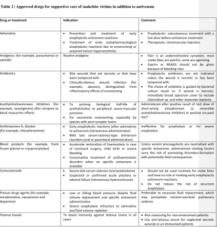
[H3] Additional supportive treatment. Organ and system failures caused by envenoming must be detected and treated (Table 2). Supraglottal or endotracheal intubation and assisted ventilation either manually or by a ventilator is vital in cases of bulbar and respiratory paralysis87.Especially in victims of species with predominantly postsynaptic neurotoxins such as cobras (Naja; family Elapidae), death adders (Acanthophis; family Elapidae) and some coral snakes (Micrurus; family Elapidae), acetylcholinesterase inhibitors such as neostigmine, given with atropine, may improve neuro-muscular transmission at least temporarily, but they are no substitute for antivenom111,125. Short-acting acetylcholinesterase inhibitors (for example, edrophonium) or the ice pack test (whereby an ice pack is applied to one upper eyelid in a patient with bilateral ptosis; the ice lowers the local temperature and inhibits endogenous acetylcholinesterase) may be used to predict response to endogenous cholinesterase inhibition126. Hypotension and shock persisting after antivenom treatment is treated with cautious fluid volume repletion and pressor drugs such as dopamine19,20. If acute kidney injury progresses despite conservative management, renal replacement therapy is needed.
improve circulation by incising fascial compartments). Fasciotomy is rarely justified as intracompartmental pressure usually remains within normal limits, and fasciotomy in patients whose anti-haemostasis has not been corrected by adequate doses of antivenom, has proved catastrophic. Unnecessary fasciotomy prolongs hospital stay and contributes to long-term morbidity128.
Rehabilitation is a rare luxury for snakebite victims but is essential for helping to restore function to the bitten limb, especially in children and agricultural workers, and to ameliorate the chronic physical handicap that blights the lives of many snakebite survivors.
[H2] Hospital discharge and follow-up
[H1] Quality of life
Snakebite envenomings occur mostly in impoverished settings11, affecting underserved rural populations that, quite often, lack the appropriate resources to confront this neglected disease. Snakebite alters the lives of victims, but also families and communities because this disease largely affects people devoted to agricultural or pastoralist activities whose economic performance, and that of their dependents, relies on their good health. The death or incapacitation of someone responsible for the basic economic support of his or her family can devastate the socioeconomic sustainability and interpersonal stability of many people. Moreover, where medical treatment and especially antivenom is not provided free of charge, the economic cost of snakebite can be catastrophic — creating debt, forcing asset liquidation, and driving families further into the cycle of poverty130,131.
Although many snake bites are fatal, far more leave behind victims who experience horrifically debilitating disfigurement and long-term disability. Without timely access to health systems with adequate resources and capacity the consequences of a snakebite envenoming may include various types of sequelae2,7,129,132,133. Where health systems falter, affected people as well as their families and communities must deal with these consequences on their own often with severe consequences, often expanding the toll of a single snakebite envenoming.
Rolling back the impact of snakebite envenoming on quality of life requires concerted and coordinated efforts spanning entire health systems. Snakebite envenoming is a ‘tool-ready’ disease, in the sense that effective therapy (that is, antivenom) exists, and other cross-cutting interventions are available to strengthen health systems, empower communities and encourage policy change. Implementing an effective action plan to control snakebite envenoming will lead to a situation where snakebite victims are treated rapidly with safe, effective antivenoms by trained health staff, followed up with ancillary treatment, together with psychological, social and economic support after hospital discharge. Public and private organizations, as well as community-based groups, should follow up and help people affected by sequelae of snakebites.
[H1] Outlook
[H2] Diagnosis
bite site, but may be less useful for detecting circulating venom as a marker of systemic envenoming105.
[H2] Management of systemic effects
Antivenom has been the primary treatment of systemic snake envenoming for over a century. Whilst life-saving, antivenoms still have therapeutic limitations137,138. Conventional antivenom has limited efficacy against some effects of envenoming, such as local tissue damage, and, when treatment is delayed, presynaptic neurotoxicity. In addition, only 10–15% of IgG in a vial of antivenom is specific to venom proteins, because the animals used for antivenom manufacture already have mature immune systems, and hyper-immunisation with venom is unable to engender higher venom-specific IgG titres. Finally, the greater the phylogenetic disparity between the snakes whose venoms are used for immunisation, the greater the number of IgG specificities generated in the venom-immunised animals. This means that the proportion of total IgG targeting the venom of any one snake is small, and consequently more vials are needed to achieve clinical cure. This creates a therapeutic paradox because each extra vial of antivenom increases both the risk of potentially severe antivenom-induced adverse effects and treatment cost. Thus, highly efficient, polyspecific antivenoms are needed, without compromising safety and affordability.
polyspecific antivenom. To address the dose–efficacy challenge, the current intent is to isolate B cells from epitope string-immunized mice, and extract, screen and manipulate the IgG-encoding genes to prepare monoclonal antibodies to produce an antivenom consisting only of IgG capable of polyspecifically neutralising the toxins present in the venoms of a defined group of snakes. Humanizing these monoclonal antibodies also offers substantial safety advantages over the current antivenoms142.
Another toxin-specific recombinant antivenom approach is based on screening bespoke high-density toxin-specific microarrays to identify the most medically-important venom toxin epitopes bound by clinically-effective antivenoms143. This toxin-focused approach aligned with the production of human or humanized IgGs (or fractions thereof, for example, single-chain variable fragments) using new biotechnology production processes may yield antivenoms at a cost similar to conventional antivenoms in the future144, but with a much-improved dose-efficacy. International collaborative efforts are underway in these research and development topics.
Chemical inhibitor research is also being promoted142 as an overlooked but potentially source of drugs to treat snakebites. For example, a recent drug repurposing study identified a potent, broad-spectrum inhibitor of the nearly-ubiquitous group of venom PLA2s145. Other efforts have been directed at the synthesis of nanoparticles which could sequester and neutralize venom toxins146. If successful, the chemical outputs of these approaches will likely have substantial cost and logistic advantages.
A pharmacological study reported that an ointment containing a nitric oxide donor, which impedes the intrinsic lymphatic pump, delays ingress of venom proteins into the systemic circulation and improved survival of venom-injected rats147. This first aid-focused application is likely to be effective in predominantly neurotoxic venoms, but needs to be excluded for envenoming by those snakes that cause local tissue destruction.
No medicinal treatment of the local tissue-destructive effects of envenoming exists. Antivenom, unless it is administered very soon after the bite, is largely ineffective in preventing the rapidly manifested destruction of tissues by venom. Chemical inhibitors have potential as a treatment for venom-induced local tissue destruction owing to their low-cost, thermostability and rapid tissue-distribution dynamics. Small peptidomimetic matrix metalloproteinase inhibitors have shown exciting, repurposed, ability to neutralize SVMP-induced haemorrhage and dermonecrosis148. It is not inconceivable that combining inhibitors of SVMPs and PLA2s145,146,148,149 may produce an affordable, rapid, polyspecific, and effective treatment of venom-induced tissue damage.
Other groups are pursuing a recombinant approach using single-domain VHH antibody fragments, based upon the demonstration that an experimental VHH antivenom (prepared from the heavy chain-only IgG3 fraction of a venom-immunised dromedary camel) proved the most dose-effective antivenom137 to neutralize various toxic effects of the West African saw-scaled viper (Echis ocellatus). The small size, thermostability and toxin-specificity of camelid VHH promote them as exciting therapeutic candidates for preventing the tissue necrotic effects of envenoming for saw-scaled vipers and other snake venoms150,151.
[H2] Treatment availability and accessibility
social science-related topics. The long-term goal is to develop knowledge-based policies ensuring that people suffering snakebite envenomings receive safe and effective antivenoms in a timely fashion. New treatments should be affordable and available to invariably remote, impoverished snakebite victims138,152. In addition, innovative treatment and diagnostic research needs to be complemented with equally important research on ways to ensure affected communities can better access effective healthcare, and indeed upon affordable and appropriate means by which they can reduce their likelihood of being bitten.
[H2] Venoms as a source of therapy
Some of the targets of toxins are also dysregulated in a number of human diseases, such as thrombotic disorders, vascular pathologies, neurodegenerative diseases and inflammatory conditions. The study of venom toxins is of growing interest for the pharmacological and biotechnological communities, as venoms are increasingly recognized as a rich source for lead compounds that can drive the development of biotechnological tools and novel biotherapeutics154. Determining the molecular landscape of snake venoms represents the necessary first step towards these basic and applied goals.
[H2] Global efforts to reduce impact
stakeholders, and to empower WHO to provide the leadership needed to coordinate a global control programme. WHO is the only organization with the political, operational and policy reach to drive an integrated global strategy involving multiple actors and a multidisciplinary approach, and this is clearly demonstrated by the success in combating other neglected tropical diseases in the five years since the London Declaration on neglected tropical diseases was enacted to support the WHO’s 2020 Roadmap.
Inclusion of snakebite envenoming in the neglected tropical disease category will focus attention on concentrated efforts to control snakebite through an integrated and intensified disease management strategy incorporating several key elements, ranging from prevention, to improved primary treatment, rehabilitation and investment in research that can unlock new diagnostic and therapeutic pathways, or enhance surveillance and resource deployment (Box 4).
The tasks included in Box 4 are by no means an exhaustive list of all the potential interventions available to immediately bring the burden of snakebite envenoming under effective control, but it exemplifies the key approaches that will drive the process. Broadening the current focus which revolves around improving treatment with antivenoms, measuring burden and assessing the consequences to take in a more holistic strategy that begins with community engagement, empowerment and education and spans all stages of treatment, reporting, therapeutic and diagnostic translational research, policy, training and research is essential.
Highlighted references
Mohapatra, B. et al. Snakebite mortality in India: a nationally representative mortality survey. PLoS Negl. Trop. Dis. 5, e1018 (2011).
A nationally representative study of snakebite mortality in India demonstrating that the magnitude of the problem in terms of mortality is much higher than previously thought.
Williams, D. et al. The Global Snakebite Initiative: an antidote for snake bite. Lancet375, 89-91(2010).
This paper describes the launching of the first organization to confront snakebite envenoming from a global perspective.
Harrison, R.A. et al. Snake envenoming: a disease of poverty. PLoS Negl. Trop. Dis. 3, e569 (2009).
The relationship between snakebite envenoming and poverty is highlighted in this study.
Habib, A.G. et al. Snakebite is under appreciated: appraisal of burden from West Africa. PLoS Negl. Trop. Dis. 9, e0004088 (2015).
This study analyzed the impact of snakebite envenoming in terms of disability-adjusted life years in 16 countries in sub-Saharan Africa.
Calvete, J.J. Venomics: integrative venom proteomics and beyond. Biochem. J. 474, 611-634 (2017).
A comprehensive view of the complexity of snake venoms and its biological and medical implications.
Montecucco C., Gutiérrez, J.M. & Lomonte, B. Cellular pathology induced by snake venom phospholipase A2 myotoxins and neurotoxins: common aspects of their mechanisms of action. Cell. Mol. Life Sci. 65, 2897-2912 (2008).
A review of the mechanisms by which venom phospholipases A2 induce myotoxicity and
Escalante, T., Rucavado, A., Fox, J.W. & Gutiérrez, J.M. Key events in microvascular damage induced by snake venom hemorrhagic metalloproteinases. J. Proteomics 74, 1781-1794 (2011).
This review summarizes the mechanisms by which zinc-dependent venom metalloproteinases induce microvascular damage and hemorrhage.
World Health Organization Regional Office for Africa. Guidelines for the Prevention and Clinical Management of Snakebite in Africa (WHO, Brazzaville, 2010) http://www.afro.who.int/pt/grupos-organicos-e-programas/2731-guidelines-for-the-prevention-and-clinical-management-of-snakebite-in-africa.html
WHO Guidelines for snakebite envenoming in Africa, to be used in the training of health staff on the correct diagnosis and management of envenomings.
Sharma, S.K. et al. Effectiveness of rapid transport of victims and community health education on snake bite fatalities in rural Nepal. Am. J. Trop. Med. Hyg. 89, 145-150 (2013).
A study describing a successful intervention at the community level aimed at improving the access of envenomed patients to health facilities.
World Health Organization. WHO Guidelines for the Production, Control and Regulation of Snake Antivenom Immunoglobulins. WHO, Geneva, 2010. http://www.who.int/bloodproducts/snake_antivenoms/en/A detailed account on the main aspects related to the production and control of antivenoms, containing valuable information for manufacturers, researchers, and regulatory agencies.
Abubakar, I.S. et al. Randomised controlled double-blind non-inferiority trial of two antivenoms for saw-scaled or carpet viper (Echis ocellatus) envenoming in Nigeria. PLoS Negl. Trop. Dis. 4, e767 (2010).
An example of a clinical trial based on a randomized controlled double-blind design for the evaluation of efficacy and safety of antivenoms in the treatment of snakebite envenomings.
Vaiyapuri, S. et al. Snakebite and its socioeconomic impact on the rural population of Tamil Nadu, India. PLoS ONE 8, e80090 (2013).
A description of socioeconomic consequences of snakebites in an impoverished rural setting.
Harrison, R.A., Cook, D.A., Renjifo, C., Casewell, N.R., Currier, R.B. & Wagstaff, S.C. Research strategies to improve snakebite treatment: challenges and progress. J. Proteomics 74, 1768-1780 (2011).
This review discusses some of the main research areas that need to be developed in order to generate novel diagnostic and therapeutic tools to confront snakebite envenoming.
Laustsen, A.H. et al. From fangs to pharmacology: The future of snakebite envenoming therapy. Curr. Pharm. Des. 22, 5270-5293 (2016).
A summary of novel therapeutic alternatives to approach snakebite envenoming, including recombinant antibodies and natural and synthetic venom inhibitors.
Acknowledgments
The authors thank Dr. Jeronimo Bravo (Instituto de Biomedicina de Valencia, CSIC) for assisting in the elaboration of the structural models shown in Figure 3.
Author contributions
Introduction (J.M.G.); Epidemiology (A.G.H.); Mechanisms/pathophysiology (J.J.C. and J.M.G.); Diagnosis, screening and prevention (D.A.W.); Management (D.A.W.); Quality of life (D.J.W.); Outlook (J.M.G., R.A.H. and D.J.W.); Overview of the Primer (J.M.G.)
Competing interests
The authors declare no competing interests.
Publisher's note
Springer Nature remains neutral with regard to jurisdictional claims in published maps and institutional affiliations.
How to cite this Primer
Box 1 | Technologies to analyse snake venom composition.
First-generation venom proteomics (venomics) relied on reversed-phase high-performance liquid chromatography separation and quantification of venom components followed by in-gel digestion of the protein bands corresponding to the chromatographic peaks. Protein bands were resolved by sodium dodecyl sulfate polyacrylamide gel electrophoresis, de novo tandem mass spectrometry sequencing of the peptides (usually produced by trypsin digestion) and database searching by Basic Local Alignment Search Tool (BLAST) analysis155. An experienced researcher typically completes this bottom-up analysis of a venom composed of 35 chromatographic fractions and 60-70 electrophoretic bands in 6-8 weeks. The bottleneck of this technique is the quality of the tandem mass spectrometry fragmentation spectra and the presence in the databases of a sequence homologous to the de novo derived amino acid sequence tag for the digested peptide ions. Peptide-centric approaches provide incomplete sequence coverage, and in the absence of a species-specific venom gland transcriptome, only provide information on which known protein families are present in the venom without distinguishing between different proteoforms (different molecular forms of a protein) or toxin isoforms (proteins encoded by closely related genes).
Box 2. Toxic activities of main protein families in snake venom
- Phospholipases A2: local and systemic myotoxicity, pain, damage to lymphatic vessels,
oedema, neurotoxicity, nephrotoxicity and haemolysis
- Snake venom metalloproteinases: haemorrhage, myonecrosis, extracellular matrix degradation, blistering, pain, oedema and cardiovascular shock, nephrotoxicity and coagulopathy
- Hyaluronidases: extracellular matrix degradation
- Three-finger toxins: cytotoxicity, necrosis and neurotoxicity
- Dendrotoxins: neurotoxicity
- Snake venom serine proteinases: coagulopathy, oedema and hypotension
- Vasoactive peptides (for example, bradykinin-potentiating peptides): hypotension
- Disintegrins: inhibition of platelet aggregation
- C-type lectin-like proteins: inhibition or promotion of platelet aggregation and thrombocytopenia
- Cysteine-rich secretory proteins: smooth muscle paralysis
- Small basic myotoxic peptides: muscle contracture
- Natriuretic peptides: hypotension
Box 3 | Initial clinical history from a snakebite victim.
The following five brief questions have proved helpful for rapid assessment of snakebite victims.
Where were you bitten? Examine puncture marks, swelling, inflammation, bruising, persistent bleeding and evidence of pre-hospital traditional treatment at the site of the bite.
When were you bitten? Note that if the bite or sting was very recent, there may not have been time for signs of envenoming to develop.
What were you doing when you were bitten? Note that the circumstances of the bite might be diagnostic.
Where is the snake that bit you or what did it look like? If it was killed but was left behind, send someone to bring it, in whatever condition. Ask if a photo of the snake was taken.
Box 4 | Recommendations for an integrated global strategy to confront snakebite envenoming8,138
Actively prevent contact and implement educational programmes that detail safe methods for carrying out chores or work that pose a high risk of encountering snakes. For example, when collecting firewood, picking up piles of raked leaves, harvesting crops or hunting, appropriate footwear should be worn and a torch or flashlight should be used to illuminate the path at night. Risk during sleeping can be minimized by snake-proofing dwellings and using protective (mosquito) bed-nets or raised sleeping platforms.
Establish effective, safe and affordable first aid that delays the evolution of clinical illness and improve transport such that victims of envenoming reach health care facilities for diagnosis and treatment as quickly as possible.
Train health professionals to implement standard treatment protocols and algorithms, and provide diagnostic tools to strengthen health systems and improve medical management of snakebite victims.
Develop a robust system for hospital and community-level surveillance of snakebite envenoming, including mandatory reporting of snake-bite victims seeking treatment, notifications of deaths and active detection of cases through community surveys and outreach programmes.
Establish standard definitions and measurements to facilitate the accurate collection and comparability of data.
Improve access and distribution of medicines, especially of safe, effective and affordable antivenoms by implementing programmes that reduce prices through collective bulk purchasing by consortia (governments, non-governmental organizations and aid donors).
Strengthen health systems to improve the treatment of snakebite envenoming in health facilities from admission through to discharge and follow-up. Follow-up is especially important in patients with local tissue injury and disability.
Invest in the improvement of existing antivenom treatments and the development of innovative future treatments.
Figure 1. Venomous snakes. A | Schematic illustration of the venom system in a snake of the family Viperidae (viperids). Venom is synthesized and stored in a specialized gland. When a bite occurs, venom is expelled through the action of a compressor muscle that surrounds the venom gland, and is delivered by a duct to the fangs through which it is injected in the tissues of the victim. Snake species responsible for a highest mortality owing to snakebite envenoming are Echis ocellatus (carpet viper; family Viperidae; part b) and Bitis arietans (puff adder; family Viperidae; part c) in Africa, Najanaja (cobra; family Elapidae; part d), Bungarus caeruleus (common krait; family Elapidae; part e), Daboia russelii (Western Russell’s viper; family Viperidae; part f) in Asia, Bothrops atrox (common lancehead; family Viperidae; part g) and Bothrops asper (terciopelo; family Viperidae; part h) in the Americas and Oxyuranus scutellatus (Papuan taipan; family Elapidae; part i) in Oceania. Many other snake species are also capable of inducing life-threatening envenomings. Photos b–h by David A. Warrell, and photo i by David J. Williams (used with permission). [Note to copy editor: we have iLTPs for these images from David Warrell; please insert the appropriate credit line as per style.]
Figure 2. Geographical distribution of the estimated number of snakebite envenomings and deaths. Data shown constitute a rough approximation of the estimated range of envenomings and deaths given that, in many countries, reliable information on snakebite morbidity and mortality are lacking, resulting in underreported data of this neglected tropical condition. The highest impact of snakebite envenomings occurs in Asia, sub-Saharan Africa, Latin America and parts of Oceania. Based on estimates from5,12.
that is, some toxins may exert one effect at low doses and another effect at high doses). The crystallographic or NMR structures of some members of each protein family are also shown. SVMP, snake venom metalloproteinases (Protein Data Bank (PDB) accession 3DSL for class PIII and PDB accession 1ND1 for class PI are shown); PLA2, phospholipases A2 (PDB accession 1TGM for the monomer and PDB accession 3R0L for the dimer); SVSP, snake venom serine proteinases (PDB accession 1OP0); 3FTx, three-finger toxins (PDB accession 1IJC); DTx, dendrotoxins (PDB accession 1DTX); CTL, C-type lectin-like proteins (PDB accession 1IXX); CRISP, cysteine-rich secretory proteins (PDB accession 3MZ8); LAO, L-amino acid oxidases (PDB accession 2IID); Myo, low molecular mass myotoxins (PDB
accession 4GV5). More information on the crystal structures shown and their source can be found in Supplementary file 2.
Figure 4. Immunoaffinity capturing antivenomics protocol. Whole venom is applied to an immunoaffinity column packed with antivenom antibodies that are immobilized onto Sepharose beads. After eluting the non-retained venom components, the immunocaptured proteins are eluted. Comparison of the reversed-phase high-performance liquid chromatographs of the components of whole venom, the fraction recovered from the affinity antivenom column (immunoretained fraction) and the non-immunocaptured venom fraction (non-immunoretained fraction) can provide qualitative and quantitative information on the set of toxins bearing antivenom-recognized epitopes and those toxins exhibiting poor immunoreactivity.
is, rhabdomyolysis). Acute kidney injury often develops in envenomings, owing to a multifactorial pathogenesis.
Table 1. Criteria for antivenom treatment
Clinical criterion Clinical evidence Additional treatment
Shock with or without hypovolaemia Low or falling blood pressure (with postural drop)
Spontaneous systemic bleeding (at sites distant from the bite) administer fresh frozen plasma and/or other blood products*
Incoagulable blood Persistent bleeding from trauma sites Positive 20WBCT
Altered laboratory blood coagulation profile
If massive or threatened (for example, pre-delivery in pregnant women or before surgery), administer fresh frozen plasma and/or other blood products*
Black urine Macroscopic or microscopic evidence of haemoglobin or myoglobin in the urine (urine reagent strip testing) Exclude haematuria
Restore fluid homeostasis Alkalinize urine
High risk of actute kidney injury Oliguria/anuria
Rising plasma creatinine and urea
Treatment with specific antivenom is indicated if one or more of the following criteria are fulfilled. *See Table 2. ‡ Application of an ice
Table 2 | Approved drugs for supportive care of snakebite victims in addition to antivenom
Drug or treatment Indication Comment
Adrenaline Prevention and treatment of early anaphylactic antivenom reactions
Treatment of early autopharmacological anaphylactic reactions due to envenoming or acquired venom hypersensitivity
Prophylactic: subcutaneous treatment with a low dose before antivenom treatment. Therapeutic: intramuscular injection. Analgesics (for example, paracetamol or
opioids)
Routine analgesia Pain is an underestimated symptom; most snake bites are painful, some are agonizing. Aspirin or NSAIDs should not be given
because of bleeding risks. Antibiotics Bite wounds that are necrotic or that have
been tampered with
Clinically-obvious wound infection (for example, abscess), distinguished from inflammatory effects of envenoming
Prophylactic antibiotics are not indicated unless the wound is necrotic or has been tampered with.
The choice of antibiotic is guided by bacterial culture result or, if wound is necrotic, immediate broad spectrum cover to include
Clostridium sp. and other anaerobic bacteria.
For neurotoxic envenoming, especially by species with post-synaptic toxins Mild late serum-sickness-type antivenom
reactions (oral or parenteral administration)
Ineffective for prophylaxis or for severe anaphylaxis
Blood products (for example, fresh frozen plasma or cryoprecipitates)
Accelerate restoration of haemostasis in case of imminent surgery, child birth or severe bleeding
Conservative treatment of antihaemostatic disorders when no specific antivenom is available
Unless venom procoagulants are neutralized with specific antivenom, administered clotting factors carry the risk of promoting thrombus-formation with potentially-fatal consequences
Corticosteroids Severe late serum-sickness (oral prednislolne) Suspected or confirmed acute pituitary or
adrenal failure (intravenous hydrocortisone)
Should not be used routinely for snake bites and have no role in treating early anaphylactic antivenom reactions. volume replacement and specific antivenom administration
Severe anaphylaxis refractory to adrenaline and fluid volume repletion
Preferable to excessive fluid replacement, which may precipitate volume-overload pulmonary oedema
Tetanus toxoid To boost immunity against tetanus toxins in all cases
Also reassuring for non-envenomed patients. Use anti-tetanus serum for neglected necrotic
wounds in un-immunised patients
*Application of an ice pack to one upper eyelid in a patient with bilateral ptosis; the ice lowers the local temperature and inhibits endogenous acetylcholinesterase; if positive, anticholinesterase can be administered.
1. Calvete, J. J. Proteomic tools against the neglected pathology of snake bite envenoming.
Expert Rev. Proteomics8, 739–758 (2011).
3. Pyron, R., Burbrink, F. T. & Wiens, J. J. A phylogeny and revised classification of
Squamata, including 4161 species of lizards and snakes. BMC Evol. Biol.13, 93 (2013).
4. Hsiang, A. Y. et al. The origin of snakes: revealing the ecology, behavior, and evolutionary
history of early snakes using genomics, phenomics, and the fossil record. BMC Evol. Biol.
15, (2015).
5. Chippaux, J. P. Snake-bites: appraisal of the global situation. Bull. World Health Organ.76,
515–24 (1998).
6. Mohapatra, B. et al. Snakebite Mortality in India: A Nationally Representative Mortality
Survey. PLoS Negl. Trop. Dis.5, e1018 (2011).
7. Chippaux, J.-P. Estimate of the burden of snakebites in sub-Saharan Africa: A meta-analytic
approach. Toxicon57, 586–599 (2011).
8. Gutiérrez, J. M., Williams, D., Fan, H. W. & Warrell, D. A. Snakebite envenoming from a
global perspective: Towards an integrated approach. Toxicon56, 1223–1235 (2010).
9. Gutiérrez, J. M., Theakston, R. D. G. & Warrell, D. A. Confronting the Neglected Problem
of Snake Bite Envenoming: The Need for a Global Partnership. PLoS Med.3, e150 (2006).
10. Williams, D. et al. The Global Snake Bite Initiative: an antidote for snake bite. Lancet375,
89–91 (2010).
11. Harrison, R. A., Hargreaves, A., Wagstaff, S. C., Faragher, B. & Lalloo, D. G. Snake
Envenoming: A Disease of Poverty. PLoS Negl. Trop. Dis.3, e569 (2009).
12. Kasturiratne, A. et al. The Global Burden of Snakebite: A Literature Analysis and Modelling
Based on Regional Estimates of Envenoming and Deaths. PLoS Med.5, e218 (2008).