Cite as: Can Urol Assoc J 2019;13(11):E361-5. http://dx.doi.org/10.5489/cuaj.5794
Published online February 26, 2019
Abstract
Introduction: Ureteral stricture (US) in the kidney transplant recipi-ent is a rare complication that can lead to morbidity and graft loss. Risk factor recognition is crucial in the prevention and management of this entity. Delayed graft function (DGF), as defined by the need for dialysis in the first week after transplantation, has been proposed as a risk factor in previous studies. Our objective is to determine the impact of DGF in US development in kidney transplant patients. Methods: We designed a matched case-control study. US cases in kidney transplant recipients were identified in the 2008–2017 period. We defined US as the rise in serum creatinine associated with findings suggesting obstruction in ultrasound, scintigraphy, or retrograde pyelogram; any other cause of graft dysfunction was excluded. Controls were defined as kidney transplant recipients from the same population and period without US, matched in a 1:2 fashion by age, sex, and donor type.
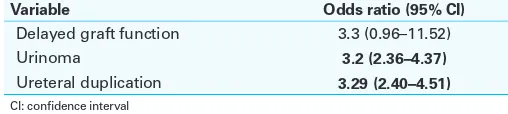
Results: From 532 kidney transplant patients, 31 cases and 62 controls were included. Cumulative US incidence was 58 per 1000 cases. When calculating for odds ratio (OR), postoperative urinoma (OR 3.2; 95% confidence interval [CI] 2.36–4.37) and ureteral duplication (OR 3.29; 95% CI 2.40–4.51) were associated with an increased risk for US, while DGF was not found to be statistic-ally significant as a risk factor (OR 3.3; 95% CI 0.96–11.52). No statistically significant differences were found between groups in other pre- and post-transplant-related factors
Conclusions: DGF was not associated with US in our cohort; how-ever, ureteral duplication and postoperative urinoma were associ-ated with an increased risk of graft ureteral stenosis development.
Introduction
Kidney transplantation is the definitive treatment for chronic kidney failure, bearing an improvement in prognosis over
dialysis.1,2 Urological complications are highly relevant, as
they may end up in graft loss.3,4 In particular, ureteral
com-plications have been reported from 4.8‒9.2%, with ureteral stenosis (US) rates from 2.4‒9.2% of the kidney transplants.5-7
Additionally, most ureteral complications occur during the first post-transplant year.5,8,9
Another feared complication is delayed graft function (DGF), which is associated with an increased risk for graft loss and acute rejection in the first post-transplant year.10
This complication rate has been reported from 2‒50% and 1.6‒10% in deceased and living donor transplants, respect-ively.11,12 The primary underlying etiology for DGF seems to
be ischemia-reperfusion damage.11
Some retrospective studies have set a role for DGF as a risk factor for US in renal grafts, along with other variables, such as donor age over 65 years and kidneys with more than two arteries.5
We hypothesized that DGF increases the risk for graft US secondary to ischemia-reperfusion damage, pro-fibrotic mol-ecule expression, and ureteral ischemia that promote aber-rant scarring.13,14 Our primary objective was to determine
the impact of DGF in US development in kidney transplant patients that underwent transplantation in the 2008‒2017 period in a tertiary care hospital.Secondarily, we wanted to calculate the graft US prevalence and describe other risk factors and treatment modalities used for the resolution of this complication.
Methods
We performed a matched case-control study in a single center in Mexico from January 2008 to January 2017. We obtained approval from the local ethics board committee. Data were retrieved from the hospital kidney transplant data-base. Cases were kidney transplant receptors diagnosed with graft US; controls were patients from the same population that did not develop US during followup.
Axel Cayetano-Alcaraz, MD
1; Juan Sebastian Rodriguez-Alvarez, MD
1; Mario Vilatobá-Chapa, MD
2;
Josefina Alberú-Gómez, MD
2; Bernardo Gabilondo-Pliego, MD
1, Francisco Rodríguez-Covarrubias, MD
1;
Luis Eduardo Morales-Buenrostro, MD
3; Carlos Enrique Méndez-Probst, MD
11Department of Urology, Instituto Nacional de Ciencias Médicas y Nutrición Salvador Zubirán, Mexico City, Mexico; 2Department of Transplants, Instituto Nacional de Ciencias Médicas y Nutrición Salvador
Zubirán, Mexico City, Mexico; 3Department of Nephrology, Instituto Nacional de Ciencias Médicas y Nutrición Salvador Zubirán, Mexico City, Mexico
Case inclusion and exclusion criteria
Cases were included as patients older than 18 years with a kidney transplant performed in this institution, coupled with a confirmed diagnosis of graft US.
We defined graft US as a rise in serum creatinine asso-ciated with hydronephrosis on ultrasound, an obstructive curve in scintigraphy, or a retrograde pyelography compat-ible with ureteral stenosis that were managed surgically (ureteroneocystostomy, Boari flap ureteroneocystostomy, or ureteroureterostomy), endoscopically (retrograde double-J stent catheterization, balloon dilation, or ureterotomy), or by interventional radiology treatment (nephrostomy or ante-grade double-J stent catheterization). Any other potential cause, such as rejection (acute or chronic) or obstructive uropathy of other etiology were excluded.
Patients that did not undergo kidney transplantation in our institution were excluded. We eliminated patients with incomplete followup or a followup period of fewer than three months. Cases were matched with controls in a 1:2 fashion by age, sex, donor type, and transplantation period (±5 years).
Control inclusion and exclusion criteria
Controls were selected as kidney transplant recipients from either living or deceased donor older than 18 years that underwent transplantation at our institution during the same followup period of the cases with no graft dysfunction unless concluded to be a DGF. We excluded patients who did not undergo transplantation at our institution. Exclusion criteria were an incomplete followup or a followup period of fewer than three months.
Clinical variables
– Donor: Age, sex, blood type, donor type (living or
deceased), extended criteria, donor relation (related or unrelated), comorbidities, and glomerular filtration rate (GFR) (calculated with the MDRD formula).
– Receptor: Age, sex, blood type, comorbidities, body
mass index, smoking status, previous transplantation, HLA mismatch, pre-transplant anti-T panel reactive antibodies (PRA %), donor-specific HLA antibodies, pre-transplant diuresis, induction and maintenance scheme, BK virus infection, CMV infection, five-day followup ultrasonography data (hydronephrosis, perigraft fluid collection, distal intrarenal resistive index), periopera-tive complications (lymphocele, hematoma, urinoma, urinary fistula, arterial anastomosis stenosis), positive urine culture, acute rejection.
– Renal graft: Ischemia time, ≥2 graft arteries, ≥2 graft veins, ureteral duplication, ureteral implant technique, ureteral stent use, surgical bleeding, surgical time, time
to stent withdrawal, ureteral stenosis diagnostic method, time to ureteral stenosis development, treatment type (surgical, endoscopic, interventional).
– Delayed graft function: Defined as the need for dialysis
in the first post-transplant week.11,15,16
Sample size
We calculated a sample size of 216 patients, 72 cases and 144 controls. The figures were calculated with a power of 80%, 95% confidence interval (CI), a two control per case proportion, with a 9% estimated control exposure and a min-imal odds ratio detection of three (Fleiss, Statistical Methods for Rates and Proportions, formulas 3.18 & 3.19; results obtained from Dean AG, Sullivan KM, Soe MM. OpenEpi: Open Source Epidemiologic Statistics for Public Health. Available at: www.OpenEpi.com [updated April 6, 2013]. Accessed April 10, 2017).
Statistical analysis
Data were analyzed with central tendency measures. Quantitative and qualitative data were compared using the student t-test and Chi-square, respectively. A p value under 0.05 was considered statistically significant. We obtained odds ratio (OR) for significant differences with the Mantel-Haenzel test. Statistical Package for the Social Sciences (SPSS®), version 20 was used for calculations.
Results
We reviewed 532 kidney transplantations in the 2008‒2017 period; 31 cases and 62 controls were included. Median followup was 60.09 months (interquartile rage [IQR] 46.7). Table 1 describes the basal recipient and donor pre-trans-plantation characteristics; table 2 describes perioperative transplant variables; and table 3 describes followup kidney receptor characteristics and immunosuppressive schemes.
A ureteral stent was placed in 28 (90.3%) cases, and 54 (87.1%) controls without significant statistical differences (p=0.206); median time for ureteral stent withdrawal was 29.68 days (standard deviation [SD] 15.9) and 34.02 days (SD 20.3) for cases and controls, respectively, also with no statistical significance (p=0.3). Immunosuppressive regimes for induction and maintenance did not show statistical sig-nificance among groups.
OR calculations
Characteristics of cases with graft ureteral stenosis
The calculated incidence of US was 5.8%, while the inci-dence rate was 0.093 person-years. The median time for US development was 62 days (IQR 64).
Imaging studies for US diagnosis were distributed as 28 (90.3%), seven (7.5%), and 13 (41.9%) cases for an ultrasound, renal scintigraphy, and retrograde pyelogram, respectively.
US management was surgical in 28 cases (90.3%), bal-loon dilation and retrograde stenting in two cases (6.4%), and one case required interventional treatment with ante-grade stenting. Surgical management included 23 cases (74.2%) with ureteral neocystostomy and five (15.1%) with the Boari flap technique.
Discussion
In our sample of 532 kidney transplants performed in the research period, the incidence of US was of 5.8%, with an incidence rate of 0.093 person-years, slightly higher than an international series; for instance, in a series of 2000 trans-plants performed in a single medical center from 1980 to 2010, the documented US prevalence was 2.7%.6 A rate of
3.5% in an 894 transplant samples from 1993‒2009 was reported by Faenza et al.5 Karam et al reported a 4.1% rate in
Table 1. Basal recipient and donor pre-transplantation characteristics CKD etiology, n (%)
Idiopathic
Pretransplant diuresis, n (%) 20 (64.5) 46 (74.2) 0.332 Dialysis to transplant time,
months, mean (SD)
36.7 (38.4) 28.8 (28) 0.9
Donor characteristics
Female gender, n (%) 15 (48.4) 35 (56.5) 0.46 Age, mean (SD) 40.2 (12.3) 38.7 (13.8) 0.58 Donor type, n (%) Expanded criteria donor, n (%) 6 (19.4) 4 (6.5) 0.58 Comorbidities, n (%)
ADPKD: autosomal dominant polycystic kidney disease; BMI: body mass index; CKD: chronic kidney disease; GFR glomerular filtration rate; HLA: human leukocyte antigen; SD: standard deviation; SLE: systemic lupus erythematosus.
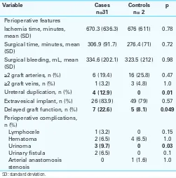
Table 2. Kidney transplant perioperative features for cases and controls
Variable Cases
n=31
Controls n= 2
p
Perioperative features Ischemia time, minutes, mean (SD)
670.3 (636.3) 676 (611) 0.78
Surgical time, minutes, mean (SD)
306.9 (91.7) 276.4 (71) 0.72
Surgical bleeding, mL, mean (SD)
334.6 (202.1) 323.5 (212) 0.98
≥2 graft arteries, n (%) 6 (19.4) 16 (25.8) 0.47
≥2 graft veins, n (%) 1 (3.2) 3 (4.8) 1.0 Ureteral duplication, n (%) 4 (12.9) 0 0.01
Extravesical implant, n (%) 26 (83.9) 49 (79) 0.57 Delayed graft function, n (%) 7 (22.6) 5 (8.1) 0.049
Perioperative complications,
Table 3. Characteristics in the post-transplant followup for cases and controls Basal post-transplant creatinine, mean (SD)
1.2 (0.35) 1.18 (0.31) 0.39
Post-transplant basal GFR by MDMR, ml/min/1.73 m2, mean
(SD)
71 (32.2) 71 (20.4) 1.0
5-day graft ultrasound anomalies Hydronephrosis, n (%) Perigraft fluid collection, n (%) Distal intrarenal resistive index, mean (SD)
Positive post-surgery urine culture 29 (29) 23 (37.1%) 0.44
1787 transplants from 1990‒2002.8 More recently, US was
reported in 3.39% from a 973 transplant sample performed in the 2004‒2014 period, with a mean time to stenosis of 10.6±23.0 months (range 0.5‒98).4
Time to stenosis occurrence is related to its etiology. Early stenosis can be associated to tissue edema, kink of the ureter, narrow ureter diameter, or extrinsic compression by hema-tomas or lymphoceles.17, 18 While the late US can be
associ-ated with inappropriate arterial blood supply that results in ischemia and fibrosis.3 Nevertheless, definitions vary among
studies, are not based on international consensus, and tend to be arbitrary.3,5,6,8 In the present study, early US (defined
as less than three months post-transplant) was more com-mon, with a proportion of 74.2% (n=23) and mean time from transplantation to US diagnosis of 62 days (IQR 64). Late US occurred in 25.8% (n=8) of cases. This proportion is similar to the one reported by Faenza et al (81.2% early stenosis vs. 18.8% late stenosis).5
DGF is a relatively common, early post-transplant com-plication, with a rate of up to 50% of deceased donor trans-plantations.19 Conversely, its occurrence in living donor
transplantations is fairly uncommon, with rates reported from 1.6‒10.0%.11,12 DGF has been associated with graft
dysfunc-tion, rejection in the first year, and decreased graft survival.20
Some studies have proposed DGF as a risk factor for US; in 2006, Karam et al in a single-center, retrospective analy-sis of 1787 patients with renal transplantation, found that US was correlated to DGF (p=0.016), donor age >65 years (p=0.001), and more than two graft arteries (p=0.009); how-ever, multivariate analysis showed only DGF (OR 1.03; 95% CI 1.01‒1.05) and >2 arteries (OR 1.45; 95% CI 1.00‒2-00) as an independent risk factors for US.8 Fontana et al found
similar results.7 Nonetheless, DGF by previous groups was
defined as the number of days to reach a GFR of >10 mL/ minute, and the study methods did not control potential confounding factors, such as donor age and type.
At the moment, there is no consensus about a superior definition for DGF, but the most commonly used is the need for dialysis in the first post-transplant week; it has been sug-gested to be adopted as the universal clinical variable for research studies.15 In our study of matched case-control
analysis, although DGF was found to have a statistically sig-nificant difference in frequency between cases and controls by Chi-square analysis, we rejected the hypothesis of DGF as a risk factor for US, considering the Mantel-Haenzel analysis
did not show a statistically significant effect of exposure (OR 3.3; 95% CI 0.96‒11.52). Despite being associated with US in previous studies, our contrasting results may owe to different study design and previous non-standardized defin-itions for DGF.7,8 However, the associated factor may not be
DGF, per se, but one of the associated mechanisms, such as ischemia-reperfusion damage, which may not always reach a clinical threshold to be considered as such.
The factors that we found for an increased risk of US were perioperative urinoma (OR 3.2; 95% CI 2.36‒4.37) and ureteral duplication (UD) (OR 3.29; 95% CI 2.40‒4.51). Previous studies did not report perioperative complications as risk factors for US.7,8 However, urinoma is commonly
associated with a ureterovesical (UV) anastomotic leak or distal ureteral ischemia, depending on the occurrence time, leading to periureteral graft fibrosis.21,22
On the other hand, UD is a common anatomy variant reported in 1 of 100‒500 cases. Mixed results have been reported in small studies, where it may have been associ-ated with an increased incidence of urological complica-tions (10.5%), mainly US;23 other studies show no difference
in perioperative complications.24 A higher technical difficulty
performing the UV anastomosis may explain the association between UD and US.
Since US is regarded as an uncommon complication in renal transplantation, the study design appropriately evalu-ated the associevalu-ated risk factors with this entity. Furthermore, the research study period is recent, and distinct renal trans-plant management aspects (such as patient selection, proto-col followup, immunosuppressive schemes, and surgical groups) were standardized since the beginning of the per-iod. Our study limitations are mainly a failure to achieve the optimal sample size owing to the rarity of this entity, as well as its single-center and retrospective nature.
Conclusions
DGF was not associated with ureteral stenosis in our cohort; however, UD and postoperative urinoma were associated with a graft ureteral stenosis development.
Competing interests: The authors report no competing personal or financial interests related to this work.
This paper has been peer-reviewed.
References
1. Wolfe RA, Ashby VB, Milford EL, et al. Comparison of mortality in all patients on dialysis, patients on dialysis awaiting transplantation, and recipients of a first cadaveric transplant. N Engl J Med 1999;341:1725-30. https://doi.org/10.1056/NEJM199912023412303
Table 4. Factors associated with an increased risk of ureteral stenosis development
Variable Odds ratio (95% CI)
Delayed graft function 3.3 (0.96–11.52)
Urinoma 3.2 (2.36–4.37)
Ureteral duplication 3.29 (2.40–4.51)
2. Schold JD, Buccini LD, Goldfarb DA, et al. Association between kidney transplant center performance and the survival benefit of transplantation vs. dialysis. Clin J Am Soc Nephrol 2014;9:1773-80. https:// doi.org/10.2215/CJN.02380314
3. Palazzetti A, Oderda M, Dalmasso E, et al. Urological consequences following renal transplantation: A review of the literature. Urologia 2015;82:211-8. https://doi.org/10.5301/uro.5000132 4. Gil-Sousa D, Oliveira-Reis D, et al. Ureteral stenosis after renal transplantation — a single-center 10-year
experience. Transplant Proc 2017;49:777-82. https://doi.org/10.1016/j.transproceed.2017.01.050 5. Faenza A, Nardo B, Fuga G, et al. Urological complications in kidney transplantation: Ureteroneocystostomy
vs. uretero-ureterostomy. Transplant Proc 2005;37:2518-20. https://doi.org/10.1016/j.transpro-ceed.2005.06.079
6. Eufrasio P, Parada B, Moreira P, et al. Surgical complications in 2000 renal transplants. Transplant Proc 2011;43:142-4. https://doi.org/10.1016/j.transproceed.2010.12.009
7. Fontana I, Bertocchi M, Rossi AM, et al. Late ureteral stenosis after kidney transplantation: A single-center experience. Transplant Proc 2010;42:1174-5. https://doi.org/10.1016/j.transproceed.2010.03.053 8. Karam G, Hetet JF, Maillet F, et al. Late ureteral stenosis following renal transplantation: Risk factors and
impact on patient and graft survival. Am J Transplant 2006;6:352-6. https://doi.org/10.1111/j.1600-6143.2005.01181.x
9. Rajpoot DK, Gomez A, Tsang W, et al. Ureteric and urethral stenosis: A complication of BK virus infection in a pediatric renal transplant patient. Pediatr Transplant 2007;11:433-5. https://doi.org/10.1111/j.1399-3046.2006.00673.x
10. Yarlagadda SG, Coca SG, Formica RN, et al. Association between delayed graft function and allograft and patient survival: A systematic review and meta-analysis. Nephrol Dial Transplant 2009;24:1039-47. https://doi.org/10.1093/ndt/gfn667
11. Perico N, Cattaneo D, Sayegh MH, et al. Delayed graft function in kidney transplantation. Lancet 2004;364:1814-27. https://doi.org/10.1016/S0140-6736(04)17406-0
12. Park HS, Hong YA, Kim HG, et al. Delayed graft function in living-donor renal transplantation: 10-year experience. Transplant Proc 2012;44:43-6. https://doi.org/10.1016/j.transproceed.2011.11.057 13. Ponticelli C. Ischemia-reperfusion injury: A major protagonist in kidney transplantation. Nephrol Dial
Transplant 2014;29:1134-40. https://doi.org/10.1093/ndt/gft488
14. Eltzschig HK, Eckle T. Ischemia and reperfusion — from mechanism to translation. Nat Med 2011;17:1391-401. https://doi.org/10.1038/nm.2507
15. Mallon DH, Summers DM, Bradley JA, et al. Defining delayed graft function after renal transplantation: Simplest is best. Transplantation 2013;96:885-9. https://doi.org/10.1097/TP.0b013e3182a19348 16. Ditonno P, Impedovo SV, Palazzo S, et al. Effects of ischemia-reperfusion injury in kidney transplantation:
Risk factors and early and long-term outcomes in a single center. Transplant Proc 2013;45:2641-4. https://doi.org/10.1016/j.transproceed.2013.07.025
17. Zagdoun E, Ficheux M, Lobbedez T, et al. Complicated lymphoceles after kidney transplantation. Transplant Proc 2010;42:4322-5. https://doi.org/10.1016/j.transproceed.2010.09.127
18. Shoskes DA, Hanbury D, Cranston D, et al. Urological complications in 1000 consecutive renal transplant recipients. J Urol 1995;153:18-21. https://doi.org/10.1097/00005392-199501000-00008 19. Schroppel B, Legendre C. Delayed kidney graft function: from mechanism to translation. Kidney Int
2014;86:251-8. https://doi.org/10.1038/ki.2014.18
20. Hall IE, Reese PP, Doshi MD, et al. Delayed graft function phenotypes and 12-month kidney transplant outcomes. Transplantation 2017;101:1913-23. https://doi.org/10.1097/TP.0000000000001409 21. Berli JU, Montgomery JR, Segev DL, et al. Surgical management of early and late ureteral compli-cations after renal transplantation: techniques and outcomes. Clin Transplant 2015;29:26-33. https://doi.org/10.1111/ctr.12478
22. Di Carlo HN, Darras FS. Urologic considerations and complications in kidney transplant recipients. Adv Chronic Kidney Dis 2015;22:306-11. https://doi.org/10.1053/j.ackd.2015.04.003
23. Haferkamp A, Dorsam J, Mohring K, et al. Ureteral complications in renal transplantation with more than one donor ureter. Nephrol Dial Transplant 1999;14:1521-4. https://doi.org/10.1093/ndt/14.6.1521 24. Ghazanfar A, Zaki MR, Pararajasingam R, et al. Outcome of kidney transplant with double ureter: A
multicenter study. Exp Clin Transplant 2015;13:152-6.