FORMULATION AND EVALUATION OF LORNOXICAM MICROSPONGES USING EUDRAGIT
RS 100 AND EUDRAGIT RSPO
BANNARAVURI THIREESHA, AYYA RAJENDRA PRASAD*, HAROLED PETER P L
Department of Pharmaceutics, Nirmala College of Pharmacy, Mangalagiri – 522 503, Andhra Pradesh, India.Email: [email protected] Received: 24 June 2018, Revised and Accepted: 14 June 2018 ABSTRACT
Objective: The objective of the present study was preparation and evaluation of lornoxicam microsponges to prolong their drug release up to 12 h for effective osteoarthritis, rheumatoid arthritis, and acute lumbar-sciatica therapy.
Methods: Lornoxicam microsponges were prepared by the quasi-emulsion solvent diffusion technique using different concentrations of polymers such as Eudragit RS 100 and Eudragit RSPO in ethanol and dichloromethane organic solvent mixture. Microsponges were evaluated for their particle size, percentage yield, entrapment efficiency, scanning electron microscopy (SEM), and in vitro drug release studies.
Results: The percentage yield, entrapment efficiency, average particle size, and in vitro drug release for optimized formulation F12 were found to be 70.23% w/w, 81.34% w/w, 172.72 µm, and 96.64% up to 8 h, respectively. From SEM, it was observed that microsponges were found to be spherical in shape with rough surface texture. The formulation F12 shows zero-order release kinetics with an r2 value of 0.961 and the value of Korsmeyer–Peppas model was found to be 0.792; it follows super case II non-Fickian diffusion. The in vitro drug release studies showed that formulations comprised varying concentrations of Eudragit RSPO in higher proportion exhibited much-retarded drug release as compared to formulations comprised a higher proportion of varying concentrations of Eudragit RS 100.
Conclusion: Among all the formulations F12 shows better results, which are released more than 80% of the drug release within 8 h; hence, it is optimized. These developed microsponges are releasing the drug for a longer period, which will be effective for osteoarthritis, rheumatoid arthritis, and acute lumbar sciatica therapy.
Keywords: Lornoxicam, Microsponges, Quasi-emulsion solvent diffusion method, Eudragit RS 100, Eudragit RSPO.
INTRODUCTION
Microsponge delivery system is porous, polymeric microspheres that can entrap broad range of active ingredients and release the drug over an extended period of time [1]. The microsponge technology was developed by won in 1987 [2]. Microsponges are porous microspheres, biologically inert particles that are made of synthetic polymers and the particles serve to protect the entrapped drug compound from physical and environmental degradation [3]. They are tiny sponge-like spherical particles that consist of innumerable of interconnecting voids within a non-collapsible structure with a large porous surface. The size of
the microsponges usually ranges from 5 to 300 μm in diameter, and a typical 25 μm sphere can have as many as 2,50,000 pores and an
internal pore structure equivalent to 10 ft in length, providing a total pore volume of about 1 ml/g [4]. They are used as a carrier system since they have the capacity to entrap a wide range of actives in their non-collapsible structures with porous surface, through which active ingredients are released in a controlled manner. These microsponges entrapped with the drug can be incorporated into formulations such as tablets, capsules, creams, gel, lotions, and powders [5]. This technology also offers entrapment of active pharmaceutical ingredients, increased elegance, improved stability enhanced formulation flexibility, and reduced side effects [6].
Lornoxicam is a nonsteroidal anti-inflammatory drug of the oxicam class with analgesic, anti-inflammatory, and antipyretic properties. Lornoxicam formulations are available in oral and parenteral formulations. Lornoxicam is a yellow or slightly yellowish powder.
It is slightly soluble in water, soluble in methanol and ethanol. Its release from the sustain release dosage form is limited to the lower gastrointestinal tract which consequently leads to a delayed onset of its analgesic action. It is prescribed for osteoarthritis, rheumatoid arthritis, acute lumbar sciatica conditions, and for post-operative pain management. Lornoxicam is given in doses of 8–16 mg daily by mouth for the treatment of pain. Doses above 8 mg should be given in divided doses. Similar doses may be given by intravenous or intramuscular injection, although in rare cases the maximum initial daily dose may be increased to 24 mg, treatment by injection should be limited to 2 days [7-9]. The aim of this study was to prepare sustained release lornoxicam microsponge based capsules using polymer such as Eudragit RS 100 and Eudragit RSPO with reduced frequency and side effects, for effective osteoarthritis, rheumatoid arthritis, and acute lumbar sciatica therapy. A comparative study of all the formulations prepared by quasi-emulsion solvent diffusion method was aimed, and the effects of drug-polymer ratios and external phase compositions used on release kinetics have also been studied (Fig. 1).
MATERIALS AND METHODS Materials
Lornoxicam was procured from Pradeep Kumar Pharma Pvt., Ltd. (Mumbai - India), Eudragit RS 100 and Eudragit RSPO are gifted by Evonik Pharma, Mumbai, India. Methanol, ethanol, polyvinyl alcohol (PVA), polyethylene glycol 400, propylene glycol, and potassium dihydrogen orthophosphate were purchased from LOBA Chemie Pvt., Ltd., Mumbai, India.
© 2018 The Authors. Published by Innovare Academic Sciences Pvt Ltd. This is an open access article under the CC BY license (http://creativecommons. org/licenses/by/4. 0/) DOI: http://dx.doi.org/10.22159/ajpcr.2018.v11i10.26861
Methods
Procedure for drug and excipients compatibility by Fourier-transform infrared (FTIR) studies
The FTIR spectra of samples of lornoxicam, Eudragit RS 100, Eudragit RSPO, physical mixture of drug and Eudragit RS 100, drug and Eudragit RSPO and drug, Eudragit RS 100 and Eudragit RSPO were recorded by using FTIR spectrometer. Spectra between 4000 and 400 cm-1 of the drug, a before mentioned polymers and for drug-polymers powder mixtures were recorded using FTIR spectrophotometer (Bruker, ATR, version1.2.4) using KBr pellet technique. In this ATR sampling technique, solid samples to be analyzed should free from moisture. Samples were dried by placing in the oven for 20 min at 40°C. One spatula of dried sample placed into mortar and pestle and properly grained. The prepared sample was placed on the crystal of ATR for recording spectrum [10].
Formulation of lornoxicam microsponges
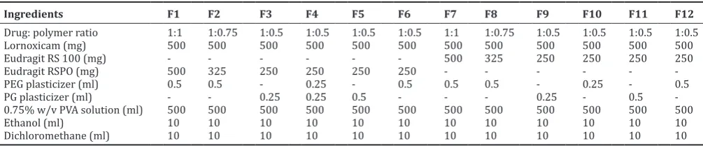
All the formulations were prepared by the quasi-emulsion solvent diffusion technique using the polymers Eudragit RS 100 and Eudragit RSPO and plasticizers polyethylene glycol 400 and propylene glycol. Drug, polymer, and plasticizer were dissolved in a mixture of ethanol and dichloromethane and then sonicated for 10 min. This solution was poured drop by drop with a syringe into 1000 ml beaker containing 0.75% w/v PVA solution, maintained at a temperature of 30–40°C with stirring at 1000–1200 rpm speed for 5 h to allow the volatile solvent for evaporation. The formulated microsponges were filtered, washed with distilled water and dried at 40°C [5,6,11]. The various formulations prepared using different polymers and plasticizers with different ratios are shown in Table 1.
Evaluation of lornoxicam microsponges Scanning electron microscopy (SEM) studies
The lornoxicam microsponges were observed under a SEM. The instrument used in this study was Hitachi S-3700N, Japan. The microsponges were mounted directly on the SEM sample stub, using double-sided sticking tape and coated with a gold film (thickness 180– 200 nm) under reduced pressure.
Particle size analysis
The particle size was measured using an optical microscope, and the mean particle size was calculated by measuring 100 particles with the help of a calibrated ocular micrometer. The slide containing microsponges were mounted on the stage of the microscope and diameter of at least 100 particles was measured using a calibrated optical micrometer.
Percentage production yield
The production yield of the microsponges was determined by calculating accurately the initial weight of raw materials and the last weight of microsponges obtained, and their percentage yield (w/w) was determined using below equation [12].
Yield Actual weight of the product Total weight of excipient and ( )% =
d
drug×100
Percentage of unentrapped drug
Formulated microsponges were filtered from PVA solution. Filtered microsponges were washed thoroughly with 0.75% w/v PVA solution, and washings were added to the above filtrate. 5 ml was taken from this mixture of filtrate and washings, and centrifuged for 10 min and filtered. Filtered sample was suitably diluted with 0.75% w/v PVA solution and analyzed spectrophotometrically at 354 nm using ultraviolet (UV)-Vis spectrophotometer. Percentage of the unentrapped drug was calculated [13].
Total amount of drug used for microsponges
×100
Entrapment efficiency
Microsponges equivalent to 16 mg of pure drug was crushed, powdered and was taken in 100 ml volumetric flask. To this, 80 ml of methanol was added and shaken for 1 h on a mechanical shaker and then sonicated for 5 min to complete removal of lornoxicam from microspheres. After sonication, volume was made up to the mark with methanol. This solution was centrifuged and filtered. Filtered sample was suitably diluted with methanol and analyzed spectrophotometrically at 353 nm using UV-VIS spectrophotometer. Entrapment efficiency was calculated as follows [14,15].
In vitro drug release studies Procedure
In vitro, drug release studies were carried out using USP type II apparatus at 100 rpm. Microsponges equivalent to 16 mg of pure drug was added to 900 ml of pH6.8 phosphate buffer which is used as the dissolution medium. The temperature of the dissolution medium was maintained at 37±0.5°C. An aliquot (5 ml) of dissolution medium was withdrawn at specific time intervals up to 12 h, filtered and suitably diluted before spectrophotometric analysis. Sink conditions were maintained by replenishing the medium with an equal amount (5 ml) of pH6.8 phosphate buffer. The absorbance of the sample was measured at 357 nm by UV-Visible spectrophotometer [16]. The concentration of lornoxicam in test samples was calculated using calibration curve. Six samples were run for each formulation in pH 6.8 phosphate buffer.
Table 1: Formula of lornoxicam microsponges
Ingredients F1 F2 F3 F4 F5 F6 F7 F8 F9 F10 F11 F12
Drug: polymer ratio 1:1 1:0.75 1:0.5 1:0.5 1:0.5 1:0.5 1:1 1:0.75 1:0.5 1:0.5 1:0.5 1:0.5
Lornoxicam (mg) 500 500 500 500 500 500 500 500 500 500 500 500
Eudragit RS 100 (mg) - - - 500 325 250 250 250 250
Eudragit RSPO (mg) 500 325 250 250 250 250 - - -
-PEG plasticizer (ml) 0.5 0.5 - 0.25 - 0.5 0.5 0.5 - 0.25 - 0.5
PG plasticizer (ml) - - 0.25 0.25 0.5 - - - 0.25 - 0.5
-0.75% w/v PVA solution (ml) 500 500 500 500 500 500 500 500 500 500 500 500
Ethanol (ml) 10 10 10 10 10 10 10 10 10 10 10 10
RESULTS AND DISCUSSION
Drug and excipient compatibility studies by FTIR
FTIR spectrum of lornoxicam was recorded, and spectral interpretation was done. The characteristics IR absorption peaks of lornoxicam C=O 1 amide stretching at 1641.30 cm-1, N-H 2o amide stretching at 3122.62 cm−1, C-Cl aromatic stretching at 670.15 cm−1, Thiazide SO
2 at 1354.60 cm−1, C-H aromatic stretching at 3068.73 cm−1, and O-H stretching at 3393.35 cm−1 were there in drug sample spectrum, which confirmed the purity of lornoxicam.
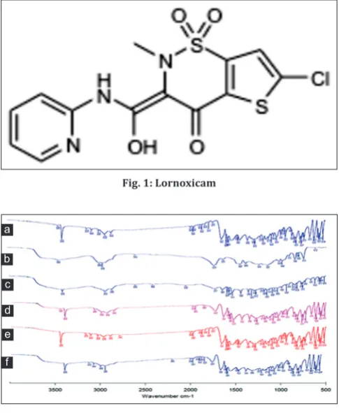
Compatibility study using FTIR was carried out to ensure any possible interaction between drug and Eudragit RS100 and Eudragit RSPO used. FTIR spectroscopic study results revealed no any new peak appearance or disappearance of existing peaks, discarding any chemical interaction probability among drug and polymers used. The characteristic C=O 1o amide stretching vibration at 1640.91 cm-1, N-H 2o amide stretching at 3123.29, C-Cl aromatic stretching at 670.05 cm-1, Thiazide SO
2 at 1354.71 cm-1, C-H aromatic stretching at 3068.71 cm-1, and O-H stretching at 3393.53 cm-1 peaks of lornoxicam were present in the physical mixture of drug and polymers. Thus, FTIR spectroscopy results showed that lornoxicam was compatible with selected polymers (Fig. 2).
SEM
Morphology of prepared microsponges was discovered by SEM analysis. SEM image of optimized formulation (F12) of lornoxicam microsponges shown in Fig. 3. SEM results indicated that microsponges formed were highly porous, spherical in shape with rough surface texture and tiny particles are adhered on the porous outer surface. Pores were induced by diffusion of solvent from the surface of microsponges.
Percentage yield, percentage of unentrapped drug, percentage entrapment efficiency, and particle size studies of lornoxicam microsponges
The percentage yield of all formulations was carried out and was found within the range between 69.58% and 75.35%. Percentage of the unentrapped drug was found to be 24.06%–30.35%. Entrapment efficiency was found to be 62.24%–70.34%. The mean particle size of the microsponges significantly increased with increase in polymer concentration. The reason may be due to the viscosity of medium which increases as the polymer concentration increases. This may be resulting in the formation of larger particles. The particle size of prepared microsponges was observed in the range of 172.72–234.44 µm. The sizes of microsponges affect the encapsulation efficiency and the release rate of the drug. It was observed that as the ratio of drug to polymer was increased, the encapsulation efficiency was decreased. This could probably be due to the fact that in high drug to polymer ratio, the amount of polymer available per microsponge was comparatively less. Probably in high drug-polymer ratios less polymer amounts surround the drug and reducing the thickness of polymer wall and microsponges with smaller size were obtained (Table 2).
In vitro drug release
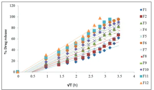
In vitro, drug release studies were performed in pH 6.8 phosphate buffer for 12 h. The cumulative percentage of drug release of prepared formulations was found to be in the following order: F12> F11> F6> F5> F10> F9> F4> F3> F8> F7>F2>F1. The percentage drug release of formulations F1, F2, F3, F4, F5, F6, F7, F8, F9, and F10 was found to be 61.21%, 65.61%, 80.68%, 83.25%, 92.57%, 95.09%, 73.46%, 86.65%, and 89.17%, in 12 h respectively. The percentage drug release of formulation F11 was found to be 95.47% in 10 h. Formulation F12 showed high release, 95.47% in 8 h. This could be due to smaller microsponges are formed at a lower polymer concentration and have a large surface area exposed to dissolution medium, giving rise to faster drug release. Hence, it is considered as an optimized formulation.
Results indicate that proportion of polymers in the formulation was the key factor governing the release of drug from microsponges. As the concentration of polymer increased, there was an increase in particle
size and diffusional path length. This may decrease the overall drug release from the polymer matrix. Formulations comprised Eudragit RSPO in higher proportion exhibited much-retarded drug release as compared to formulations comprised Eudragit RS 100 in higher proportion. The drug release profile from microsponges for all the formulations is shown in Figs. 4-7 and Table 3.
Release of lornoxicam from the microsponges for the optimized formulation F12 was found to follow zero-order kinetics (correlation
Fig. 1: Lornoxicam
Fig. 2: Fourier-transform infrared Spectras of (a) Lornoxicam, (b) Eudragit RSPO, (c) Eudragit RS 100, (d) physical mixture
of lornoxicam and Eudragit RSPO, (e) physical mixture of lornoxicam and Eudragit RS 100, (f) physical mixture of
lornoxicam, Eudragit RS 100, and Eudragit RSPO
Fig. 3: Scanning electron microscopy photograph of optimised formulation (F12) of lornoxicam microsponges a
b
c
d e
coefficient, r2 value 0.961). Higuchi plot showed an r2 value of 0.971 for optimized formulation F12 suggesting that the diffusion plays an important role in the controlled release. The data were fitted to Korsmeyer–Peppas equation; the value of diffusion exponent “n” for optimized formulation F12 is 1.731, indicated that the drug release follows super case II diffusion.
CONCLUSION
Microsponges of lornoxicam were prepared by the quasi-emulsion solvent diffusion method using polymers such as Eudragit RS 100 and Eudragit RSPO. As the polymer concentration is increasing, the particle size of microsponges was increased, and the drug release was decreased.
Among all the formulations, F12 shows better results, which are released more than 80% of the drug release within 8 h. Hence, lornoxicam loaded microsponges prepared by this quasi-emulsion solvent diffusion method are potential for prolong the release of the drug, which will be effective for osteoarthritis, rheumatoid arthritis, and acute lumbar sciatica therapy.
ACKNOWLEDGMENT
The authors are thankful to management, Nirmala College of Pharmacy, Managalagiri, Guntur (Dist.), A.P., for providing necessary facilities to carry out the research work.
Table 2: Percentage yield, Percentage of unentrapped drug, percentage entrapment efficiency, and particle size of lornoxicam microsponges
Formulation Percentage yield
(%w/w)* Percentage of unentrapped drug (%w/w)* Percentage of Entrapment efficiency (%w/w)* Particle size (µm)*
F1 75.08±0.965 30.35±1.036 62.24±1.729 234.44±0.965
F2 72.21±1.034 30.27±1.873 63.50±1.137 221.24±1.211
F3 69.58±0.928 29.23±2.589 64.41±2.645 208.44±1.325
F4 70.16±2.147 29.54±2.764 65.29±2.749 203.24±2.564
F5 71.43±2.138 28.48±1.773 69.65±1.211 189.78±3.034
F6 73.12±1.917 28.82±1.248 67.75±1.765 187.96±3.252
F7 75.35±1.346 28.57±2.432 67.75±1.629 218.54±2.954
F8 73.92±3.270 27.34±2.032 68.27±1.375 214.26±2.342
F9 71.28±2.164 27.83±1.258 68.57±2.125 193.2±2.456
F10 73.65±1.560 26.67±1.045 69.21±1.432 190.5±3.348
F11 70.45±1.317 26.19±2.845 69.68±2.327 175.1±3.587
F12 70.23±1.305 24.06±2.035 70.34±2.034 172.72±3.623
*(n=3), (average±SD)
Table 3: Correlation coefficient (r2) values of different formulations of lornoxicam microsponges (F1-F12) Formulation
Code r
2 values
Zero‑order (r2) First‑order (r2) Higuchi Model (r2) Korsmayer–Peppas r2 value n Value
F1 0.981 0.928 0.881 0.954 1.408
F2 0.991 0.948 0.912 0.927 1.410
F3 0.995 0.966 0.970 0.895 1.487
F4 0.991 0.968 0.949 0.880 1.489
F5 0.970 0.970 0.978 0.880 1.504
F6 0.961 0.961 0.983 0.840 1.487
F7 0.994 0.963 0.935 0.817 1.415
F8 0.994 0.971 0.937 0.905 1.442
F9 0.986 0.970 0.958 0.893 1.509
F10 0.976 0.975 0.970 0.858 1.511
F11 0.973 0.906 0.980 0.809 1.558
F12 0.961 0.807 0.971 0.792 1.731
Fig. 4: Zero-order release profile of lornoxicam microsponges
AUTHOR’S CONTRIBUTIONS
The first author carried out the experimental part of the work. The second author guided and monitored the experimental design, data compilation, critical revision of the article, and corrected the manuscript and third author data analysis, interpretation, and drafting the article.
CONFLICTS OF INTEREST
All the authors hereby declare that there are no conflicts of interest.
REFERENCES
1. Jain N, Sharma PK, Banik A. Recent advances on microsponge delivery system. Int J Pharm Pharm Sci 2011;2:13-23.
2. Kaity S, Maiti S, Ghosh AK, Pal D, Ghosh A, Banerjee S, et al.
Microsponges: A novel strategy for drug delivery system. J Adv Pharm Technol Res 2010;1:283-90.
3. Kumar R, Sharma SK, Jaimini M, Alam N. Microsponge drug delivery systems for novel topical drug delivery. Int J Pharm Sci Lett 2014;3:384-90.
4. Ravi R, Kumar SK, Parthiban S. Formulation and evaluation of the microsponges gel for an anti acne agent for the treatment of acne. Indian J Pharma Sci 2013;1:32-8.
5. Osmani RA, Aloorkar NH, Thaware BU, Kulkarni PK, Moin A, Hani U, et al. Microsponge based drug delivery system for augmented gastroparesis therapy. Asian J Pharm Sci 2015;5:442-51.
6. Osmani RA, Aloorkar NH, Ingale DJ, Kulkarni PK, Hani U,
Fig. 6: Higuchi plot of lornoxicam microsponges (F1-F12) (n=6)
Fig. 7: Korsmeyer–Peppas plots of lornoxicam microsponges (F1‑F12) (n=6)
Bhosale RR, et al. Microsponges based novel drug delivery system for augmented arthritis therapy. Saudi Pharm J 2015;5:562-72.
topical drug delivery of mometasone furoate. Int J Pharm Pharm Sci 2011;4:133-7.
11. Harsh S, Patel K, Upadhyay UM. Formulation and evaluation of controlled release colon targeted micro sponge of aceclofenac. Pharma Innov J 2014;10:81-7.
12. Rajab NA, Jawad MS. Formulation and in vitro evaluation of piroxicam microsponge as a tablet. Int J Pharm Pharm Sci 2015;2:104-14. 13. Zia R, Nazir A, Khan MK, Maan AA, Rashid A. Preparation of ascorbic
acid and cholecalciferol microsponges for topical application. Int J Pharm Pharm Sci 2017;10:280-7.
14. Thireesha B, Prasad AR. Development and validation of a simple UV spectrophotometric method for the determination of lornoxicam in 0.75% w/v PVA solution. Indo Am J Pharm Res 2016;9:6470-7. 15. Prasad AR, Thireesha B. UV-Spectrophotometric method development
and validation for the determination of lornoxicam in microsponges. Int J App Pharm 2018;1:74-8.
16. Prasad AR, Thireesha B. Development and validation of a simple UV spectrophotometric method for the determination of lornoxicam in pH 7.4 phosphate buffer. Indo Am J Pharm Res 2016;9:6478-85. lornoxicam.htm.