R E V I E W
Open Access
An alternative pathway for alphavirus entry
Joseph P Kononchik Jr, Raquel Hernandez and Dennis T Brown
*Abstract
The study of alphavirus entry has been complicated by an inability to clearly identify a receptor and by experiments which only tangentially and indirectly examine the process, producing results that are difficult to interpret. The mechanism of entry has been widely accepted to be by endocytosis followed by acidification of the endosome resulting in virus membrane-endosome membrane fusion. This mechanism has come under scrutiny as better purification protocols and improved methods of analysis have been brought to the study. Results have been obtained that suggest alphaviruses infect cells directly at the plasma membrane without the involvement of endocytosis, exposure to acid pH, or membrane fusion. In this review we compare the data which support the two models and make the case for an alternative pathway of entry by alphaviruses.
Alphaviruses
Alphaviruses are transmitted to vertebrates by hemato-phagic insects such as mosquitoes and ticks [1]. These insects are the vectors in the enzootic cycle. Reptiles, small mammals and birds are primary reservoirs. Humans and larger mammals are largely a dead end in the virus life cycle due to the low levels of viremia pro-duced. Symptoms of alphavirus infection vary from asymptomatic to encephalitis or arthritis [2]. Of those that cause disease, Eastern, Western and Venezuelan encephalitis viruses contribute significantly to disease in large mammals and humans[3]. Chikungunya is reemer-ging as a threat to humans and has been gaining ground in Africa, Asia, the Philippines and Italy with imported cases in France and in international travelers returning to the United States and South America [4-8].
Sindbis Virus (SINV), the prototype virus of genus alphavirusin the familyTogaviridaeis a group IV mem-brane containing virus with a positive sense RNA gen-ome [9]. It is widely used in laboratory studies due to its non-pathogenic phenotype in humans and biosafety level 2 containment status. SINV can be grown to high titer in both mammalian and insect cells [10,11] and has been shown to infect diverse cell types. Certain strains of SINV, specifically the heat resistant SINV (SVHR) are ideal for Alphavirus entry studies. Purified SVHR, in contrast to other membrane containing viruses, can have a particle to plaque-forming-unit (pfu) ratio that
approaches unity [11]. Knowing that every particle is infectious ensures that all observations of cell-virus interactions are of productive virus infections. These features of SINV are optimal for the study of entry of this class of membrane containing viruses including stu-dies involving direct observation by electron microscopy. The term virus entry refers specifically to the mechan-ism by which the virus binds to the host cell receptor, penetrates the cell membrane barrier and releases the infectious RNA into the cell initiating the infection.
Alphavirus Structure
Alphaviruses are small 70 nm viruses that have 240 copies each of three structural proteins, E1, E2 and capsid (C) assembled in a 1:1:1 stoichiometry. These three proteins create two nested T = 4 icosahedral shells that sandwich a host derived lipid bilayer [12] (Figure 1). The outer protein shell is composed of E1 and E2 heterodimers that assem-ble into aggregates of three producing the three pronged spike which protrudes from the virus surface. The inner protein shell is made of only C, encapsulating the 49S RNA. This inner protein shell is the shape determining component of the virus [13,14]. The organization of the E1/E2 complexes on the cell membrane likely exist in a two-dimensional 6-fold symmetry sheet prior to capsid envelopment [15]. When the membrane glycoproteins begin the process of encapsulation, the nucleocapsid recruits the E1-E2 trimers into the developing outer shell by specifically binding the E2 endodomains. Through its repeated 240 interactions between a hydrophobic cleft on C and the E2 endodomain this process organizes the * Correspondence: [email protected]
Department of Molecular and Structural Biochemistry N C State University, Raleigh, NC 27695, USA
glycoproteins into the 6-fold and requisite 5-fold rotational arrays necessary to form a three-dimensional icosahedral structure [16]. Mutations which disrupt this process result in structurally misshaped particles [13,17,18] The resulting virus particle has 80 spikes that are primarily made of E2 (colored blue in Figure 1 pH 7.0) with a protein skirt that is primarily E1 (colored green in Figure 1 pH 7.0) which covers the incorporated membrane [19,20]. The two pro-tein shells with their significant level of lateral and trans-membrane interactions [21,22] result in a very rigid and precisely organized particle that is unlike that of other membrane containing viruses. It has been shown recently, that the particles of mammalian and insect grown SVHR are not structurally identical [23]. The cellular response to infection by insect and mammalian derived virus has also been shown to be different [24]. Particles produced from insect cells are more compact, lacking some RNA interca-lation in the capsid protein shell seen in mammalian grown virus. The thickness of the membrane of virus pro-duced from mosquito and mammalian cells does not solely account for the difference in the structure of the virus particles; however the outer protein shell seems to be extended in the mammalian grown virus suggesting that the protein organization between the two particles may be in slightly different functional conformations.
The highly organized structure of the alphaviruses with the many protein-protein interactions stabilizing the structure and the membrane bilayer occluded by the outer protein shell have important implications for the process of entry of the viral genome into host cells. Whereas many membrane containing viruses such as
influenza can be described as a membranous structure with embedded proteins the alphaviruses are protein shells with an associated membrane. This protein shell must be compromised if the virus is to transfer its RNA to the cell cytoplasm. The structure of the virus protein shell may be critical for the mechanism of genome deliv-ery to work properly. It is hypothesized that the E1 pro-tein in the mature alphavirus exists in a metastable configuration poised to use the energy stored during virus assembly for the entry process upon encountering an appropriate trigger [25,26]. The energy stored for this conformational change is the result of folding the E1 pro-tein though a series of disulfide bonded intermediates as the protein is compacted into the metastable structure during the assembly of the spike heterotrimer [25]. The metastable nature of the spike complex is revealed when the E1 protein is released from the mature virus using detergent because the protein reorganizes into several disulfide bridged configurations which can be separated on non-denaturing gels[25].
The current molecular model for how the virus trans-fers its RNA into the cell is hypothesized to be by low pH mediated membrane fusion, after endocytosis of the virus attached to its receptor. E2 contains the receptor binding sequence while E1 is known to contain the properties necessary for membrane fusion [9]. It is pre-dicted that as the endosome is acidified, the virus mem-brane fuses with the endosome and releases the nucleocapsid into the cell cytoplasm. There are some reports which implicate ribosomes in the process of releasing RNA from its association with the capsid pro-tein [27-29]. The process by which E1 mediates mem-brane fusion can theoretically be supported by a crystal structure of E1* (PDB code: 1I9W) which displays the position and the conformation of the putative fusion domain [30] and flexible regions within the protein. This structure was obtained by proteolytic cleavage from purified Semliki Forest Virus (SFV). A second structure was obtained by expression of the E1 ectodo-main inE. coli. [31-33]. This crystal structure of E1 was subsequently fit into a cryoEM-reconstruction of SINV (PDB code: 1Z8Y) [34]. The crystal structure of the E1 protein has three primary domains I, II and III. Domain I is the NH proximal domain that contains the putative fusion loop. Domain II is the central domain and domain III is the most distal domain. One important caveat when interpreting structural data is that the data represents a native structure or structural intermediate of the entity in question. This condition would require that sound biochemical data confirm assumptions about the structural data without imposing other methods of confirmation to the analysis. A second condition would be that the fit of the higher resolution crystal structure not be distorted in the larger density landscape of the
second much more diffuse cryoEM density. One such analysis put in question the number and location of the disulfide bonds in the native, infectious form of E1. In the crystal structure, there are a number of disulfide bonds identified; supporting the hypothesis that disulfide bonds play a role in protein assembly [35]. However some cystines identified as participating in disulfide bridges in the crystal structure were identified as free cystines using protein modification and mass spectrome-try in the intact infectious virus [36]. These conflicts are not unexpected because the protein crystal structure is dependent on how the protein is purified, the crystals are grown, and the structure is refined. It is the nature of the crystal analysis process that the structure is of a protein in its lowest energy conformation. This means that the crystal structure could be any one of the var-ious intermediate conformations assumed when E1 is extracted by detergent (described above) or expressed without its membrane spanning domain in an environ-ment other than the endoplasmic reticulum.
A second caveat which seriously affects the quality of the data is that the entity to be studied not be distorted by the ability of proteins to be manipulated and expressed in some form inE. coli. There are currently crystal structures of an E1-E2 fusion protein (PDB codes 3MUU), chikungunya glycoproteins (PDB codes: 3N40;3N41;3N42;3N43;3N44;2XFB;2XFC) and their fit into the SINV and/or Semliki Forest Virus cryo-recon-struction (PDB codes: 3MUW;2XFB;2XFC). These struc-tures were produced by expressing the ectodomains of the E1 and E2 glycoproteins connected together by a lin-ker (58). This fusion protein was constructed due to the inability of expressing E2 inE. coli. While it is common practice to remove troublesome domains from proteins to allow for crystallization to occur, how is it determined when there has been too much manipulation of the sequence? These structures have the same problems as E1*. The assumption that the transmembrane domains, which have been removed in the c DNA clone, and inte-gration into the ER membrane play no role in the correct folding and assembly of the spike structure is an unpro-ven assumption. For virus entry of the macromolecular SINV particle, structure is critical. The overall alphavirus icosahedral structure is unique in that it is a membrane containing virus that does not have the membrane exposed. The membrane itself is not the form determin-ing factor as with influenza, and because it is beneath the outer protein shell is not readily available to fuse with host membranes. The essential role of the membrane may be to provide the scaffold upon which the virus is assembled. To fully explore the mechanism of virus pene-tration and entry other methods of analysis other than interpretations of structural fit can provide the missing events required for SINV infection.
Adsorption and receptor recognition
The process of virus attachment/absorption to the host cell is probably a multistep event. It is not a new propo-sal that virus infection begins with scanning of the cell surface that begins with a general“sticking”to the cell via one or more proteins (or the cell membrane itself) followed by“rolling”on the cell surface as it locates the proper receptor to initiate the penetration step of virus entry. This type of interaction has been identified recently using single particle fluorescence resonance energy transfer (FRET). SINV saturated with a mem-brane intercalated fluorescent self-quenching dye can be visualized when the dye is excited with a specific wave-length light [37]. Fusion of virus with the cell membrane is detected when lipid mixing (fusion) releases the pro-teins and dequenching of the dye occurs causing it to fluoresce. Using this technique it has been shown that SINV moves on the cell surface in a neutral pH environ-ment. If the pH of the medium bathing the cells is low-ered the virus does not fuse with the cell surface as is the case with influenza, rather the virus “freezes” in place (K. Wenniger, unpublished observation). These observations indicate that viruses probe the cell surface for the proper receptor molecule.
As arboviruses, alphaviruses infect insect and verte-brate hosts. Since alphaviruses need to infect cells which provide widely divergent biochemical and genetic envir-onments, it is likely that they either use a ubiquitous receptor, or are able to use multiple proteins as a recep-tor. The receptor/s has not been identified. Many pro-teins and polysaccharides have been implicated as being part of the receptor complex. The list includes, heparin sulfate [38-40], the major histocompatability complex (MHC) [41,42], the major laminin receptor [43], DC sign, L sign [44], heat shock 70 protein [45], an uniden-tified 110 kDa nerve cell protein [46], and a 63 kDa pro-tein in chicken cells [47]. The length of the list of possible receptors strongly suggests that there are multi-ple proteins that alphaviruses can use, and that the spe-cific receptor is both cell and virus spespe-cific. If, however, there is one widely used receptor it would have to be a fundamental piece of genetic and biochemical machinery that has been conserved throughout evolution. The alphaviruses have evolved to use highly conserved pro-teins that span the large breath of species that serve as hosts and/or multiple proteins as a receptors.
number of additional studies that employed various techniques to determine the receptor. One such study used soluble glycoproteins from Semliki Forest Virus (SFV) and showed that the MHC bound the glycopro-teins and that detergent soluble MHC protein was able to inhibit SFV infection of HeLa cells [41]. Since then, a number of questions about the validity of this argument have arisen since cells that lack the MHC complex are not resistant to SFV infection [49]. Additionally, mos-quito cells, which fundamentally lack a human immune system and thus do not express MHC, are also readily infected by alphaviruses.
The use of anti-idiotypic antibodies as receptor loca-tors has been used as well to determine the receptor for alphaviruses. This approach is responsible for the dis-covery of the 63 kDa chicken protein [47] and even-tually led to the implication of the high-affinity laminin receptor [43]. While this is considered a major receptor for alphaviruses, reexamination of the original experi-ment that identified the 63 kDa chicken protein revealed that this protein was not the chicken laminin receptor [47]. Additionally antibody against the chicken laminin receptor did not inhibit infection significantly (< 10%). This suggests that although the laminin receptor is con-served across many species, it is not the only virus receptor. It is entirely likely that there are multi-protein complexes that are not required for, but enhance infec-tion. Other investigations of the laminin receptor used anti-idiotype antibodies to examine the laminin receptor as a possible virus receptor in mosquito cells. In these studies a 32 kDa protein was discovered in mosquito cells to which Venezuelan Equine Encephalitis Virus (VEE) bound as did laminin and SINV [9]. Antibodies were also used to investigate virus binding with a strain of SINV which was selected to be a rapid penetrating virus [50]. Upon binding to the cell at neutral pH assu-mingly to its receptor, SFV was shown to go through conformational changes as new antibody epitopes were exposed on the surface of the virus [51]. This is presum-ably related to the conformational change that is seen in reconstructed virus particles which were exposed to low pH [12].
A measure of successful virus entry
There are numerous techniques that can be used to examine the interactions between the virus and the cell. These techniques can generally fall into two categories: direct and indirect observation. Direct observation uses familiar techniques such as thin-section microscopy, cryo-electron microscopy, tomography, and other less familiar techniques like freeze-fracture immunolabeling to examine virus-cell interactions. When properly pre-pared and processed, the interactions between the virus and cell seen in the microscope are the actual
interactions that occurred at the time of fixation. By examining different times during the infection process, the possible mechanism and pathway of virus entry can be elucidated. Unfortunately, thin-sections and other direct observation techniques cannot distinguish between a successful infection process, and that of an unproductive interaction. This makes microscopy very subjective and is the primary problem when observing virus-cell interactions, as even with a relatively low par-ticle/pfu ratio of 1:10, most observations are of virus-cell interactions that do not lead to a successful infec-tion. For SINV, the SVHR strain can be purified to a particle/pfu ratio of 1:1, (28) and this virus was used in EM studies of virus infection that led to the direct cell penetration hypothesis of virus entry [12]. Many of the previous observations that have been made using EM are of viruses preparation made with strains or samples with high or undetermined particle/pfu ratio and there-fore are difficult to interpret.
When direct observation is not possible, or cannot answer the question being asked, techniques that take advantage of indirect or secondary reporters have been used. Some indirect reporters that have been used as a measure of successful virus entry include virus RNA production [52], virus protein synthesis [53] and virus production [54]. While these tools are useful and have shed light on many virus-cell interactions, they are at a disadvantage insomuch that they cannot determine if a virus has not entered the cell as the events assayed are not entry. Many of these biochemical reporters are sig-nificantly downstream of the initial events of virus adsorption and entry. As a result, each step between entry and the reporter has the potential to be inhibited giving a false negative. To give a time-scale to RNA translation and protein synthesis, super infection inhibi-tion is detectable 15 minutes post infecinhibi-tion [55,56]. This implies that entry is a very fast process, and that the genome is quickly unpacked and rapidly available for processing. The speed of infection, the problems of indirect reporters, and the limits of direct observation need to be carefully considered when building an assay to measure successful virus entry.
Alphavirus entry
conformational change of SINV was visualized using cyro-electron microscopy reconstruction of particles exposed to low pH [12]. The change resulted in a pro-truding spike from the 5-fold axis of the virus, com-pared to the lack of this density in the untreated particles (Figure 1, green arrow). The location of the spike suggests that it is both 5-fold in symmetry, and that it is likely composed of E1, the glycoprotein that expresses the putative fusion loop. Interpretation of these data in the context of the prevailing infection by fusion model would predict that the virus and cell mem-branes fuse and the nucleocapsid is delivered to the cell cytoplasm. However, evident from these reconstructions is that no lipid is exposed at high pH, or upon return of the particles to low pH. This observation forms the basis of the following hypothesis which suggests that there is noa priori reason to conclude that because a virus has a membrane, penetration must be by fusion. Discussed here are two suggested modes of entry for the alphavirus and the supporting data.
Viral Endocytosis followed by membrane fusion
The most commonly accepted mechanism of entry for alphaviruses is via endocytosis and the subsequent acidi-fication of the vesicle leading to membrane fusion. This is similar to the mechanism accepted for influenza. The studies in support of this mechanism are extensive, and rely on the observations that alphaviruses have mem-brane fusion capabilities, and that inhibition of the acidi-fication of endosomes inhibits the entry of the virus. The original fusion assays were done with cultured cells, and it was shown that treating cells with adsorbed virus to a brief low pH environment, resulted in cell-cell fusion (fusion from without) [62]. This suggested the acid environment of an endosome could provide the requirements for fusion and penetration. However in these studies the cells were always returned to neutral pH before fusion was seen and it was subsequently shown that this return to neutral pH was required to induce fusion of the virus and cell membranes[63]. The pH of the endosome does not return to neutrality. The analysis of fusion was then moved from a live cell cul-ture to an in vitro assay including liposomes and other artificial membranes [64]. In these assays, the fusion process did not require a return to neutral pH, but required a significant amount of cholesterol in the target membrane [65]. This cholesterol requirement was sup-ported by a report that cells grown in serum treated with Cab-O-Sil, a silicate that removes cholesterol from the serum could not be infected as measured by immu-nofluorescent antibody labeling of newly folded virus protein [66]. Here a third caveat must be add must be made, that is how far afield from the native system can the experimental design be taken before the results are
valid biochemically but cannot reproduce the biological environment? This should especially be considered in the context of a biological macromolecule expressing multiple required functions. There are two specific con-cerns regarding the cholesterol requirement for infection that have been neglected. First, the liposome is protein free and therefore receptor free and second the amount of cholesterol required to induce fusion of SINV to lipo-somes is high, up to 50 mol%. However, the require-ment for such a high concentration of cholesterol is in direct conflict with the fact that alphaviruses infect and assemble in mosquitoes, which are cholesterol auxo-trophs [67,68]. Second, Cab-O-Sil, while removing cho-lesterol also removes a significant number of other lipids, which may be required for proper function of the cells and virus stability [68]. In fact, growing the same cell type used in the Cab-O-Sil experiment in serum-free media resulted in no significant change in their sen-sitivity to infection [68]. Together, these data imply that a large amount of cholesterol is not required for alpha-virus infection but is required for fusion with liposomes. Thus, if cholesterol is not required for infection and fusion of living cells does not occur at acid pH the lipo-some model for studying virus penetration may not be valid.
shown to reduce the amount of virus produced in mam-malian cells, however in insect cells the virus titer was enhanced [73,74]. Chloroquine did increase the pH of mosquito cell endosomes as the cells were protected from diphtheria toxin which is known to require an acid environment to enter cells. Also in mosquito cells ammonium chloride was shown to inhibit steps in the non-structural protein processing after initial translation of the incoming RNA; many steps from the initial virus entry which had to have occurred [74]. While chemical inhibitors have been invaluable in the study of cellular metabolic pathways, it is imperative that the design of the experiment incorporate appropriate controls for sec-ondary effects.
Using a SINV construct that contained a GFP repor-ter, a thorough study was done to assess the effect of Bafilomycin A1 (BafA1) on the entry of alphaviruses into mammalian and mosquito cells [75]. The results of this study showed that BafA1 did not prevent virus entry into mammalian or mosquito cells or inhibit RNA synthesis. It was also shown that BafA1treatment pro-duced the same results when cells were transfected with virus RNA a process that bypassed the entry events. Additionally, the effect of BafA1 in mammalian cells was reversible and the authors concluded that BafA1 was inhibiting the proper folding of the newly synthe-sized proteins and that the functioning V-type ATPase was required for proper protein assembly. When exam-ining the effect of BafA1 on mosquito cells, no observa-ble inhibition of virus entry was observed, even at high concentrations of BAfA1. It has been demonstrated that the insect V-ATPase is sensitive to BAFA1 at nanomolar concentrations [76]. All of the data supporting entry at the plasma membrane correlate well with a recent publi-cation showing SINV replipubli-cation complexes at the plasma membrane [77] as it would be easiest to explain the presence of the replication complexes here if the mechanism of entry were at the plasma membrane.
Entry in the absence of membrane fusion
An alternative mechanism of entry that is gaining sup-porting evidence is virus entry at the plasma membrane in the absence of endocytosis, exposure to low pH and membrane fusion. This mode of entry is proposed to employ a pore complex made from virus and host pro-teins that connects the interior of the virus to the host cell cytoplasm via a protein channel. Supporting this mechanism of infection is that infection by alphaviruses has been shown to be a leaky process allowing passage of ions and small molecules across the compromised plasma membrane [78-81]. The infected cell membrane becomes leaky early in the infection prior to protein expression and also late during infection [82,83]. Mem-brane fusion by contrast is a non-leaky process [84].
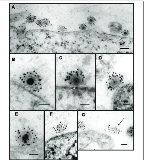
Were the viruses merely to bind to its receptor and then be taken into the cell, as is the case in the low pH -endocytosis model there would be no loss of continuity in the plasma membrane. It was shown that infection with alphavirus created pores in the plasma membrane that could be blocked with rare earth ions which allowed investigators to assign a size to the pores [81]. It has also been shown that the pores created in the infection process are large enough to allow the passage of the toxin alpha-sarcin into the cell (42). The 17 kDa, 150 amino acid protein was shown to co-enter during infection. Interestingly, it has been shown that the 6 k protein which links PE2 to E1 in the structural polypro-tein can also produce pores in the plasma membrane late in infection [85] In 2004, Paredeset al.were able to show full and empty particles (Figure 2) on the surface of mammalian cells using thin-sections of BHK cells infected with SVHR which had a particle/pfu ratio of ≈1. Empty particles were identified by using anti-SINV antibodies with secondary antibodies conjugated to gold beads, as they would have been easily overlooked other-wise (Figures 2A, D, F). From the thin-sections, multiple particles with varying electron dense cores were shown and suggested that the virus was losing its RNA to the cell cytoplasm through a proteinaceous connection between the virus and the cell. The virus subsequently appeared to lose structural stability and was seen released from the cell leaving the proteinaceous pore behind. (Figure 2B, E, F, G). The process of cell penetra-tion was found to be extremely fast requiring a complex process of attaching virus at 4° 0 and then adding warm media that contained fixative to capture the image. It was found that even at low temperature 4-5% of the attached particles had lost their electron dense RNA core during the binding and fixation period. Cells trea-ted with warm fixative had 25-26% of particles which had lost their RNA. Alphavirus entry in the absence of endocytosis was also shown by infecting cells at low temperature a condition under which membranes can-not fuse and endocytosis is inhibited [86]. These experi-ments demonstrated that entry could occur when fusion was not possible showing the two processes to be unre-lated. These data support the mechanism proposed in Paredeset al.which show physical evidence that alpha-viruses use a proteinaceous pore to infect cells.
Figure 2Electron micrographs of thin sections of Sindbis virus-cell complexes at pH 7.2. (A) Low magnification showing‘’full’’and
modification of the FRIL (freeze-replicate-immunolabel) technique developed by Fujimoto [87,88]. In brief, the cross-linked samples were flash frozen in liquid ethane, fractured at low temperature and pressure. The fracture plane that goes through the sample follows the path of least resistance, which is the middle of lipid bilayers making two replicas: the inner leaflet, or P-face, and the outer leaflet, the E-face. As a result, when the fracture hits a cell, it tends to follow the plasma membrane. This makes it ideal for examining proteins and interactions that occur on the cell surface. In this experiment, the replicas had cross-linked virus proteins attached to the outside of the E-face. Using immunolabeling, these pro-teins were labeled, and the organization of the propro-teins was examined by electron microscopy. The data col-lected showed labeled virus particles adsorbed to the cells’ surface. These particles were observed as discrete rings of gold bead tags surrounding a structure that tra-versed the membrane bilayer. We have suggested that this transmembrane structure comprises part of the pore complex.
Summary
Direct observation of alphavirus-cell interaction by elec-tron microscopy and through the use of chemical inhibi-tors and interaction with artificial membranes has created seemingly conflicting data. In many direct obser-vations, virus can be seen in endosomes inside the cell [89]. These data suggested that the virus particles enter through an endocytic pathway. Coupled with the confor-mational changes that occur upon exposure to low pH and the fusion capabilities shown by SINV, the sug-gested route of entry is similar to that of influenza. However, other observations including thin-sections and freeze-fracture have shown that SINV can infect without endocytosis and acidification. These studies also found that as the particle to PFU ratio of the virus employed improved to unity no virus was seen in endosomes and empty particles were seen on the cell surface. These data suggest that the mechanism for virus entry is at the cell surface and involves direct release of the virus gen-ome into the cytoplasm by a pore-like structure.
The use of chemical inhibitors of endosome formation and acidification has relied on reporter events which occur many steps away from the process of entry. Many of the agents have secondary effect on events that occur after entry which have led to erroneous conclusions.. Likewise studies of virus interaction with artificial mem-branes have produced a set of requirements for the fusion event that are not related to those occurring with living cells. It appears that, although alphaviruses pos-sess the ability to fuse membranes this is is an event that occurs under special laboratory conditions and may be unrelated to the true entry process.
Arthropod borne viruses are significant sources of dis-ease in man and domestic animals and some are poten-tial agents of bioterrorism. Strategies to control infections by these agents include the development of compounds which will block critical steps in the entry pathway. An accurate image of this pathway and the identification of the cell receptor participating in the entry process is critical for the development of agents which can block infection.
Acknowledgements
The authors are supported by a grant from The Foundation for Research, Carson City NV and by the North Carolina Agricultural Research Service.
Authors’contributions
JK, RH and DB contributed to and edited the manuscript. All authors read and approved the final manuscript.
Competing interests
The authors declare that they have no competing interests.
Received: 3 June 2011 Accepted: 15 June 2011 Published: 15 June 2011
References
1. Adamczyk M, Moore JA, Yu Z:Application of surface plasmon resonance toward studies of low-molecular-weight antigen-antibody binding interactions.Methods2000,20:319-328.
2. Leckband DE, Kuhl TL, Wang HK, Muller W, Herron J, Ringsdorf H:Force probe measurements of antibody-antigen interactions.Methods2000,
20:329-340.
3. Gould EA, Coutard B, Malet H, Morin B, Jamal S, Weaver S, Gorbalenya A, Moureau G, Baronti C, Delogu I,et al:Understanding the alphaviruses: Recent research on important emerging pathogens and progress towards their control.Antiviral Research2010,87:111-124.
4. CDC - Chikungunya [[http://www.cdc.gov/ncidod/dvbid/chikungunya/]].. 5. Update: chikungunya fever diagnosed among international travelers–
United States, 2006. MMWR Morb Mortal Wkly Rep2007,56:276-277. 6. Angelini R, Finarelli AC, Angelini P, Po C, Petropulacos K, Macini P,
Fiorentini C, Fortuna C, Venturi G, Romi R,et al:An outbreak of chikungunya fever in the province of Ravenna, Italy.Euro Surveill2007,
12:E070906-070901.
7. Ledrans M, Quatresous I, Renault P, Pierre V:Outbreak of chikungunya in the French Territories, 2006: lessons learned.Euro Surveill2007,12: E070906-070903.
8. Solignat M, Gay B, Higgs S, Briant L, Devaux C:Replication cycle of chikungunya: A re-emerging arbovirus.Virology2009,393:183-197. 9. Strauss JH, Strauss EG:The Alphaviruses: Gene Expression,
Replication, and Evolution. Micro. Rev. 58:491-562.Micro Rev1994,
58:491-562.
10. Brown DT, Condreay LD:Replication of Alphaviruses in Mosquito Cells.In
The Togaveriridae and Flaviviridae.Edited by: Schlesinger MJ. Plenum Publishing Corporation; 1986:171-207.
11. Hernandez R, Sinodis C, Brown DT:Sindbis virus: propagation, quantification, and storage.Curr Protoc Microbiol2010, Chapter 15: Unit15B 11.
12. Paredes AM, Ferreira D, Horton M, Saad A, Tsuruta H, Johnston R, Klimstra W, Ryman K, Hernandez R, Chiu W, Brown DT:Conformational changes in Sindbis virions resulting from exposure to low pH and interactions with cells suggest that cell penetration may occur at the cell surface in the absence of membrane fusion.Virology2004,
324:373-386.
13. Tuttle PVt, Rundell AE, Webster TJ:Influence of biologically inspired nanometer surface roughness on antigen-antibody interactions for immunoassay-biosensor applications.Int J Nanomedicine2006,1:497-505. 14. Ferreira D, Hernandez R, Horton M, Brown DT:Morphological variants of
Sindbis virus produced by a mutation in the capsid protein.Virology
15. VonBonsdorff CH, Harrison SC:Sindbis virus glycoproteins form a regular icosahedral surface lattice.J Virol1978,28:578.
16. Brown DT, Waite MR, Pfefferkorn ER:Morphology and morphogenesis of Sindbis virus as seen with freeze-etching techniques.J Virol1972,
10:524-536.
17. Hernandez R, Ferreira D, Sinodis C, Litton K, Brown DT:Single amino acid insertions at the junction of the sindbis virus E2 transmembrane domain and endodomain disrupt virus envelopment and alter infectivity.J Virol
2005,79:7682-7697.
18. Nelson S, Hernandez R, Ferreira D, Brown DT:In vivo processing and isolation of furin protease-sensitive alphavirus glycoproteins: a new technique for producing mutations in virus assembly.Virology2005,
332:629-639.
19. de Wildt RM, Mundy CR, Gorick BD, Tomlinson IM:Antibody arrays for high-throughput screening of antibody-antigen interactions.Nat Biotechnol2000,18:989-994.
20. Zhang W, Mukhopadhyay S, Pletnev SV, Baker TS, Kuhn RJ, Rossmann MG:
Placement of the structural proteins in sindbis virus.J Virol2002,
76:11645-11658.
21. Hernandez R, Lee H, Nelson C, Brown DT:A single deletion in the membrane-proximal region of the Sindbis virus glycoprotein E2 endodomain blocks virus assembly.J Virol2000,74:4220-4228. 22. Whitehurst CB, Willis JH, Sinodis CN, Hernandez R, Brown DT:Single and
multiple deletions in the transmembrane domain of the Sindbis virus E2 glycoprotein identify a region critical for normal virus growth.Virology
2006,347:199-207.
23. He L, Piper A, Meilleur F, Myles DA, Hernandez R, Brown DT, Heller WT:The structure of Sindbis virus produced from vertebrate and invertebrate hosts as determined by small-angle neutron scattering.J Virol 84:5270-5276.
24. Shabman RS, Morrison TE, Moore C, White L, Suthar MS, Hueston L, Rulli N, Lidbury B, Ting JP, Mahalingam S, Heise MT:Differential induction of type I interferon responses in myeloid dendritic cells by mosquito and mammalian-cell-derived alphaviruses.J Virol2007,81:237-247. 25. Mulvey M, Brown DT:Formation and rearrangement of disulfide bonds
during maturation of the Sindbis virus E1 glycoprotein.J Virol1994,
68:805-812.
26. Carleton M, Lee H, Mulvey M, Brown DT:Role of glycoprotein PE2 in formation and maturation of the Sindbis virus spike.J Virol1997,
71:1558-1566.
27. Wengler G, Wurkner D:Identification of a sequence element in the alphavirus core protein which mediates interaction of cores with ribosomes and the disassembly of cores.Virology1992,191:880-888. 28. Cooper MA, Williams DH:Kinetic analysis of antibody-antigen interactions
at a supported lipid monolayer.Anal Biochem1999,276:36-47. 29. Scicluna LA, McCullough KC:Rapidity of specific antibody-antigen
interactions.Scand J Immunol1999,50:167-176.
30. Levy-Mintz P, Kielian M:Mutagenesis of the putative fusion domain of the Semliki Forest virus spike protein.J Virol1991,65:4292-4300. 31. Gibbons DL, Reilly B, Ahn A, Vaney MC, Vigouroux A, Rey FA, Kielian M:
Purification and crystallization reveal two types of interactions of the fusion protein homotrimer of Semliki Forest virus.J Virol2004,
78:3514-3523.
32. Lescar J, Roussel A, Wien MW, Navaza J, Fuller SD, Wengler G, Rey FA:The Fusion glycoprotein shell of Semliki Forest virus: an icosahedral assembly primed for fusogenic activation at endosomal pH.Cell2001,
105:137-148.
33. Tsai HF, D’Avirro N, Selsing E:Gene conversion-like sequence transfers in a mouse antibody transgene: antigen selection allows sensitive detection of V region interactions based on homology.Int Immunol2002,
14:55-64.
34. Gibbons DL, Vaney MC, Roussel A, Vigouroux A, Reilly B, Lepault J, Kielian M, Rey FA:Conformational change and protein-protein interactions of the fusion protein of Semliki Forest virus.Nature2004,
427:320-325.
35. Abell BA, Brown DT:Sindbis virus membrane fusion is mediated by reduction of glycoprotein disulfide bridges at the cell surface.J Virol
1993,67:5496-5501.
36. Whitehurst CB, Soderblom EJ, West ML, Hernandez R, Goshe MB, Brown DT:
Location and role of free cysteinyl residues in the sindbis virus e1 and e2 glycoproteins.J Virol2007,81:6231-6240.
37. Wessels L, Elting MW, Scimeca D, Weninger K:Rapid membrane fusion of individual virus particles with supported lipid bilayers.Biophys J2007,93:526-538. 38. Byrnes AP, Griffin DE:Binding of Sindbis virus to cell surface heparan
sulfate.J Virol1998,72:7349-7356.
39. Smit JM, Waarts BL, Kimata K, Klimstra WB, Bittman R, Wilschut J:
Adaptation of alphaviruses to heparan sulfate: interaction of Sindbis and Semliki forest viruses with liposomes containing lipid-conjugated heparin.J Virol2002,76:10128-10137.
40. Zhang W, Heil M, Kuhn RJ, Baker TS:Heparin binding sites on Ross River virus revealed by electron cryo-microscopy.Virology2005,332:511-518. 41. Helenius A, Morein B, Fries E, Simons K, Robinson P, Schirrmacher V,
Terhorst C, Strominger JL:Human (HLA-A and HLA-B) and murine (H-2K and H-2D) histocompatibility antigens are cell surface receptors for Semliki Forest virus.Proc Natl Acad Sci USA1978,75:3846-3850. 42. Maassen JA, Terhorst C:Identification of a cell-surface protein involved in
the binding site of Sindbis virus on human lymphoblastic cell lines using a heterobifunctional cross-linker.Eur J Biochem1981,115:153-158. 43. Wang KS, Kuhn RJ, Strauss EG, Ou S, Strauss JH:High-affinity laminin
receptor is a receptor for Sindbis virus in mammalian cells.J Virol1992,
66:4992-5001.
44. Klimstra WB, Nangle EM, Smith MS, Yurochko AD, Ryman KD:DC-SIGN and L-SIGN can act as attachment receptors for alphaviruses and distinguish between mosquito cell- and mammalian cell-derived viruses.J Virol2003,
77:12022-12032.
45. Ryan C, Stevens TH, Schlesinger MJ:Inhibitory effects of HSP70 chaperones on nascent polypeptides.Protein Sci1992,1:980-985. 46. Ubol S, Griffin DE:Identification of a putative alphavirus receptor on
mouse neural cells.J Virol1991,65:6913-6921.
47. Wang KS, Schmaljohn AL, Kuhn RJ, Strauss JH:Antiidiotypic antibodies as probes for the Sindbis virus receptor.Virology1991,181:694-702. 48. Smith AL, Tignor GH:Host cell receptors for two strains of Sindbis virus.
Arch Virol1980,66:11-26.
49. Oldstone MB, Tishon A, Dutko FJ, Kennedy SI, Holland JJ, Lampert PW:
Does the major histocompatibility complex serve as a specific receptor for Semliki Forest virus?J Virol1980,34:256-265.
50. Olmsted RA, Meyer WJ, Johnston RE:Characterization of Sindbis virus epitopes important for penetration in cell culture and pathogenesis in animals.Virology1986,148:245-254.
51. Ahn A, Klimjack MR, Chatterjee PK, Kielian M:An epitope of the Semliki Forest virus fusion protein exposed during virus-membrane fusion.J Virol1999,73:10029-10039.
52. Glomb-Reinmund S, Kielian M:fus-1, a pH shift mutant of Semliki Forest virus, acts by altering spike subunit interactions via a mutation in the E2 subunit.J Virol1998,72:4281-4287.
53. DeTulleo L, Kirchhausen T:The clathrin endocytic pathway in viral infection.Embo J1998,17:4585-4593.
54. Perez L, Carrasco L:Involvement of the vacuolar H(+)-ATPase in animal virus entry.J Gen Virol1994,75(Pt 10):2595-2606.
55. Johnston RE, Wan K, Bose HR:Homologous interference induced by Sindbis virus.Journal of Virology1974,14:1076-1082.
56. Karpf AR, Lenches E, Strauss EG, Strauss JH, Brown DT:Superinfection exclusion of alphaviruses in three mosquito cell lines persistently infected with Sindbis virus.J Virol1997,71:7119-7123.
57. Harrison SC:Viral membrane fusion.Nat Struct Mol Biol2008,15:690-698. 58. Melikyan GB:Common principles and intermediates of viral
protein-mediated fusion: the HIV-1 paradigm.Retrovirology2008,5:111. 59. Kielian M, Jungerwirth S:Mechanisms of enveloped virus entry into cells.
Mol Biol Med1990,7:17-31.
60. Meyer WJ, Gidwitz S, Ayers VK, Schoepp RJ, Johnston RE:Conformational alteration of Sindbis virion glycoproteins induced by heat, reducing agents, or low pH.J Virol1992,66:3504-3513.
61. Anthony RP, Paredes AM, Brown DT:Disulfide bonds are essential for the stability of the Sindbis virus envelope.Virology1992,190:330-336. 62. White J, Kartenbeck J, Helenius A:Fusion of Semliki Forest Virus with the
plasma membrane can be induced by low pH.Journal of Cell Biology
1980,87:264-272.
63. Edwards J, Brown DT:Sindbis virus-mediated cell fusion from without is a two-step event.J Gen Virol1986,67:377-380.
65. Kielian MC, Helenius A:Role of cholesterol in fusion of Semliki Forest virus with membranes.J Virol1984,52:281-283.
66. Lu YE, Cassese T, Kielian M:The cholesterol requirement for sindbis virus entry and exit and characterization of a spike protein region involved in cholesterol dependence.J Virol1999,73:4272-4278.
67. Mitsuhashi J, Nakasone S, Horie Y:Sterol-free eukaryotic cells from continuous cell lines of insects.Cell Biol Int Rep1983,7:1057-1062. 68. Kourentzi K, Srinivasan M, Smith-Gill SJ, Willson RC:Conformational
flexibility and kinetic complexity in antibody-antigen interactions.J Mol Recognit2008,21:114-121.
69. Helenius A, Marsh M, White J:Inhibition of Semliki forest virus penetration by lysosomotropic weak bases.J Gen Virol1982,58(Pt 1):47-61.
70. Marsh M, Wellsteed J, Kern H, Harms E, Helenius A:Monensin inhibits Semliki Forest virus penetration into culture cells.Proc Natl Acad Sci USA
1982,79:5297-5301.
71. Talbot PJ, Vance DE:Biochemical studies on the entry of sindbis virus into BHK-21 cells and the effect of NH4Cl.Virology1982,118:451-455. 72. Talbot PJ, Vance DE:Evidence that Sindbis virus infects BHK-21 cells via a
lysosomal route.Can J Biochem1980,58:1131-1137.
73. Coombs K, Mann E, Edwards J, Brown DT:Effects of chloroquine and cytochalasin B on the infection of cells by Sindbis virus and vesicular stomatitis virus.J Virol1981,37:1060-1065.
74. Hernandez R, Luo T, Brown DT:Exposure to low pH is not required for penetration of mosquito cells by Sindbis virus.J Virol2001,75:2010-2013. 75. Hunt SR, Hernandez R, Brown DT:The Role of the Vacuolar-ATPase in
Sindbis Virus Infection.J Virol2011.
76. Nawa M:Effects of bafilomycin A1 on Japanese encephalitis virus in C6/ 36 mosquito cells.Arch Virol1998,143:1555-1568.
77. Frolova EI, Gorchakov R, Pereboeva L, Atasheva S, Frolov I:Functional Sindbis virus replicative complexes are formed at the plasma membrane.J Virol2010,84:11679-11695.
78. Madan V, Sanz MA, Carrasco L:Requirement of the vesicular system for membrane permeabilization by Sindbis virus.Virology2005,332:307-315. 79. Wengler G, Koschinski A, Dreyer F:Entry of alphaviruses at the plasma
membrane converts the viral surface proteins into an ion-permeable pore that can be detected by electrophysiological analyses of whole-cell membrane currents.J Gen Virol2003,84:173-181.
80. Wengler G, Koschinski A, Repp H:During entry of alphaviruses, the E1 glycoprotein molecules probably form two separate populations that generate either a fusion pore or ion-permeable pores.J Gen Virol2004,
85:1695-1701.
81. Koschinski A, Wengler G, Repp H:Rare earth ions block the ion pores generated by the class II fusion proteins of alphaviruses and allow analysis of the biological functions of these pores.J Gen Virol2005,
86:3311-3320.
82. Madan V, Sanz MA, Carrasco L:Requirement of the vesicular system for membrane permeabilization by Sindbis virus.Virology2005,332:307-315. 83. Nieva JL, Sanz MA, Carrasco L:Membrane-permeabilizing motif in Semliki
forest virus E1 glycoprotein.FEBS Lett2004,576:417-422.
84. Smit JM, Li G, Schoen P, Corver J, Bittman R, Lin KC, Wilschut J:Fusion of alphaviruses with liposomes is a non-leaky process.FEBS Lett2002,
521:62-66.
85. Sanz MA, Madan V, Carrasco L, Nieva JL:Interfacial Domains in Sindbis Virus 6 K Protein. DETECTION AND FUNCTIONAL CHARACTERIZATION.J Biol Chem2003,278:2051-2057.
86. Wang G, Hernandez R, Weninger K, Brown DT:Infection of cells by Sindbis virus at low temperature.Virology2007,362:461-467.
87. Fujimoto K:Freeze-fracture replica electron microscopy combined with SDS digestion for cytochemical labeling of integral membrane proteins. Application to the immunogold labeling of intercellular junctional complexes.J Cell Sci1995,108(Pt 11):3443-3449.
88. Fujimoto K:SDS-digested freeze-fracture replica labeling electron microscopy to study the two-dimensional distribution of integral membrane proteins and phospholipids in biomembranes: practical procedure, interpretation and application.Histochem Cell Biol1997,
107:87-96.
89. Marsh M, Bolzau E, Helenius A:Penetration of Semliki Forest virus from acidic prelysosomal vacuoles.Cell1983,32:931-940.
doi:10.1186/1743-422X-8-304
Cite this article as:Kononchiket al.:An alternative pathway for
alphavirus entry.Virology Journal20118:304.
Submit your next manuscript to BioMed Central and take full advantage of:
• Convenient online submission
• Thorough peer review
• No space constraints or color figure charges
• Immediate publication on acceptance
• Inclusion in PubMed, CAS, Scopus and Google Scholar
• Research which is freely available for redistribution