DOI: 10.1534/genetics.104.029355
Caenorhabditis elegans atx-2
Promotes Germline
Proliferation and the Oocyte Fate
Eleanor M. Maine,*
,1Dave Hansen,
†,2Deborah Springer* and Valarie E. Vought*
*Department of Biology, Syracuse University, Syracuse, New York 13244 and†Department of Genetics,
Washington University School of Medicine, Saint Louis, Missouri 63110
Manuscript received March 26, 2004 Accepted for publication June 8, 2004
ABSTRACT
In theCaenorhabditis elegansgermline, proliferation is induced by Notch-type signaling. Entry of germ cells into meiosis is triggered by activity of the GLD-1 and GLD-2 pathways, which function redundantly to promote meiosis and/or inhibit proliferation. Activation of the germline Notch-type receptor, GLP-1, ultimately inhibits the activities of the GLD-1 and GLD-2 pathways. We previously identified severalego
(enhancer ofglp-1) genes that promote germline proliferation and interact genetically with the GLP-1 signaling pathway. Here, we show thatatx-2is anegogene. Our data suggest that ATX-2 is not a positive regulator of the GLP-1 signaling pathway and GLP-1 signaling is not the sole positive regulator of ATX-2 activity. Moreover, our data indicate that GLP-1 must have an additional function, which may be to repress activity of a third meiotic entry pathway that would work in parallel with the GLD-1 and GLD-2 pathways. In addition to its role in proliferation, ATX-2 acts downstream of FOG-2 to promote the female germline fate.
M
ANY animal tissues contain populations of prolif- aldandGreenwald 1995). Interaction between LAG-2 and GLP-1 is thought to trigger proteolytic cleavage of erating and differentiating cells (Spradlinget al.2001). The presence of proliferating (stem) cells ensures the GLP-1 intracellular domain, GLP-1intra, for transport to the nucleus where it forms a transcriptional regula-that differentiation can continue without depleting the
tissue. TheCaenorhabditis elegans adult germline contains tory complex with the LAG-1 and LAG-3/SEL-8 proteins (seeMummandKopan2000). LAG-1 is a CSL-type tran-proliferating cells, which ensure the continued production
of gametes. Germ cell precursors begin to proliferate in scriptional regulator, and LAG-3/SEL-8 is a glutamine-rich protein hypothesized to tether LAG-1 and GLP-early larval development (L1 stage), and proliferation
con-tinues throughout adulthood (Schedl 1997; Hubbard 1intra(Doyleet al. 2000;PetcherskiandKimble2000;
SeydouxandSchedl2001). In the absence of GLP-1 andGreenstein2000;SeydouxandSchedl2001).
Pro-liferation in the larva is maintained by signaling from signaling, germ cells undergo only 1 or 2 rounds of mitosis in L1 stage before prematurely entering meiosis (Austin
cells in the somatic gonad, the distal tip cells (DTCs),
and the anchor cell (AC;KimbleandWhite1981;Pep- and Kimble1987;Lambie andKimble1991). In con-trast, constitutive GLP-1 activity prevents proliferating
per et al. 2003a). During midlarval development (L3
stage), proximal germ cells enter meiosis, and mitosis germ cells from entering meiosis (Berry et al. 1997;
Pepperet al. 2003b;Hansenet al. 2004b). Instead, germ becomes confined to the distal germline (Hansenet al.
2004b). From this point onward, DTC signaling is solely cells overproliferate, forming a tumor.
The GLD-1 and GLD-2 pathways act redundantly to responsible for maintaining proliferation (Pepperet al.
2003a). promote germ cell entry into meiosis and/or inhibit
proliferation and function independently to regulate Induction of germline proliferation is mediated by
Notch-type signaling (seeSchedl1997;Seydoux and other aspects of germline development (Franciset al. 1995a,b; Kadykand Kimble1998). Thegld-2(null) gld-1
Schedl 2001; Pepper et al. 2003a). The germline
ex-presses a Notch-type receptor, GLP-1, that is activated (null) double mutant has an overproliferation/tumor-ous germline phenotype that is caused by a defect in by the somatic DSL-type signal, LAG-2 (Crittendenet
al. 1994;Hendersenet al. 1994;Taxet al. 1994;Fitzger- meiotic entry (KadykandKimble1998;Hansenet al. 2004b). Genetic data indicate that the GLD-1 and GLD-2 pathways are repressed by GLP-1 signaling (Franciset
Sequence data from this article have been deposited with the al. 1995b; Kadyk and Kimble 1998; Hansen et al. EMBL/GenBank Data Libraries under accession no. AY571963. 2004a). GLD-1 expression is inhibited in the distal end
1Corresponding author:Department of Biology, Syracuse University,
of the germline by the FBF translational inhibitor (
Crit-108 College Place, Syracuse, NY 13244. E-mail: emmaine@syr.edu
tendenet al. 2002) and rises as cells move proximally 2Present address:Department of Biological Sciences, University of
Calgary, Calgary, AB T2N 1N4, Canada. away from the DTCs (Joneset al. 1996). The GLD-1 and
GLD-2 pathways are both thought to regulate expression tion/meiosis choice also function in the sperm/oocyte choice. For example, GLD-1 promotes the male fate, while of target genes at a post-transcriptional level on the
basis of their molecular identities. GLD-1 is a STAR/ NOS-3 and the FBFs promote the female fate (Francis
et al. 1995b; Zhang et al. 1997; Kraemer et al. 1999). KH domain translational repressor (JonesandSchedl
1995; Jan et al. 1999; Clifford et al. 2000; Lee and Interestingly, two proteins may work in opposition with respect to one fate choice and together in another choice.
Schedl2001;Xuet al. 2001;MarinandEvans2003).
The GLD-1 pathway also includes NOS-3 (Hansenet al. For example, although GLD-1 and NOS-3 both promote meiotic entry, they promote different sexual fates (
Fran-2004a), which shows similarity to the Drosophila
transla-tional regulator, Nanos (Kraemer et al. 1999;Subra- ciset al. 1995a,b;Kraemeret al. 1999).
We previously identified a set of genes that promote
maniamandSeydoux1999). GLD-2 is the catalytic
do-main of poly(A) polymerase and presumably regulates germline proliferation by screening for genetic en-hancers of a weakglp-1loss-of-function mutation (Qiao
gene expression at the post-transcriptional level (Wanget
al. 2002). GLD-2 physically interacts with GLD-3, a KH et al. 1995; Smardon et al. 2000; J. Spoerke and E.
Maine, unpublished data). These screens yielded alleles domain RNA-binding protein (Eckmannet al. 2002) that
may direct GLD-2 to the target mRNA. Both the GLD-1 of lag-1 as well as several novel ego (enhancer of glp-1) genes. Enhancement of a weak glp-1 loss-of-function and the GLD-2 pathways are hypothesized to increase
expression of genes required for meiosis and/or de- phenotype by theegomutations suggests that these genes could be general positive regulators of Notch signaling crease expression of genes required for proliferation.
In addition to the GLD-1 and GLD-2 pathways, evi- (i.e., tissue nonspecific) or might function downstream of Notch signaling in a germline-specific manner. While dence suggests a third pathway promotes meiotic entry in
adults (Hansen et al. 2004b). The basic observation in lag-1encodes a component of the GLP-1 signaling path-way (Christensenet al.1996), the phenotypes of most support of a third pathway is that some germ cells enter
meiosis even in the absence of GLD-1 and GLD-2 activity egogenes suggest that they have other functions in addi-tion to promoting GLP-1 signaling (Qiaoet al. 1995). (Hansen et al. 2004b). Meiotic entry is relatively late
and infrequent in these animals, suggesting that, at least Indeed,ego-1encodes an RNA-directed RNA polymerase (RdRP) that promotes not only proliferation but also under standard laboratory conditions, the third pathway
has lower activity and becomes active later than the specific aspects of meiosis and gametogenesis as well as RNA interference (RNAi;Smardonet al. 2000). GLD-1 and GLD-2 pathways. Although poorly defined,
this third pathway is downstream of GLP-1 signaling and Here, we introduce a new regulator of germline devel-opment,atx-2. ATX-2 protein is the soleC. elegans rela-positively regulated by NOS-3 (Hansen et al. 2004b).
Consequently, NOS-3 activity apparently promotes mei- tive of mammalian ataxin-2, a protein implicated in RNA metabolism (Shibataet al. 2000) and nervous system otic entry via two different pathways.
A second cell fate choice in theC. elegansgermline function (Imbertet al. 1996;Pulstet al. 1996;Sanpei
et al. 1996). Functional genomic studies have previously is the sperm/oocyte choice. In XX animals
(hermaphro-dites), germ cells produce sperm during larval develop- shown that depletion of ATX-2 protein by RNAi causes embryonic lethality (Gonczyet al. 2000;Kamathet al. ment and switch to oocyte production in the L4 stage
(seeJoneset al. 1996; reviewed bySchedl1997;Good- 2003) and sterility (Maedaet al. 2001); the former was also shown by Kiehlet al.(2000). Here, we show that
win and Ellis 2002; Stothard and Pilgrim 2003).
Sex determination inC. elegansdepends on a well-char- ATX-2 functions in the germline to promote germ cell proliferation and the oocyte fate. We show that ATX-2 acterized genetic regulatory cascade (Meyer2000;
Sto-thardandPilgrim2003). A set of “global” sex determi- may function independently of GLP-1 signaling, as it is not a positive regulator of the GLP-1 signaling pathway nation genes act in both the soma and the germline to
promote either the male or the female fate, depending and GLP-1 signaling cannot be the sole positive regula-tor of atx-2. Our data indicate that GLP-1 has a third on the X chromosome:autosome (X:A) ratio. Additional
regulators function in the XX germline to ensure the function beyond suppression of the GLD-1 and GLD-2 pathways, providing support for a third meiotic entry production of sperm despite the “female” X:A ratio.
One set of regulators promotes the male fate during pathway that acts in parallel with GLD-1 and GLD-2. Finally, we show that ATX-2 acts downstream of FOG-2 larval development (fog-2 andgld-1) and a second set
of regulators promotes the switch to oogenesis in late in the sex determination process to promote the sperm-oocyte switch. We discuss the mechanisms by which L4 stage (thenos,fbf, andmoggenes). Loss-of-function
(lf) mutations in genes that promote the male fate can ATX-2 may regulate cell fate and the implications of our data for the role of GLP-1 signaling in the germline. feminize the XX germline (a Fog phenotype) such that
it produces only oocytes. Similarly,lfmutations in genes that promote the female fate can masculinize the XX
MATERIALS AND METHODS germline (a Mog phenotype), such that it fails to switch
to oogenesis and instead continues to produce sperm Nematode strains:Standard culture conditions were used
(EpsteinandShakes1995). Wild-type strainC. elegansvariant
prolifera-Bristol (N2) and mutant phenotypes are as described byHodg- (bn18ts) ego-4(⫹) unc-49. Using single nucleotide polymor-phism (SNP) mapping, we placedego-4(om30)to the right of
kinandMartinelli(1999) or as indicated. Nomenclature
fol-lows standard guidelines (HodgkinandMartinelli1999). Mu- an SNP at position 29778 in clone R01H10. We assayed this polymorphism in 32 Unc-32 non-Ego-4 recombinants recov-tations used were as follows:
ered from anunc-32 glp-1(ts) ego-4(om30)/⫹⫹⫹strain where
LGI:gld-1(q485),gld-2(q497),hT2; theego-4chromosome is from the N2 wild-type background
LGII:fog-2(q71andoz40),nos-3(oz231, q650); and the type chromosome is from the polymorphic wild-LGIII: dpy-19(e1259), unc-32(e189), glp-1(bn18ts, q175), ego-4 type strain, CB4856. In 2/32 cases, recombination had oc-(om30),unc-49(e382),unc-69(e587),hT2. curred to the right of the SNP, indicating theego-4lies to the right of the SNP. We used RNAi to assay the 40 predicted The following alleles are known to be null:gld-1(q485),gld-2
open reading frames in the region between the R01H10 SNP
(q497),glp-1(q175), andnos-3(oz231)(seeHansenet al. 2004b).
andunc-49foregoactivity. Depletion of one predicted gene Developmental analysis:TheC. eleganshermaphrodite
con-product, D2045.1/ATX-2, enhancedglp-1(bn18ts)at 20⬚.
tains two gonad arms with distinct, independently regulated
RT-PCR:Total RNA was isolated from a group of 30dpy-19
populations of germ cells. Therefore, our data are reported
(e1259) ego-4(om30) hermaphrodites and a group of 30dpy-19
in terms of numbers of gonad arms evaluated rather than
(e1259)control animals, each grown to 1 day past L4 stage, using numbers of animals. Proliferatingvs.meiotic germ cells were
a TRIZOL-based method. The RNA was resuspended in 30l distinguished on the basis of nuclear morphology and
expres-of DEPC-treated water for each genotype, and RT-PCR was per-sion of marker proteins. Nuclei were visualized by staining
formed using 1, 2, and 5l of the RNA as template. The with the DNA dye, DAPI, using standard methods (e.g.,Qiao
different amounts of template were used to ensure that
ampli-et al. 1995). The meiotic marker, HIM-3 (Zetkaet al. 1999),
fication was in the linear range. RT-PCR was performed using and REC-8 (Pasierbeket al. 2001), which under our staining
the manufacturer’s instructions (Superscript One Step RT-PCR, conditions is a marker for mitotic cells (Hansenet al. 2004b),
Invitrogen, Carlsbad, CA). The region amplified by RT-PCR were visualized by indirect immunofluorescence as described
contained five exon splice sites so that genomic DNA
contamina-byHansenet al. (2004a,b).
tion, if present, could be detected. RNA interference assays:RNAi was performed by the
feed-ing method ofTimmonset al. (2001) or the injection method ofFire et al. (1998). In the former case, we used a feeding
RESULTS construct containing a (message-coding) portion ofatx-2
ge-nomic DNA cloned into the L4440 vector and transformed
Identification ofatx-2 as anego gene:We previously intoEscherichia colistrain HT115 (Timmonset al. 2001). This
identified several genes that promote germline prolifer-construct was a kind gift of Julie Ahringer. Cells were seeded
onto NGM-Lite plates (SunandLambie1997) that contained ation and/or inhibit meiotic entry by screening for ge-appropriate concentrations of Ampicillin and IPTG and al- netic enhancers of a weak glp-1 loss of function (ego
lowed to grow at room temperature (ⵑ20⬚–22⬚) forⵑ48 hr. mutations;Qiao et al. 1995). Specifically, we used the Seeded plates were stored at 15⬚ and used withinⵑ2 weeks
temperature-sensitive glp-1(bn18ts) mutation (
Kodoyi-of seeding. To ensure consistent expression Kodoyi-of the dsRNA,
anniet al.1992) that, at 20⬚, has nearly wild-type germ-the plasmid was routinely retransformed into HT115 ragerm-ther
than restreaked. L4 or adult animals were placed onto the line proliferation (Qiaoet al. 1995). In these mutants, plates and their progeny were raised continuously in the pres- germ cell number is reduced (e.g.,ⵑ50% of wild type ence of the dsRNA. For injections, a portion of theatx-2cDNA at young adult stage), but⬎99% of germlines maintain was cloned into pBluescript to generate pEL80. The cDNA
proliferation (Qiaoet al. 1995). We screened for recessive insert was amplified by PCR, transcribedin vitro, annealed to
enhancer mutations that would produce a severe Glp-1 produce dsRNA for injection, and injected at a final
concentra-tion of 500 ng/l. loss-of-function phenotype at 20⬚in theglp-1(bn18ts) back-The efficacy ofatx-2RNAi varied from one experiment to ground.
another, as is typical for RNAi of most genes (seeMaine2001). To initiate molecular studies of the ego-4 gene, we To control for this variation, (1) wild-type animals were treated
mapped it relative to genetic markers and SNPs and in parallel with mutants in each experiment (e.g., using the
then used RNAi to test each gene in the interval for same batch of feeding plates or dsRNA and the same culture
condition) and (2) the effect of atx-2(RNAi) on any given enhancement ofglp-1(seematerials and methods.) genotype was tested several times. We consistently observed a By analogy with our genetic screen, we asked whether reduced number of proliferating cells inatx-2(RNAi) germ- RNAi-mediated knockdown of each gene product en-lines, as evidenced by a smaller mitotic “zone” at the distal
hancedglp-1(bn18ts) at 20⬚. We raisedglp-1(bn18ts) ani-end and, in some cases, a complete loss of proliferation.
mals on the appropriate dsRNA feeding strain at 20⬚ cDNA analysis:Partialatx-2cDNA sequence is available from
the C. elegans EST (Expressed Sequence Tag) Project. We and monitored their germline development. One ORF, obtained two cDNAs, yk1040b12 and yk63e6, from theC. eleg- D2045.1 (also calledatx-2), behaved like anegogene in ansEST Project and sequenced them to determine the rest this assay. Mitotic proliferation was not maintained into of theatx-2cDNA sequence. The cDNA sequence differs from
adulthood in mostglp-1(bn18ts) atx-2(RNAi) germlines, the predicted D2045.1 open reading frame (ORF) as described
but instead all germ cells prematurely entered meiosis in the text and Figure 1 and has been deposited in GenBank
(accession no. AY571963). (Table 1; Figure 2D; data not shown). For example,
ego-4 mapping: Previous mapping placed ego-4 within ⵑ1 proliferation was absent from 84% ofglp-1(bn18ts) atx-map unit to the right ofglp-1on LGIII (Qiaoet al. 1995). 2(RNAi)germlines (on average) atⵑ24 hr into adult-Using three-factor mapping, we placedego-4(om30)just to the
hood (Table 1). In these germlines, the proliferating left ofunc-49.Of 29 non-Dpy-19 Unc-49 recombinants picked
germ cells had all entered meiosis at an earlier point from adpy-19⫹⫹unc-49/⫹glp-1(bn18ts) ego-4(om30)⫹strain,
TABLE 1 3619 nucleotides, including an SL1trans-spliced leader, and differs from the predicted D2045.1 ORF at the 5⬘
atx-2promotes germline proliferation
end and in exon 10 (see materials and methods). On the basis of comparison of the cDNA and genomic % proliferating
Genotype Temp (⬚) germlines Range n sequences, theatx-2gene contains 12 exons and spans 6430 nucleotides from the trans-splice acceptor
se-Wild type 20, 25 100a NA —
quence through the polyadenylation site (Figure 1A).
atx-2(RNAi) 20 95 90–100 119
The SL1 leader sequence serves as a reliable indicator
atx-2(RNAi) 25 93 86–98 186
of the 5⬘end of the mRNA.
glp-1(bn18ts) 20 16 11–25 199
atx-2(RNAi) The predicted ATX-2 protein contains 959 amino
glp-1(bn18ts) 20 ⬎99b NA —
acids and, as previously described bySatterfieldet al. (2002), shares two regions of sequence conservation RNAi was done by feeding. Animals were raised on feeding
with mammalian ataxin-2 (Figure 1, B–D). In addition, plates and scored at ⵑ48 hr postadult molt. n, number of
gonad arms examined; NA, not applicable; % proliferating ATX-2 and ataxin-2 are both glutamine rich. Proteins germlines, the percentage of germlines containing a popula- that contain the two conserved regions are also present tion of proliferating germ cells. The percentage listed is the in insects and plants (Figure 1B). We will refer to these average of three or more independent experiments. The
proteins, collectively, as “ataxin-2 related proteins.” The range of percentage of proliferating germlines in the
indepen-conserved regions have been named the ATX2-N dent experiments is listed. Wild type and glp-1(bn18ts)were
treated in parallel in each trial. (amino-terminal) and ATX2-C (carboxy-terminal)
do-aAustinandKimble(1987).
mains (Satterfieldet al. 2002). We point out that these
bQiaoet al. (1995).
names reflect the relative positions of the domains within the protein and that the ATX2-C domain is typi-cally located in the middle of the protein. The ATX2-N We next examined whetherego-4is in fact the same domain is related in sequence to a portion of a Saccharo-gene as atx-2/D2045.1. ego-4 is identified by a single myces cerevisaeprotein, PBP1 [protein thatbindsp oly(A)-allele,om30(Qiaoet al. 1995). Theego-4(om30)pheno- binding protein;Mangus et al. 1998; Figure 1C]. The type is similar to the atx-2(RNAi) phenocopy, but not ATX2-C domain essentially contains a PAM2 motif (Fig-identical (see below). Therefore, we considered two ure 1D), which has been shown to mediate binding of possibilities: (1) om30may be a partial loss-of-function certain mammalian (Paip) proteins to poly(A)-binding mutation inatx-2or (2)atx-2andego-4may be different protein (PABP;Khaleghpouret al. 2001;Royet al. 2002). genes with related functions that happen to be located Interestingly, yeast PBP1 does not contain a PAM2 motif, close together. To investigate these possibilities, we se- despite its interaction with PABP (Manguset al. 1998). quenced the atx-2 gene from glp-1(bn18ts) ego-4(om30) atx-2activity promotes germline proliferation:We
char-animals. We did not detect a sequence change within acterized the effect of atx-2(RNAi)in a wild-type genetic the atx-2 transcribed region or within ⵑ1 kb up- or background using the feeding and injection methods downstream of the gene (seematerials and methods; (Fireet al. 1998; Timmons et al.2001; see materials data not shown). We also asked whether atx-2mRNA and methods). In the first case, wild-type animals were levels are reduced inego-4(om30) mutants. To test this raised on the bacterial feeding strain and monitored possibility, we did RT-PCR with RNA isolated from ego-4 for germline developmental defects. In the second case, (om30)and control animals (seematerials and meth- animals were injected with dsRNA and their progeny ods).atx-2mRNA levels were equivalent in the two RNA were monitored. The progression of germ cells from populations (data not shown). Therefore, the ego-4 mitosis through meiotic stages and into gametogenesis (om30) mutation does not lower expression of atx-2 was monitored on the basis of chromosome morphology
mRNA. Overall,atx-2 andego-4 appear to be different and expression of the REC-8 and HIM-3 proteins (see genes that interact at the genetic level to promote germ- materials and methods). REC-8, a cohesin compo-line proliferation; however, genetic analysis ofatx-2will nent, is strongly expressed in mitotic germ cells (
Pasier-resolve this point. beket al. 2001;Hansenet al.2004b). HIM-3, an outer
atx-2 cDNA sequence: The D2045.1 ORF was origi- component of the synaptonemal complex, is strongly nally annotated by the C. elegans genome sequencing expressed in meiotic germ cells, beginning in the transi-project and predicted to encode a protein distantly re- tion zone (Zetkaet al. 1999;Hansenet al. 2004b).atx-2
lated to mammalian ataxin-2 (Kiehlet al. 2000;Satter- (RNAi)consistently produced a smaller “mitotic zone”
fieldet al. 2002). D2045.1 was subsequently namedatx-2 than did controls: at 24 hr after L4 stage, the mitotic (ataxin-2 related) by Kiehl et al. (2000). To facilitate zone extended 11 cell diameters from the distal tip cell molecular studies and better understand the relation- (n⫽ 7 gonad arms; SE⫽ 0.5 cell diameters)vs. 19.5 ship betweenC. elegansATX-2 and mammalian ataxin- cell diameters for control animals treated with gfp 2, we determined theatx-2cDNA sequence (Figure 1; dsRNA and wild-type germlines (Figure 2, A–C;Hansen
Figure1.—Structure of theatx-2gene. (A) Theatx-2message contains 12 exons (boxes) and a SL1trans-spliced leader.
5⬘-and 3⬘-UTRs are indicated in black. ATX-2 is predicted to contain 959 amino acids. It differs from the D2045.1 prediction in containing 87 fewer amino acids at the amino terminus and an additional 20 amino acids in exon 10. The PBP1 homology domain is shaded; exon 7 contains the PAM2 motif (*). See text for more details. (B) Schematic of ataxin-2 related proteins from several organisms. Homology is limited to the PBP1 (hatched box) and PAM2 (heavy line) regions. (C and D) Alignment of conserved domains from ataxin-2-like proteins. (C) ATX2-N domain and (D) PAM2 motifs from mouse ataxin-2 (MAtx2; accession no. NP_033151), human ataxin-2 (HAtx2; accession no. NP_002964),Drosophila melanogaster Datx2 (accession no. NP_732033), Anopheles gambiae ataxin-2 related protein (AAtx2; accession no. EAA05947), C. elegans ATX-2, and Arabidopsis thalianaataxin-2 related protein (AtAtx2; accession no. NP_566471). The Sm and PBP-1 homology domains within ATX2-N are underlined.
mals eventually lost the mitotic zone altogether. For mutants are resistant to RNAi in the soma, but have normal sensitivity in the germline (Sijen et al. 2001). example, byⵑ48 hr after the adult molt (ⵑ60 hr after
L4), the mitotic zone was absent fromⵑ5% of germlines Therefore, this result suggests that ATX-2 promotes germline development by functioning in the germline (on average) at 20⬚ (Table 1). This effect was slightly
stronger at 25⬚ (Table 1), as is typical for the RNAi itself rather than in somatic cells.
atx-2(RNAi) enhances the ego-4 proliferation defect: response of many genes (Maine2001). The distal end
ofatx-2(RNAi)gonad arms is often spade-like in shape In a glp-1(⫹) background, ego-4(om30) mutants have a reduced number of germ cells, although young adults compared with wild-type gonad arms; the reason for
this morphological difference is unknown. typically retain a mitotic zone (Qiao et al. 1995). We now report that the mitotic zone is subsequently lost by We observed the same set of atx-2(RNAi) germline
defects in both wild-type (N2) andrrf-1 mutant back- a substantial proportion ofego-4(om30) adults (ⵑ20% of germlines atⵑ48 hr into adulthood;n⫽61), with all grounds.rrf-1encodes an RdRP (Smardonet al. 2000)
Figure2.—atx-2(RNAi)produced proliferation and sex determination defects. One arm of the hermaphrodite gonad in (A) wild-type, (B) atx-2 (RNAi), and (D) 20⬚glp-1(bn18ts) atx-2(RNAi) ani-mals is shown. Asterisk indicates the distal end of each germline. (A) Mitotic cells are present at the distal end. Meiotic cells are present; the transition (leptotene-zygotene) and pachytene “zones” are indicated (TZ and PZ). Spermatogenesis is com-plete, and mature sperm (sp) are present at the proximal end of the gonad. Oocytes are present. (B) Spermatogenesis is ongoing; germ cells have failed to switch to oogenesis. 1⬚, primary sperma-tocytes. (C) The distal region of wild-type and atx-2(RNAi)germlines is shown. The region of strong nucleoplasmic REC-8 staining corresponds to the mitotic germ cell nuclei (Hansenet al. 2004b). The region of REC-8 staining is consistently smaller inatx-2(RNAi)germlines than in the wild-type germlines. (D) No proliferating germ cells are present. Sperm (sp), primary spermatocytes (1⬚), and a few pachytene nuclei (PZ) are visible. Asterisk indicates the distal end of the germline. Photos in A, B, and D were taken at the same magnification.
meiosis. Moreover, when a mitotic zone is present, it extensive meiotic entry (Table 2; Figure 3D). All arms contained a distal mitotic region and most had some typically is smaller than wild type (data not shown).
Given thatatx-2(RNAi)andego-4(om30)both produce proliferating cells in the proximal end of the gonad, while the remaining arms completely lacked proximal a proliferation defect and interact withglp-1, we asked
whether atx-2 RNAi enhances the ego-4(om30) pheno- proliferating cells. Therefore,atx-2(RNAi) at least par-tially suppresses thegld-2 gld-1meiotic entry defect. We type. We raisedatx-2(RNAi)andego-4(om30) atx-2(RNAi)
animals in parallel and examined them for proliferating also note that these germlines are somewhat underpro-liferative compared with wild type. The requirements germ cells.ego-4(om30) atx-2(RNAi)animals consistently
had a more severe proliferation defect than did either forgld-1andgld-2activity later in meiotic prophase pro-gression were apparently not suppressed byatx-2(RNAi),
ego-4(om30) or atx-2(RNAi) animals. For example, the
mitotic zone was present in onlyⵑ40% ofego-4(om30) becausegld-2 gld-1;atx-2(RNAi)germ cells did not com-plete meiosis (Figure 3D).
atx-2(RNAi)germlines (on average) atⵑ48 hr into
adult-hood; in the remaining ⵑ60% of germlines, all cells We know that ATX-2 cannot simply be a positive regu-lator of GLP-1 signaling, becausegld-2 gld-1; atx-2(RNAi)
had prematurely entered meiosis (n⫽40). In contrast,
a mitotic zone was present in 98% of atx-2(RNAi)con- andgld-2 gld-1; glp-1animals do not have equivalent germ-lines. Instead, we observed significantly more meiotic trols that were done in parallel (n ⫽ 56) and 80% of
ego-4(om30) animals (above). Therefore, depletion of entry ingld-2 gld-1; atx-2(RNAi)germlines thanHansen
et al. (2004b) observed in gld-2 gld-1; glp-1 germlines. ATX-2 enhanced theego-4(om30)proliferation defect.
atx-2(RNAi) suppresses the gld-2 gld-1 meiotic entry By the same logic, GLP-1 signaling cannot be the sole positive regulator ofatx-2expression (although it could defect:We investigated the relationship between ATX-2
and GLD-1/GLD-2 by testing whether atx-2(RNAi) can be a positive regulator). One possibility is that ATX-2 may promote proliferation and/or inhibit meiosis inde-suppress the gld-2 gld-1 meiotic entry defect. gld-2 gld-1
germlines are tumorous because few germ cells enter mei- pendent of GLP-1 signaling. For example, ATX-2 may act in parallel with GLP-1 signaling to negatively regulate osis. They contain predominantly mitotic cells, which are
REC-8 positive and HIM-3 negative. However, a few mei- targets downstream of the GLD-1 and/or GLD-2 pathways, or, alternatively, a third meiotic entry pathway.
otic nuclei are often present, which are REC-8 negative
and HIM-3 positive (Hansenet al. 2004a; compare Fig- Given the genetic interaction betweenego-4andatx-2, we asked whetherego-4(om30)suppresses thegld-2 gld-1
ure 3, A and C). We asked whether atx-2(RNAi)
Figure 3.—Depletion of ATX-2 promotes meiotic entry. One arm of the adult hermaphrodite germline is shown in each panel. Chromosomes were colabeled with the DNA dye, DAPI, and antibodies against HIM-3 and REC-8. In (A) wild-type and (B)atx-2(RNAi)germlines, extensive meiotic cells are present and HIM-3 expression is strong. Note that oocytes are not present in B, and ectopic sperm are located distal to the loop. (C) The tumorousgld-2 gld-1germline has very few meiotic germ cells as evidenced by extensive REC-8 staining and little HIM-3 staining. (D)gld-2 gld-1; atx-2(RNAi)animals contain extensive meiotic germ cells as evidenced by extensive HIM-3 staining. REC-8 staining indicates the presence of a mitotic zone in the distal germline.
gld-2 gld-1; dpy-19 ego-4progeny for the presence of pa- and GLD-2 pathways. For example, GLP-1 signaling may repress a third meiotic entry pathway. In the absence chytene cells and suppression of the tumor. We found
no suppression of the meiotic entry defect (n ⫽40). of GLP-1 signaling, this pathway would be more active. Hyperactivation of this third pathway in combination A third meiotic entry pathway?To further investigate
the relationship between atx-2 and meiotic entry, we with atx-2(RNAi) would result in a greater degree of meiotic entry thanatx-2(RNAi)alone.
tested whetheratx-2(RNAi)increases the level of meiotic
entry ingld-2 gld-1; glp-1triple mutants. In the absence The existence of a third meiotic entry pathway that is active in late L4 larvae/adults has previously been of GLD-1/GLD-2 pathway activities, GLP-1 is not
re-quired to maintain germ cell proliferation (Kadykand hypothesized on the basis of two lines of evidence: (1) meiosis occurs ingld-2 gld-1double mutants (albeit
de-Kimble1998). We find a substantial increase in meiotic
entry in gld-2 gld-1; glp-1 atx-2(RNAi) germlines com- layed and at a low level) whereas meiosis is completely absent in germlines with ligand-independent, constitu-pared withgld-2 gld-1; atx-2(RNAi)germlines (Figure 4,
A–C; Table 2). In most cases, the distal mitotic region tive GLP-1 signaling, and (2) thegld-2 gld-1 tumorous phenotype is enhanced by a weakglp-1gain-of-function is completely absent as evidenced by the lack of REC-8
staining and the presence of HIM-3 staining extending allele (Hansenet al. 2004b; see below). Our data pro-vide independent epro-vidence that a third pathway is likely all the way to the distal end.
This result further demonstrates that ATX-2 is active to exist. Our data are consistent with two general alterna-tives for how ATX-2 might interact with meiotic entry in the absence of GLP-1 activity, confirming that ATX-2
is not a positive regulator of GLP-1 signaling and GLP-1 pathways. ATX-2 might act directly downstream of GLD-1 and/or GLD-2 or might act in parallel with GLD-1 signaling cannot be the sole positive regulator of atx-2
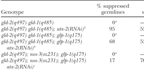
TABLE 2 to determine the extent of suppression byatx-2(RNAi)
becausegld-2 ; nos-3germlines have substantial meiotic Tests for suppression of meiotic entry
entry on their own (data not shown). Therefore, we tested defects byatx-2(RNAi)
whetheratx-2(RNAi)could suppress thegld-2; nos-3; glp-1
meiotic entry defect. Indeed, we found that some gonad % suppressed
Genotype germlines n arms showed strong suppression by atx-2(RNAi)as evi-denced by the lack of proliferating cells (Table 2; Figure
gld-2(q497) gld-1(q485) 0a —
4D). We take this result as an indication that ATX-2
gld-2(q497) gld-1(q485); atx-2(RNAi)b 95 55
may not be a negative regulator of the third pathway,
gld-2(q497) gld-1(q485); glp-1(q175) 0a —
gld-2(q497) gld-1(q485); glp-1(q175) 68 53 and instead model 1 or 2 (Figure 5, A and B) would be
atx-2(RNAi)b
more likely to be correct. However, two caveats to this
gld-2(q497); nos-3(oz231); glp-1(q175) 0a —
conclusion are that nos-3(null) mutants do not
com-gld-2(q497); nos-3(oz231); glp-1(q175) 17 70 pletely eliminate the activity of the third pathway (
Han-atx-2(RNAi)c
senet al. 2004b) andatx-2(RNAi)may not fully eliminate Consistent results were obtained in independent RNAi ex- ATX-2 activity. Therefore, we cannot eliminate the pos-periments; 100% ofgld-2 gld-1andgld-2; nos-3double mutants sibility that ATX-2 represses the third meiotic entry have a meiotic entry defect (tumorous phenotype) that, sur- pathway.
prisingly, is partially enhanced by glp-1 null mutations,e.g.,
atx-2(RNAi) does not suppress glp-1(oz112g f): We
glp-1(q175) null (Hansen et al. 2004b). Although all three
wanted to investigate whether GLP-1 signaling might be genotypes cause a severe meiotic entry defect, they can be
ranked with respect to the proportion of meiotic nuclei, as fol- a positive regulator of ATX-2 activity or, alternatively, lows:gld-2; nos-3; glp-1⬎gld-2 gld-1⬎gld-2 gld-1; glp-1(Hansenet ATX-2 activity is completely independent of GLP-1. To al.2004b).n, number of gonad arms examined; % suppressed investigate this question, we asked whetheratx-2(RNAi) germlines, the percentage of germlines with a decrease in
can suppress a ligand-independent, constitutive glp-1
the proportion of proliferating nuclei (REC-8 positive, HIM-3
gain-of-function mutation. Theglp-1(oz112gf )allele en-negative), a corresponding increase in the proportion of
mei-otic nuclei (REC-8 negative, HIM-3 positive; seeHansenet al. codes a ligand-independent receptor that produces a 2004b), and the presence of pachytene nuclei, which are never germline tumor (Berryet al. 1997). In the most extreme observed in gld-2 gld-1, gld-2 gld-1; glp-1orgld-2; nos-3; glp-1 case, animals carrying twoglp-1(oz112)alleles plus a third
synthetic tumorous mutants.
wild-type allele [glp-1(oz112/oz112/⫹)], germ cells never
aDefined here as zero, on the basis of criteria inHansen
enter meiosis (Hansenet al. 2004b). If GLP-1 signaling
et al. (2004b), Table 1.
bExperiments were done by feeding at 25⬚. positively regulates ATX-2 activity, then we reasoned cExperiments were done by injection; animals were raised
that atx-2(RNAi) should at least partially suppress glp-1
at 20⬚. (oz112gf). Alternatively, if ATX-2 is regulated indepen-dently of GLP-1, thenatx-2(RNAi)may not suppressglp-1 (oz112gf).
partially offset the loss of GLD-1 and GLD-2 activity, and We tested whether atx-2(RNAi) can restore meiosis more meiotic entry would occur. Alternatively, ATX-2 to glp-1(oz112/oz112) ; glp-1(⫹) germlines. We found might repress a third meiotic pathway. If so, then deple- that glp-1(oz112/oz112/⫹) atx-2(RNAi) and glp-1(oz112/ tion of ATX-2 would increase the activity of this third oz112/⫹)animals have a similar phenotype, indicating pathway, in turn increasing the level of meiotic entry that atx-2(RNAi) does not suppress glp-1(gf) (data not
(Figure 5C). shown). This result suggests that ATX-2 activity is not
We investigated the relationship between ATX-2 and regulated by GLP-1 signaling. We verified that oz112 the hypothetical third pathway by asking whether atx-2 animals are sensitive to RNAi by treating them withncc-1 (RNAi) suppresses the meiotic entry defect/tumorous dsRNA. Loss ofncc-1function prevents cell division in all phenotype of gld-2; nos-3andgld-2; nos-3; glp-1animals tissues (Boxem et al. 1999). We found that glp-1(oz112) (Hansen et al. 2004b). Genetic data suggest that NOS-3 animals responded properly toncc-1(RNAi), indicating that promotes meiotic entry by regulating not only GLD-1 but they are sensitive to RNAi (data not shown). Our results also a second target, which presumably is a component suggest that ATX-2 may function independent of GLP-1 of the third pathway (Hansenet al.2004b). For example, signaling, a hypothesis consistent with other data pre-the meiotic entry defect is greater in gld-2 gld-1; nos-3 sented above. However, the caveat remains that atx-2
than in gld-2 gld-1 animals. We sought to determine (RNAi)may not fully deplete ATX-2. Thus, ifglp-1(oz112)
whether ATX-2 might repress this hypothetical third is suppressed only in the complete absence of ATX-2
pathway targeted by NOS-3. activity, then we may not see suppression under our
Hansenet al. (2004b) have shown that the different conditions (seediscussion).
synthetic tumorous genotypes are associated with differ- atx-2promotes the oocyte fate:In addition to its role ent amounts of meiotic entry, as follows:gld-2 ; nos-3⬎ in proliferation,atx-2functions in sex determination by
Figure4.—Extensive meiosis occurs ingld-2 gld-1; glp-1 atx-2(RNAi)andgld-2; glp-1 atx-2(RNAi); nos-3germlines. One arm of the adult hermaphrodite germline is shown in each panel. (A) The characteristicgld-2 gld-1; glp-1tumorous phenotype (Kadyk
andKimble1998;Hansenet al. 2004b). (B) Suppression of thegld-2 gld-1; glp-1tumorous phenotype byatx-2(RNAi). The mitotic
region is absent, consistent with the absence of GLP-1 signaling. Note that the extent of meiotic entry is higher here than in
gld-2 gld-1; atx-2(RNAi)animals (Figure 3D). (C) Another example of suppression of thegld-2 gld-1; glp-1tumor byatx-2(RNAi). (D) Suppression of thegld-2; glp-1; nos-3meiotic entry defect byatx-2(RNAi). Extensive meiosis is visible. The distal mitotic region is absent.
hood. Consequently, sperm and/or primary spermato- masculinization of the XX soma; therefore we conclude that ATX-2 functions specifically in germline sex deter-cytes eventually extend up around the loop region,
rather than being confined to the spermatheca (Figures mination (data not shown). Sex determination in the XX germline involves tissue-specific regulatory mecha-2B and 3B). These defects are similar to those produced
by mutations in previously identified genes that pro- nisms that repress the global feminizing mechanism during larval development and then allow the germline mote the oocyte fate, including themog(m
asculiniza-tionof thegermline) genes (GrahamandKimble1993; to become female at approximately the L4/adult molt (Schedl 1997). The first known step in
germline-spe-Graham et al. 1993). The continued production of
sperm during adulthood indicates a defect in the sperm- cific regulation is repression oftra-2expression by the FOG-2/GLD-1 complex (Clifford et al. 2000). In the oocyte switch rather than a defect in oogenesis (Graham
andKimble1993). Moreover, sperm counts indicated that absence of TRA-2 activity, the germline is male. To switch to oogenesis, the FOG-2/GLD-1 complex must these germlines contained more sperm [average 390⫾
66 (SE) at 24–48 hr postadult molt;n ⫽7 gonad arms] be inactivated (by a mechanism that is unclear at pres-ent). In mutants that lack fog-2 activity, tra-2activity is than theⵑ150 sperm produced by wild-type germlines.
Inatx-2RNAi feeding experiments, an average ofⵑ72% thought to be abnormally high in the XX larval line, which precludes the male fate and causes the germ-of germlines at 25⬚andⵑ44% of germlines at 20⬚were
Mog (Table 3). In atx-2 RNAi injection experiments, line to instead be female (Schedl and Kimble 1988;
Jan et al. 1999;Cliffordet al. 2000). ⵑ77% of germlines were Mog at 20⬚(Table 3).
Figure5.—Models for the reg-ulation of germline proliferation and meiotic entry. In each case, GLP-1 signaling is shown to re-press three meiotic entry path-ways. For simplicity, the targets of the third pathway are not shown. (A) Model 1. ATX-2 activity is re-pressed by GLD-1 and GLD-2. ATX-2 may positively regulate genes required for proliferation (Y) and/or negatively regulate genes required for meiosis (Z). (B) Model 2. ATX-2 acts in paral-lel with the GLD-1 and GLD-2 pathways to regulate common tar-gets. For example, ATX-2 upregu-lates Y, a negative target of GLD-1, and represses Z, a positive target of GLD-2. Other possibilities exist. (C) Model 3. ATX-2 represses the third meiotic entry pathway. Com-ponents of the third pathway are not known, therefore we simply indicate it as lying downstream of NOS-3. Since ATX-2 can suppressgld-2 gld-1double mutants, we have shown ATX-2 as interacting with the third pathway at a point downstream of NOS-3. See text.
mutations infog-2. We raisedfog-2(oz40)andfog-2(q71) way to promote proliferation and/or repress meiotic entry. On the basis of our data, GLP-1 signaling has null mutants in the presence ofatx-2dsRNA and found
that most of them produced sperm. Someatx-2(RNAi); another function beyond repression of GLD-1 and GLD-2 activity, suggesting that these two pathways are
fog-2(⫺) animals produced both sperm and oocytes
whereas others produced only sperm (Table 3). There- not the only targets of GLP-1 signaling and substantiat-ing the idea of a third meiotic entry pathway. In addition fore, depletion of ATX-2 suppressed the Fog-2
pheno-type. On the basis of this result, atx-2 may act down- to its role in proliferation,atx-2functions in sex determi-nation to promote the sperm-oocyte switch in XX ani-stream offog-2 to promote the female fate. In animals
that produce sperm and oocytes (rather than only mals. ATX-2 appears to act downstream of the XX germ-line masculinizing gene,fog-2, to either promote activity sperm), we suspect that the RNAi was less effective at
reducing ATX-2 protein levels. In fact, the efficacy of of a feminizing gene (e.g., tra-2) or limit activity of a masculinizing gene (e.g.,fem-3). The dual role of ATX-2 the RNAi was relatively weak in this particular set of
experiments (see Table 3 controls). in the proliferation/meiosis and male/female choices is consistent with the pattern observed for other regula-We next tested whether atx-2(RNAi) could suppress
feminizing mutations in genes that act downstream of tors in theC. elegansgermline. For example, NOS-3, FBF,
fog-2. We found thatatx-2(RNAi)could not suppress the GLD-1, and GLD-2 function in later aspects of
develop-tra-2(q122)gain-of-function allele (which is deleted for ment subsequent to meiotic entry (Franciset al. 1995a,b; one GLD-1 binding site in the 3⬘-UTR;Janet al. 1999) KadykandKimble1998;Kraemeret al. 1999).
at 25⬚(n ⫽164) or loss-of-function mutations infem-1 Mammalian ataxin-2 was first studied because it is (n⫽24). The simplest interpretation of these data is that associated with the human neurodegenerative disease,
atx-2acts upstream of tra-2 or as a positive regulator of spinocerebrellar ataxia (Imbertet al. 1996;Pulstet al.
tra-2. This hypothesis needs to be tested using an atx-2 1996; Sanpei et al. 1996). Neurodegeneration is
trig-(null)mutation (seediscussion). gered by polyglutamine expansions that lead to forma-tion of protein plaques. On the basis of molecular fea-tures of the ataxin-2 protein, as well as its ability to bind DISCUSSION
to RNA-binding proteins (Shibata et al. 2000; Kha-leghpouret al. 2001;Royet al. 2002), ataxin-2 has been TheC. elegans atx-2gene promotes germline
prolifera-hypothesized to function in RNA metabolism (
Satter-tion and the oocyte fate. We have examinedatx-2
func-field et al. 2002). We propose that C. elegans ATX-2 tion using RNAi to deplete the ATX-2 protein. We find
acts at the post-transcriptional level to regulate gene that atx-2 activity prevents premature meiotic entry.
expression for germline proliferation and female sex ATX-2 does not upregulate GLP-1 signaling activity, and
determination. A role for ATX-2 in theC. elegansnervous GLP-1 signaling cannot be the sole positive regulator
system is not known given that RNAi (by feeding and of ATX-2 activity. Therefore, atx-2may work
TABLE 3
atx-2promotes the oocyte fate
% sperm⫹
Genotype Temp (⬚) oocytes % Mog % Fog % othera n
atx-2(RNAi)feedingb 20 56 44 0 0 424
atx-2(RNAi)injection 20 23 77 0 0 75
atx-2(RNAi)feedingb 25 28 72 0 0 186
fog-2(q71oroz40) NA 0 0 100c 0 —
atx-2(RNAi)d 20 82 18 0 0 50
fog-2(oz40); atx-2(RNAi)d 20 56 6 25 12.5 16
fog-2(q71); atx-2(RNAi)d 20 62 38 0 0 16
fog-2mock RNAid 20 0 0 100 0 40
n, number of gonad arms examined; NA, not applicable.
aNo gametes were produced.
bData are the average of four or more independent feeding experiments; % Mog germlines ranged from
18 to 83% at 20⬚and 50 to 97% at 25⬚.
cSchedlandKimble(1988);Cliffordet al. (2000).
dWild type andfog-2were treated in parallel, with wild type serving as a control for efficacy of the RNAi in
this specific set of experiments. Mock RNAi was done by feeding HT115 cells containing the L4440 vector to
fog-2mutants to make sure that this treatmentper sedid not suppress the Fog phenotype.
ATX-2 identifies another regulatory mechanism that ulating expression of genes that are required for prolif-eration and/or repressing expression of genes required promotes germline proliferation:GLP-1 activity restricts
expression of the GLD-1 meiotic entry pathway and is for meiosis. If they regulate common targets, then pre-sumably GLD-1 and GLD-2 activity would override ATX-2 absolutely required for maintenance of germline
prolif-eration in an otherwise wild-type background (Austin activity to allow meiotic entry. Third, ATX-2 may nega-tively regulate a third meiotic entry pathway, as discussed andKimble 1987; Franciset al. 1995a; Hansen et al.
2004a,b). GLP-1 activity is also suspected to restrict activ- below (Figure 5C).
Analysis of ATX-2 suggests an additional function for ity of the GLD-2 pathway (KadykandKimble1998). We
suggest that ATX-2 may promote germline proliferation GLP-1 signaling:One striking observation is the signifi-cantly higher proportion of meiotic germ cells ingld-2
and/or prevent meiotic entry via a mechanism that is
partially or completely independent of GLP-1 signaling. gld-1; glp-1 atx-2(RNAi)germlines than ingld-2 gld-1; atx-2 (RNAi) germlines. On the basis of these results, GLP-1 For example, ATX-2 may function in parallel with GLP-1
signaling in the germline. ATX-2 clearly does not pro- signaling must have (at least) one other function in addition to repressing the GLD-1 and GLD-2 pathways. mote GLP-1 activity and, if GLP-1 signaling is a positive
regulator of ATX-2 activity, then it cannot be the sole One obvious possibility is that GLP-1 activity represses the proposed third meiotic entry pathway (Hansenet al. positive regulator. Moreover, the inability ofatx-2(RNAi)
to suppress glp-1(oz112) suggests that GLP-1 may have 2004b). Two previous lines of evidence suggested the exis-tence of a third pathway. First,gld-2 gld-1tumorous germ-no role in regulating ATX-2. We germ-note, however, that
atx-2(RNAi)may not fully eliminate ATX-2 activity. An lines have some meiotic germ nuclei whereasglp-1(oz112gf)
tumorous germlines do not; second, a weakglp-1(gf)allele
atx-2(null) mutation would allow us to know with
cer-tainty that the loss of ATX-2 function does or does not enhances the tumorous phenotype ofgld-2 gld-1animals [gld-2 gld-1; glp-1(ar202gf )animals show less meiotic en-suppressglp-1(gf ).
Suppression of the tumorousgld-2 gld-1meiotic entry try thangld-2 gld-1animals;Hansenet al. 2004b]. Our data provide independent evidence for the existence of defect byatx-2(RNAi)is consistent with several models.
First, ATX-2 activity may be directly repressed by the a third pathway insofar as they reveal a third function for GLP-1. By this logic, GLP-1 signaling represses the GLD-1 and/or GLD-2 pathways (Figure 5A). Second,
ATX-2 may act in parallel with GLD-1 and/or GLD-2 to third meiotic entry pathway in gld-2 gld-1; atx-2(RNAi)
animals, thereby allowing proliferation to occur at the regulate common target genes (Figure 5B). GLD-1 and
GLD-2 presumably upregulate expression of genes re- distal end. The third pathway is no longer repressed in
gld-2 gld-1; glp-1 atx-2(RNAi)animals and is free to pro-quired for meiotic entry and/or repress expression of
genes required for proliferation. The identities of these mote meiotic entry throughout the germline.
The relationship betweenatx-2andego-4:atx-2RNAi target genes are unknown, although evidence suggests
that the GLD-2 pathway also promotesgld-1translation strongly enhanced the proliferation defect associated withego-4(om30). This result is consistent with enhance-(Hansenet al. 2004a). ATX-2 would presumably work
indi-cating that all three genes promote germline prolifera- line and is consistent with ATX-2 protein being present tion. atx-2 and ego-4 are also both required for embry- throughout the germline (as has been observed for ogenesis (Qiaoet al. 1995;Gonczyet al. 2000;Kiehlet NOS-3;Kraemeret al. 1999), although other
explana-al. 2000;Kamathet al. 2003). Although phenotypic and tions are possible.
mapping data suggest that atx-2 andego-4 may be the Other functions ofatx-2:Previous studies have dem-same gene, we could not locate theom30lesion within onstrated an essential role for atx-2 in embryogenesis
atx-2. We considered that om30might lie in a distant (Gonczy et al. 2000; Kiehl et al. 2000;Kamath et al. regulatory site that influencesatx-2transcription. How- 2003). We likewise note substantial embryonic lethality ever, we did not find any reduction inatx-2 transcript associated withatx-2(RNAi) (data not shown). The re-levels inego-4(om30)animals. Our data are most consis- quirement for ATX-2 activity in the early embryo is likely tent with the hypothesis thatego-4andatx-2are distinct to be satisfied by maternal expression. The presence of genes that promote germline proliferation. atx-2mRNA in the proximal germline is consistent with
atx-2and sex determination:To promote the switch incorporation of atx-2mRNA and/or protein into oo-from spermatogenesis to oogenesis, ATX-2 might posi- cytes. Analysis of the role of ATX-2 in embryogenesis is tively regulate a feminizing gene, such astra-2, or repress likely to be a fruitful avenue of research.
a masculinizing gene, such as fem-3 (see reviews by We thank Tim Schedl, John Belote, and members of the Maine lab
Schedl1997;GoodwinandEllis2002;Stothardand for insightful discussions; Xingyu She for assistance with the figures
Pilgrim 2003). Regulation of bothtra-2andfem-3 has and construction of pEL80; Julie Ahringer for theatx-2L4440 con-struct; Yuji Kohara and colleagues foratx-2cDNA clones; Pavel
Pasier-been described in some detail (see Pouti et al. 2001;
bek and Joseph Loidl for anti-REC-8 antibodies; and Monique Zetka
Goodwin and Ellis2002). Alternatives are that ATX-2
for anti-HIM-3 antibodies. Some strains used in this study were
pro-may promote the activity of another feminizer,e.g.,tra-3
vided by theCaenorhabditis elegansGenetics Center, which is supported
or one of themog,nos, orfbfgenes, or repress the activity by the National Institutes of Health (NIH) Center for Research Re-of a masculinizer,e.g., one of thefemor downstreamfog sources. This study was supported by funding from the National Sci-ence Foundation and Syracuse University (to E.M.); work done by
genes (seeStothardandPilgrim2003). Indeed,fem-3
D.H. at Washington University was supported by NIH GM-63310 to
is under particularly tight regulation, being controlled
Tim Schedl.
at the level of RNA metabolism (by the MOG proteins), translation (by the NOS and FBF proteins), and at the level of protein function (by TRA-2; seeGoodwinand
Ellis2002). Anatx-2(null)mutation would allow us to LITERATURE CITED
determine true epistasis relationships betweenatx-2and Austin, J., andJ. Kimble, 1987 glp-1is required in the germ line these genes. In any event, ATX-2 appears to act in the for regulation of the decision between mitosis and meiosis inC.
elegans.Cell58:565–571.
same “direction” as NOS-3 to promote the oocyte fate
Berry, L. W., B. WestlundandT. Schedl, 1997 Germline tumor
while it acts in opposition to NOS-3 to promote
prolifer-formation caused by activation ofglp-1, a member of theNotch
ation. family of receptors. Development124:925–936.
Boxem, M., D. G. SrinivasanandS. van den Heuvel, 1999 The
Regulation ofatx-2activity in the germline:Our
obser-Caenorhabditis elegans gene ncc-1 encodes a cdc2-related kinase
vations raise the question of how atx-2activity is
regu-required for M phase in meiotic and mitotic cell divisions, but
lated. Ifatx-2 lies directly downstream of GLD-1 and/ not for S phase. Development126:2227–2239.
Christensen, S., V. Kodoyianni, M. Bosenberg, L. Friedmanand
or GLD-2 in the regulation hierarchy, thenatx-2activity
J. Kimble, 1996 lag-1, a gene required forlin-12andglp-1
signal-may be directly repressed by GLD-1 and/or GLD-2 in
ing inC. elegans, is homologous to human CBF1 andDrosophila the transition zone. Alternatively, ATX-2, GLD-1, and Su(H).Development122:1373–1383.
Clifford, R., M. H. Lee, S. Nayak, M. Ohmachi, F. Giorginiet al.,
GLD-2 may converge to regulate common targets. If so,
2000 FOG-2, a novel F-box containing protein, associates with
then GLD-1 and GLD-2 activity may simply override
the GLD-1 RNA binding protein and directs male sex
determina-ATX-2 to allow meiotic entry (see above). In this case, tion in theC. eleganshermaphrodite germline. Development127: ATX-2 activity per se may not decrease as cells enter 5265–5276.
Crittenden, S. L., E. R. Troemel, T. C. EvansandJ. Kimble, 1994
meiosis. Instead, regulation of target mRNAs by GLD-1/
GLP-1 is localized to the mitotic region of theC. elegansgerm
GLD-2 may supersede their regulation by ATX-2. Finally, line. Development190:2901–2911.
if ATX-2 represses a third meiotic pathway, then its Crittenden, S. L., D. D. Bernstein, J. L. Bachorik, B. E. Thompson, M. Gallegoset al., 2002 A conserved RNA-binding protein
target(s) would presumably be distinct from GLD-1 and
controls germline stem cells inCaenorhabditis elegans.Nature417:
GLD-2 targets (since this pathway would be active in 660–663.
gld-2 gld-1double mutants). Doyle, T. G., C. WenandI. Greenwald, 2000 SEL-8, a nuclear protein required for LIN-12 and GLP-1 signaling inCaenorhabditis Kohara and colleagues have described theatx-2mRNA
elegans.Proc. Natl. Acad. Sci. USA97:7877–7881.
expression pattern as part of their expressed sequence Eckmann,C. R., B.Kraemer, M.Wickens, and J.Kimble,2002 GLD-tag project. (Data are available at the Nematode Expres- 3, a bicaudal-C homolog that inhibits FBF to control germline
sex determination in C. elegans. Dev. Cell3:697–710.
sion Pattern Database, http://nematode.lab.nig.ac.jp.)
Epstein, H. F., andD. C. Shakes, 1995 Caenorhabiditis elegans: Biologi-They detectatx-2mRNA throughout the larval and adult
cal Analysis of an Organism(Methods in Cell Biology, Vol. 48).
germline. This broad distribution of atx-2 transcripts Academic Press, San Diego.
germ-al., 1998 Potent and specific genetic interference by double- interact to control the sperm-oocyte switch inCaenorhabditis eleg-ans.Curr. Biol.9:1009–1018.
stranded RNA inCaenorhabditis elegans.Nature391:806–811.
Lambie, E. J., and J. Kimble, 1991 Two homologous regulatory
Fitzgerald, K., and I. Greenwald, 1995 Interchangeability of
genes,lin-12andglp-1, have overlapping functions. Development
Caenorhabditis elegansDSL proteins and intrinsic signalling activity
112:231–240. of their extracellular domains in vivo. Development121:4275–
Lee, M.-H., andT. Schedl, 2001 Identification of in vivo mRNA 4282.
targets of GLD-1, a maxi-KH motif containing protein required
Francis, R., M. K. Barton, J. KimbleandT. Schedl, 1995a gld-1,
forC. elegansgerm cell development. Genes Dev.15:2408–2420. a tumor suppressor gene required for oocyte development in
Maeda, I., Y. Kohara, M. YamamotoandA. Sugimoto, 2001
Large-Caenorhabditis elegans.Genetics139:579–606.
scale analysis of gene function inCaenorhabditis elegansby
high-Francis, R., E. MaineandT. Schedl, 1995b Analysis of the multiple
throughput RNAi. Curr. Biol.11:171–176. roles ofgld-1in germline development: interactions with the sex
Maine, E. M., 2001 RNAi as a tool for understanding germline determination cascade and theglp-1signaling pathway. Genetics
development in Caenorhabditis elegans: uses and cautions. Dev.
139:607–630.
Biol.239:177–189.
Gonczy, P., C. Echeverri, K. Oegema, A. Coulson, S. J. Joneset
Mangus, D. A., N. AmraniandA. Jacobson, 1998 Pbp1p, a factor
al., 2000 Functional genomic analysis of cell division inC. elegans
interacting withSaccharomyces cerevisiaepoly(A)-binding protein, using RNAi of genes on chromosome III. Nature408:331–336.
regulates polyadenylation. Mol. Cell. Biol.18:7383–7396.
Goodwin, E. B., andR. E. Ellis, 2002 Turning clustering loops.
Marin, V. A., andT. C. Evans, 2003 Translational repression of a Curr. Biol.12:R111–R120.
C. elegansNotch mRNA by the STAR/KH domain protein GLD-1.
Graham, P. L., andJ. Kimble, 1993 Themog-1gene is required for
Development130:2623–2632. the switch from spermatogenesis to oogenesis inCaenorhabditis
Meyer, B. J., 2000 Sex in the worm: counting and compensating
elegans.Genetics133:919–931.
X-chromosome dose. Trends Genet.16:247–253.
Graham, P. L., T.Schedland J.Kimble,1993 Moremoggenes that
Mumm, J. S., andR. Kopan, 2000 Notch signaling: from the outside influence the switch from spermatogenesis to oogenesis in the
in. Dev. Biol.228:151–165. hermaphrodite germ line ofCaenorhabditis elegans. Dev. Genet.
Pasierbek, P., M. Jantsch, M. Melcher, A. Schleiffer, D. Schweizer
14:471–484.
et al., 2001 ACaenorhabditis eleganscohesion protein with
func-Hansen, D., L. Wilson-Berry, T. DangandT. Schedl, 2004a
Con-tions in meiotic chromosome pairing and disjunction. Genes Dev. trol of the proliferation versus meiotic development decision in
15:1349–1360.
C. elegans through regulation of GLD-1 protein accumulation.
Pepper, A. S., T. W. Lo, D. J. Killian, D. H. HallandE. J. A.
Development131:93–104.
Hubbard, 2003a The establishment of Caenorhabditis elegans
Hansen, D., E. J. A. HubbardandT. Schedl, 2004b Multi-pathway
germline pattern is controlled by overlapping proximal and distal control of the proliferation versus meiotic development decision
somatic gonad signals. Dev. Biol.259:336–350. in theC. elegansgermline. Dev. Biol.268:342–357.
Pepper, A. S., D. J. KillianandE. J. Hubbard, 2003b Genetic
Hendersen, S. T., D. Gao, E. LambieandJ. Kimble, 1994 lag-2may
analysis ofCaenorhabditis elegans glp-1mutants suggests receptor encode a signaling ligand for the GLP-1 and LIN-12 receptors
interaction or competition. Genetics163:115–132. ofC. elegans.Development120:2913–2924.
Petcherski, A. G., andJ. Kimble, 2000 LAG-3 is a putative
transcrip-Hodgkin, J., andS. Martinelli, 1999 1999 Genetic Map of
Caenorhab-tional activator in the C. elegansNotch pathway. Nature 405:
ditis elegans. Caenorhabditis elegans Genetics Center, St. Paul,
364–368. MN.
Pouti, A., P. Pugnale, M. Belfiore, A. C. SchlappiandS. Saudan,
Hubbard, E. J. A., andD. Greenstein, 2000 TheC. elegansgonad:
2001 RNA and sex determination inCaenorhabditis elegans. Re-a test tube for cell Re-and developmentRe-al biology. Dev. Dyn.218:
view. EMBO Rep.2:899–904. 2–22.
Pulst, S. M., A. Nechiporuk, T. Nechiporuk, S. Gispert, X. C. Imbert, G., F. Saudou, G. Yvert, D. Devys, Y. Trottieret al., 1996
Chen et al., 1996 Moderate expansion of a normally biallelic Cloning of the gene for spinocerebellar ataxia 2 reveals a locus
trinucleotide repeat in spinocerebellar ataxia type 2. Nat. Genet. with high sensitivity to expanded CAG/glutamine repeats. Nat.
14:269–276. Genet.14:285–291.
Qiao, L., J. L. Lissemore, P. Shu, A. Smardon, M. Gelberet al.,
Jan, E., C. K. Motzny, L. E. GravesandE. B. Goodwin, 1999 The
1995 Enhancers ofglp-1, a gene required for cell-signaling inC.
STAR protein, GLD-1, is a translational regulator of sexual
iden-elegans, define a set of genes required for germline development. tity inCaenorhabditis elegans.EMBO J.18:258–269. Genetics
141:551–569.
Jones, A. R., andT. Schedl, 1995 Mutations in gld-1, a female- Roy, G., G. D. Crescenzo, K. Khaleghpour, A. Kahvejian, M. specific tumor suppressor gene inCaenorhabditis elegans, affect a
O’Connor-McCourtet al., 2002 Paip1 interacts with poly(A) conserved domain also found in Src-associated protein, Sam68. binding protein through two independent binding motifs. Mol. Genes Dev.9:1491–1504. Cell. Biol.22:3769–3782.
Jones, A. R., R. FrancisandT. Schedl, 1996 GLD-1, a cytoplasmic Sanpei, K., H. Takano, S. Igarashi, T. Sato, M. Oyakeet al., 1996 protein essential for oocyte differentiation, shows stage and sex- Identification of the spinocerebellar ataxia type 2 gene using a specific expression duringC. elegansgermline development. Dev. direct identification of repeat expansion and cloning technique, Biol.180:165–183. DIRECT. Nat. Genet.14:277–284.
Kadyk, L., and J. Kimble, 1998 Genetic regulation of entry into Satterfield, T. F., S. M. Jackson and L. J. Pallanck, 2002 A meiosis inC. elegans.Development125:1803–1813. Drosophila homolog of the polyglutamine disease geneSCA2is
Kamath, R. S., A. G. Fraser, Y. Dong, G. Poulin, R. Durbinet al., a dosage-sensitive regulator of actin filament formation. Genetics 2003 Systematic functional analysis of theCaenorhabditis elegans 162:1687–1702.
genome using RNAi. Nature421:231–237. Schedl, T., 1997 Developmental genetics of the germ line, pp. 241–
Khaleghpour, K., A.Kahvejian, G.De Crescenzo, G.Roy, Y. V. 269 inC. elegans II, edited by D. L.Riddle, T.Blumenthal, B. J.
Svitkinet al., 2001 Dual interactions of the translational repres- Meyerand J. R.Priess. Cold Spring Harbor Laboratory Press, sor Paip2 with poly(A) binding protein. Mol. Cell. Biol.21:5200– Cold Spring Harbor, NY.
5213. Schedl, T., andJ. Kimble, 1988 fog-2, a germ-line-specific sex
deter-Kiehl, T. R., H. ShibataandS. M. Pulst, 2000 The ortholog of mination gene required for hermaphrodite spermatogenesis in human ataxin-2 is essential for early embryonic patterning inC. Caenorhabditis elegans.Genetics119:43–61.
elegans.J. Mol. Neurosci.15:231–241. Seydoux, G., andT. Schedl, 2001 The germline inC. elegans:
ori-Kimble, J., andJ. White, 1981 On the control of germ cell develop- gins, proliferation, and silencing. Int. Rev. Cytol.203:139–185. ment inCaenorhabditis elegans.Dev. Biol.81:208–219. Shibata, H., D. P. HuynhandS. M. Pulst, 2000 A novel protein
Kodoyianni, V., E. M. MaineandJ. Kimble, 1992 The molecular with RNA-binding motifs interacts with ataxin-2. Hum. Mol. basis of loss-of-function mutations in theglp-1gene ofC. elegans. Genet.9:1303–1313.
Mol. Biol. Cell3:1199–1213. Sijen, T., J. Fleenor, F. Simmer, K. L. Thijssen, S. Parrishet al.,
Kraemer, B., S. Crittenden, M. Gallegos, G. Moulder, R. 2001 On the role of RNA amplification in dsRNA-triggered gene silencing. Cell107:465–476.


![[eBook] Windows Assembly Language & Systems Programming, 2nd edition (R&D Books) pdf](data:image/gif;base64,R0lGODlhAQABAIAAAP///wAAACH5BAEAAAAALAAAAAABAAEAAAICRAEAOw==)