ABSTRACT
HANSEN, STEPHANIE LAURA. The Effect of Dietary Manganese on Growth,
Reproductive Performance, and Manganese Status of Beef Heifers. (Under the direction of Jerry W. Spears.)
Three trials were conducted to examine the effects of dietary manganese (Mn) on growth, reproductive performance, and Mn status of beef heifers. In Experiment 1, 80 Angus and Simmental heifers, approximately 10 mo of age, were supplemented with 0 (control), 10, 30, or 50 mg Mn/kg DM from MnSO4 in addition to a diet containing 15.8 mg Mn/kg DM for 196 d. Performance of heifers was not affected by treatment. Liver Mn concentration increased (P=0.04) as dietary Mn level increased. Serum cholesterol was greater (P=0.001), in Angus compared to Simmental heifers for the 196-d period, but was not affected by treatment. Reproductive performance was not affected by treatment. Numerically, the number of heifers cycling at 12 mo of age, heifers bred at first service, and pregnancy rate was higher in heifers supplemented with 50 mg Mn/kg DM, compared to control heifers.
Experiment 3 used 70 Angus and Angus-Simmental cross heifers, approximately 9 mo of age, to examine the effect of dietary Mn source and level on growth, reproductive performance, and Mn status of beef heifers. Diets consisted of a corn-silage diet (analyzed 36 mg Mn/kg DM) supplemented with 0, 30 mg Mn/kg DM from MnSO4 (SO4), or 30 mg Mn/kg DM from a chelated Mn source (Mn Bioplex). Performance and liver Mn
concentration of heifers was not affected by source or level of dietary Mn. Serum cholesterol was affected by a treatment by breed interaction (P = 0.06), with cholesterol in Angus heifers increasing over time in response to treatment. Whole blood Mn concentration was not affected by source or concentration of dietary Mn. Dietary Mn source and concentration did not affect the percent of heifers cycling. Pregnancy rate was affected by dietary treatment (P = 0.01). Fewer SO4 heifers were bred than control (P = 0.01) and Bioplex heifers (P = 0.04). Results suggest that a diet containing 15.8 mg Mn/kg DM is sufficient for growth of beef heifers. Supplementation of Mn to the control diet tended to improve reproductive
THE EFFECT OF DIETARY MANGANESE ON GROWTH, REPRODUCTIVE PERFORMANCE, AND MANGANESE STATUS OF BEEF HEIFERS.
By
STEPHANIE LAURA HANSEN
A thesis submitted to the Graduate Faculty of North Carolina State University
in partial fulfillment of the requirements for the Degree of
Master of Science
ANIMAL SCIENCE
Raleigh 2005
APPROVED BY:
_________________________________ _________________________________
ii DEDICATION
This thesis is dedicated to all those who have loved and supported me through my education, but most particularly to…
My parents, Dennis and Susan Hansen, for the sacrifices they have made to give my sister and I such a wonderful life, and for having the strength to let me chase my dreams, even if it meant leaving them behind, for a while anyway.
iii BIOGRAPHY
Stephanie Laura Hansen was born in Sioux City, Iowa on March 13, 1981, to Dennis and Susan Hansen. She was raised in Sergeant Bluff, Iowa with her younger sister, Stacie, and attended high school at Sergeant Bluff-Luton High School. In the fall of 1999 she began her journey in higher education at Iowa State University in Ames, Iowa. Stephanie graduated with Honor and Distinction in December of 2002, receiving a Bachelor of Science degree in Animal Science, as well as the Academic Achievement Award for the College of
iv ACKNOWLEDGEMENTS
The author would like to thank Dr. Jerry Spears, for his infinite patience, guidance, and wisdom through her time at North Carolina State University. She is immensely grateful for the opportunity to work with him. The author would also like to extend her appreciation to Dr. Scott Whisnant and Dr. Matthew Poore, for agreeing to serve on her advisory
committee. Their time and advice are greatly valued. Thanks also to Alltech for their assistance and financial support of the author during her second year at North Carolina State University.
Thanks to Karen “Missy” Murphy for her immeasurable help and instruction in the lab. Those of us in Dr. Spears’ lab are very spoiled to have such an intelligent technician and wonderful friend in our midst everyday. Without her help, the author would probably still be blowing things up in the fume hood on a daily basis.
It was an honor to work alongside the group of dedicated people at the Butner Beef Cattle Unit, and the author extends great appreciation for all of their help in animal care and sampling to Dean Askew, Greg Schaeffer, Barbara Matthews, Jay Woodlief, and Joey Dickerson.
v TABLE OF CONTENTS
Page
LIST OF TABLES... vii
LIST OF FIGURES ... viii
LITERATURE REVIEW...1
Introduction ...1
Function of manganese...2
Manganese and glycosyltransferases...2
Manganese and cholesterol synthesis...3
Manganese in superoxide dismutase………... ...4
Manganese transport mechanism... ...5
Interactions between manganese and other minerals…………...5
Absorption of manganese... ...8
Excretion of manganese………...9
Manganese distribution in the body...10
Manganese deficiency…...13
Manganese and reproduction in ruminants...15
Manganese toxicity………...17
Sources and bioavailability of manganese...19
Manganese requirements in ruminants………..19
Literature cited……….……..20
CHAPTER 1. GROWTH, REPRODUCTIVE PERFORMANCE, AND MANGANESE STATUS OF HEIFERS FED VARYING CONCENTRATIONS OF MANGANESE…... 30
Abstract...31
Introduction... ...32
Materials and Methods... ...33
Results... ...36
Discussion………... ...36
Implications………... 42
Literature cited…………...42
CHAPTER 2. EFFECTS OF DIETARY MANGANESE CONCENTRATION ON PERFORMANCE AND MANGANESE STATUS OF PREGNANT BEEF HEIFERS AND THEIR OFFSPRING………... 51
Abstract...52
vi
Materials and Methods... ...54
Results and Discussion... ...56
Implications………... 62
Literature cited…………...63
CHAPTER 3. EFFECT OF DIETARY MANGANESE SOURCE AND CONCENTRATION ON GROWTH, REPRODUCTIVE PERFORMANCE, AND MANGANESE STATUS OF HEIFERS………. 70
Abstract...71
Introduction...72
Materials and Methods... ...73
Results and Discussion... ...76
Literature cited…………... ...79
vii LIST OF TABLES
Page
Chapter 1
Table 1. Ingredient and calculated chemical composition of growing basal diet
fed to beef heifers...46 Table 2. Effect of supplemental manganese on heifer performance…... 47 Table 3. Effects of manganese supplementation on plasma and liver manganese
concentrations and serum cholesterol concentrations of growing beef heifers... ..48 Table 4. Effects of supplemental manganese on reproductive performance of
heifers... 49 Chapter 2
Table 1. Ingredient composition of growing and gestating basal diets fed to
heifers……… .66
Table 2. Effect of dietary manganese concentration on whole blood manganese and serum cholesterol concentrations of beef heifers and their offspring……… 67 Table 3. Effect of dietary manganese concentration fed to beef heifers on
performance of their offspring………... ....68 Chapter 3
Table 1. Ingredient and chemical composition of basal diet fed to heifers………... 82 Table 2. Effect of supplemental manganese concentration and source on heifer
performance……….……….….….83 Table 3. Effects of manganese supplementation level and source on liver
manganese concentrations of beef heifers………...……...84
Table 4. Effects of supplemental manganese source and concentration on
viii LIST OF FIGURES
Page Chapter 1
Figure 1. Effect of breed on serum cholesterol level in heifers...50 Chapter 2
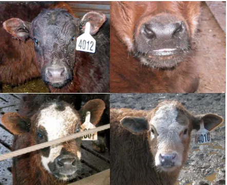
Figure 1. Calves born to heifers receiving a low manganese diet exhibiting varying degrees of superior brachygnathism...69 Chapter 3
Figure 1. Effect of treatment and breed on serum cholesterol levels of beef
1 Introduction
Manganese (Mn) is a brittle, luminous metal that is steel gray in color, with an atomic number of 25, and an atomic weight of 54.938 (McDowell, 1992). It was isolated in 1774 by Johann Gahn of Sweden (International Manganese Institute, 2005). The word manganese is of Latin origin, from the word magnes, meaning magnet. The name comes from the
magnetic properties of manganese oxide, although not all Mn is magnetic (Winter, 2003). Approximately 0.10% of the earth’s crust is composed of Mn, with concentrations in the soil averaging 500 to 600 mg Mn/kg DM. Of the heavy metals, only iron (Fe) is more abundant (McDowell, 1992). Manganese has 11 valence states, though +2 and +3 are the most important ones in biological systems (Leach and Harris, 1997).
Industrial uses for Mn increased when the organomanganese compound methylcyclo-pentadienyl manganese tricarbonyl (MMT) replaced lead as the antiknock agent in gasoline. Canada began using MMT in 1976, and by 1990 MMT had completely replaced lead in gasoline (ATSDR, 2000). Also used as an octane booster, MMT can improve oil
combustion, reducing soot levels and boiler clogging (International Manganese Institute, 2005). Typical industrial uses for Mn include the production of batteries, matches, fireworks, and glass-making. It’s most important contribution to industry is in the steel making process, where the addition of Mn adds to the resistance of steel to impact (ATSDR, 2000).
2 gastrocnemium muscle from the intercndyloid groove, and enlargement of the intertarsal joint. Perotic birds often gave birth to young that suffered from neonatal ataxia (Cotzias, 1958). The first Mn deficiency in ruminants was induced by Bentley and Phillips in 1951, and subsequent research has shown that Mn serves an important role in the reproductive performance and proper skeletal development of adult and young ruminants, respectively.
Function of Manganese
Manganese has been found to have several functions in the body, though it probably affects an animal most greatly through its involvement with enzymatic complexes.
Manganese acts as a cofactor in a number of enzymes, including pyruvate carboxylase, mitochondrial superoxide dismutase, and arginase, the final enzyme in the urea cycle (Leach and Harris, 1997). Several kinases, hydrolases, decarboxylases, and transferases are
activated by Mn in the body, although when Mn is lacking the element magnesium can often take its place. Manganese is also thought to be involved in cholesterol formation in the body, as well as insulin synthesis in the pancreas (Leach and Harris, 1997).
Manganese and glycosyltransferases
3 Manganese and cholesterol synthesis
Several studies have observed that a Mn deficiency decreases cholesterol levels in the liver (Curran and Azarnoff, 1961; Kawano et al., 1987; Davis et al., 1990; Jenkins and Kramer, 1991). Early work (Curran, 1954) looked at the effects of transition metals on synthesis of cholesterol in the liver of rats. Using chemically pure salts of vanadium (V), Fe, chromium (Cr), and Mn, the author injected 0.5 ml of dissolved salts in isotonic saline or 0.5 ml saline intraperitoneally in rats and sacrificed them one hour later. The researcher found that V and Fe decreased the incorporation of acetate into cholesterol by over 50 percent, while Cr and Mn increased synthesis of liver cholesterol by greater than 100 percent (Curran, 1954).
Manganese deficiency has been shown to reduce plasma cholesterol by decreasing the amount of cholesterol in the high density lipoprotein (HDL) fraction (Davis et al., 1990). Rats fed 0.4 mg Mn/kg DM had lower plasma cholesterol concentrations, specifically lower HDL cholesterol, than rates fed an adequate Mn diet (56 mg Mn/kg DM; Davis et al., 1990). Taylor et al. (1997) examined the effect of Mn deficiency in rats on two subclasses of HDL, HDL1 and HDL2. Using Sprague-Dawley rats and feeding diets containing 0.48 mg Mn/kg DM or 90 mg Mn/kg DM, they found that Mn deficiency caused significantly lower cholesterol and protein characteristics in HDL2 only, while no differences were seen in the HDL1 subclass. The results of this study led the authors to suggest that Mn has specific effects in HDL metabolism, and this may be how Mn exerts its effects on cholesterol synthesis (Taylor et al., 1997).
4 DM or 56 mg Mn/kg DM. This led the authors to speculate that a lack of Mn was affecting an enzyme further along the cholesterol biosynthesis pathway. They theorized that Mn may act as a cofactor for two enzymes in cholesterol synthesis, mevalonate kinase, and farnesyl pyrophosphate synthase (Davis et al., 1990). Both of these enzymes are involved in the
production of squalene, which is a precursor of cholesterol (Curran and Azarnoff, 1961). Manganese in Superoxide Dismutase
Manganese is a component of Mn-dependent superoxide dismutase (SOD), which catalyzes the reaction of O2- + O2- + 2H+ → H2O2 +O2 (Weisiger and Fridovich, 1973). Mitochondria consume more than 90% of the oxygen used by cells, and are thus more susceptible to oxygen toxicity than other cellular components (Weisiger and Fridovich, 1973). Manganese SOD is primarily found in mitochondria where it acts as an antioxidant, scavenging O2-, and in general protecting the cell from oxygen toxicity (Leach and Harris, 1997).
5 Manganese transport mechanism
Manganese is absorbed by the mucosa of the small intestine, transferred to the blood bound to albumin, taken up by the liver, and transported to other tissues via a transport protein, or excreted into the bile (Leach and Harris, 1997). For many years it was unclear as to what particular carrier was responsible for movement of Mn in the body. Among the proposed transporters were serum albumin, transferrin, and transmanganin (Davidsson et al., 1989). Davidsson et al. (1989) reported that transferrin was the major transport protein in plasma for Mn in rats when dosed either orally or intravenously with radiomanganese. Previous research had encountered difficulties distinguishing between transferrin and serum albumin, since they have very similar molecular weights. Davidsson et al. (1989) used a combination of several procedures to determine which of these two proteins the actual transporter of Mn in plasma was. Using fast protein liquid chromatography, SDS-polyacryl-amide gradient gel electrophoresis, and Western blot they observed that only transferrin appeared when analyzing the 54Mn-containing fractions. The researchers also observed that more 54Mn was bound to transferrin when 54Mn3+ was added to plasma in vitro than when 54
Mn2+ was added. This supports work (Aisen et al., 1969) showing that Mn bound to transferrin is exclusively in the trivalent state.
Interactions between manganese and other minerals
6 the similar configuration of their ionic complexes in water (Rossander-Hulten, 1991). Research in humans by Rossander-Hulten et al. (1991) examined the competitive inhibition of Fe absorption by Mn. Using healthy human subjects with variable Fe status and aged 19-50 years, the authors observed that the addition of 2 mg Mn to radiolabeled ferrous sulfate in either solution or as a hamburger meal caused a reduction of Fe absorption. Iron absorption was decreased by 21% and 34% with the addition of 7.5 or 15 mg Mn to 3 mg Fe in solution, respectively. Similarly, the addition of 15 mg Mn to 3 mg Fe in a hamburger meal caused a reduction of absorbed Fe of 40%. Rossander-Hulten et al. (1991) also found that the
absorption rates for 18 mg Fe in solution and 3 mg Fe plus 15 mg Mn in solution were 18% and 15.3%, respectively. The authors speculated that this similarity was due to a failure of the intestinal mucosa to tell the difference between Fe and Mn at the absorption site.
The interaction between Mn and Fe does not only affect Fe absorption, high Fe has been shown to have a depressive effect on Mn absorption (Davis et al., 1992). When male weanling Sprague-Dawley rats were fed 0.9, 48, or 188 mg Mn/kg diet, respectively, and a marginal or high Fe level (19 or 276 mg Fe/kg diet) Mn absorption was affected. Rats fed the diet low in Mn and Fe had much higher absorption than rats fed the other treatments, and increasing dietary Mn resulted in increasing endogenous Mn losses when Fe was marginal. However, when Fe was fed at the high level, endogenous Mn losses decreased across treatments and true absorption of Mn was significantly reduced (57.3% and 27.3%, true Mn absorption for low Mn/marginal Fe and low Mn/high Fe, respectively). Manganese
7 Calcium (Ca) and phosphorus (P) have also been shown to affect Mn absorption. Early work in poultry indicates that cases of perosis were due to high dietary Ca and P causing an increased demand for Mn by reducing the Mn bioavailability to the bird (Wilgus et al., 1936). Which mineral, Ca or P, is actually causing the reduction in Mn bioavailability is still the source of some debate. Some research has shown that high levels of both Ca and P are required to interfere with Mn absorption (Wilgus et al., 1936). More recent work by Wedekind and Baker (1990) examined the effects of varying levels of Ca and P on Mn bioavailability in male chicks. Feeding a corn-soybean meal based diet that contained 37 mg Mn/kg DM, and measuring Mn bioavailability as total tibia Mn regressed on supplemental Mn intake, Wedekind and Baker (1990) found that a greater accumulation of Mn in the tibia occurred in those treatments not receiving excess P. They also determined that feeding 0%, 0.4%, and 0.8% P to chicks resulted in a relative Mn bioavailability of 100%, 77.6%, and 62.4%, respectively, when pooled across levels of 0%, 0.5%, and 1% added Ca. A second study conducted by Wedekind and Baker (1990) found similar effects of high P on Mn when pooled across Ca levels of 0%, 0.25%, 0.5%, and 1%. With 0%, 0.22%, 0.44%, and 0.88% added P, the calculated relative Mn bioavailability was 100%, 83.6%, 78.2%, and 69.3%, respectively.
McDermott and Kies (1987) conducted a human study comparing different
8 measure bioavailability of Mn in the presence of each source of Ca. They observed that milk had the least impact on Mn bioavailability, with calcium carbonate having the most negative impact on availability of Mn. Since calcium carbonate also functions as an antacid its
presence would affect the pH of the gastrointestinal tract, thus interfering with the absorption of Mn, which is more soluble in the +2 state than the +4 state and would prefer a more acidic environment in the intestine for maximum absorptive capacity (Kies, 1994).
Absorption of Manganese
Absorption of Mn is uniformly low across species (Miller, 1973). In 1940, Greenberg and Campbell showed that orally administering Mn to rats resulted in
approximately 3-4% absorption of the dose. Work in swine by Gamble et al. (1971) showed that sows in late gestation absorbed 28% of an oral dose of 54Mn when consuming a diet containing 87.3 mg Mn/kg DM. More recently, growing pigs fed a typical diet of 42 mg Mn/kg DM and given an oral dose of 54Mn were found to have an apparent absorption of 1.7% and a true absorption of 0.5% (Finley et al., 1997).
In ruminants, the cow has an intestinal absorption rate of about 1% of dietary Mn, regardless of the level of Mn in the diet (Gibbons et al., 1976). Shortly after this report it was observed by Sansom et al. (1978) that cattle fed 1000 mg Mn/kg DM absorbed
approximately 0.5% of the Mn in the diet and that the excess was removed from the portal vein by the liver.
9 crates for 7 days, and received 0.4 mg Mn/kg DM or 14.51 mg Mn/kg DM, respectively, in their daily diet. The authors recorded nearly twice the level of Mn excreted in the feces and urine (76.7 vs. 43.2% in feces and 13.6 vs. 7.8% in urine) from the Mn-supplemented calf verses the non-supplemented calf, indicating that the Mn-supplemented calf absorbed considerably less 54Mn than the non-supplemented calf. With only one calf per treatment group, the results of the study by Howes and Dyer (1971) do not have any statistical weight, but their findings are useful. After sacrificing the two calves on their radiomanganese study, Howes and Dyer (1971) found the liver, kidney, and spleen, in that order, had the highest concentrations of radiomanganese in both the supplemented and unsupplemented calves. The authors also reported that the percent of 54Mn dose found in the liver of the
unsupplemented calf was 5.5 times higher than that of the supplemented calf; similarly, the activity of radiomanganese in bone was 23.5 times higher in the unsupplemented calf (Howes and Dyer, 1971). Their findings, collectively, suggest that Mn absorption increases as
dietary Mn decreases, and that Mn retention in tissue is higher under conditions of low Mn intake. Supporting work by Carter et al. (1974) found that young calves that received a Mn-unsupplemented milk diet had higher absorption and retention of Mn than calves
supplemented with 15 mg Mn/kg DM.
Excretion of Manganese
10 gallbladders, while Mn-supplemented cows had an average of 511 ml of bile. Several years later it was shown by Carter et al. (1974) that increasing dietary Mn by 30-fold from 0.5 to 15 mg Mn/kg DM caused a 30-fold increase in bile Mn content in calves. This same level caused an increase of two-fold in liver and gall bladder Mn concentrations, but no
measurable difference in other tissues (Carter et al., 1974).
Abrams et al. (1977) observed that young calves fed 1032 mg Mn/kg DM excreted 29.2% of a intravenous dose of 54Mn in bile compared to 2.3% excretion in bile from calves fed 32 mg Mn/kg DM. They also reported that this noticeably different excretion rate declined rapidly for the first 2 days and by the 6th day of measurement the excretion rate of 54
Mn in the bile was similar for both groups of calves (Abrams et al., 1977).
Britton and Cotzias (1966) suggested that variable excretion rather than variable absorption is the most important factor in Mn homeostasis. These findings have been
supported in subsequent research from numerous sources (Miller, 1973; Watson et al., 1973). Conversely, research conducted by Carter et al. (1974) suggests that both excretion and absorption are important factors in Mn homeostasis. They observed that the 54Mn retention rate of low Mn calves (0.5 mg Mn/kg DM) was 60.1% verses a 16.3% retention rate for calves supplemented with 15 mg Mn/kg DM when intravenously dosed with 54Mn. Similarly, absorption of orally dosed 54Mn was 9 times higher (18.2 vs. 2.2%) in non-supplemented calves than in Mn-non-supplemented calves (Carter et al., 1974).
Manganese distribution in the body
11 body. Bentley and Phillips (1951) observed no notable increase in liver Mn concentration in cows fed 30 mg Mn/kg DM verses 7-10 mg Mn/kg DM, with liver Mn concentrations of 10.2 and 9.8 mg Mn/kg DM, respectively. However, Rojas et al. (1965) observed a striking difference in liver storage of Mn in newborn calves. A calf born to a dam fed 15.8 mg
Mn/kg DM had 6.60 mg Mn/kg DM in its liver while a calf born to a dam fed 25.1 mg Mn/kg DM had 11.84 mg Mn/kg DM in its liver (Rojas et al., 1965). Again, these measurements were taken on just one animal per treatment, and a variety of factors could have caused the difference in Mn concentration in the calves’ livers. Other studies have found similar results, with liver Mn concentration increasing or decreasing depending on dietary Mn level (Ivan and Hidiroglou, 1980; Howes and Dyer, 1970).
Hidiroglou et al. (1978) found that six hours after an intravenous dose of 54Mn was given to nonpregnant ewes the highest concentration of the radiomanganese was in the pancreas, with the liver having the next highest concentration. Conversely, when 15
nonpregnant ewes fed the same diet were given an oral dose of 54Mn and sacrificed five days later, the highest concentrations of radiomanganese were found in the liver with slightly lower levels in the pancreas.
12 tissues for Mn revealed that the low-Mn diet did not reduce the Mn content of the liver, kidney, heart, pancreas or adrenal glands; however, they did find that the ovaries of the low-Mn cows were reduced by more than 50 percent compared to the low-Mn-supplemented cows (0.85 vs. 2.0 ng Mn/g DM; Bentley and Phillips, 1951).
Hidiroglou (1975) injected ewes intravenously with 54MnCl2 at a rate of 3.64 mCi per kilogram of bodyweight, at varying points in the estrous cycle. Ewes were slaughtered 6 hours post-injection and several tissues in the reproductive tract were analyzed for the
radiomanganese. It was observed that the corpus luteum and Graafian follicle appeared to be the main tissues for Mn uptake in the ovary. Hidiroglou (1975) postulated that these tissues may contain more Mn due to the high mitochondrial population of these tissues, as compared to the low-Mn containing corpus albicans, which is high in collagen content and low in mitochondrial content. Manganese uptake was higher in ewes dosed and killed on day 11 of the estrous cycle verses those dosed and killed on day 4 of the cycle (Hidiroglou, 1975).
Collectively, these studies (Bentley and Phillips, 1951; Hidiroglou, 1975) suggest that the ovary is the major tissue for Mn uptake in the ruminant reproductive tract. Although Bentley and Phillips (1951) found a significant increase in Mn content of the ovaries in their Mn-supplemented cows, they did not report what stage of the estrous cycle the cows were in. Hidiroglou (1975) indicated that a difference of only 7 days in the stage of estrous caused a two-fold increase in Mn content of ovarian tissues, so it is difficult to ascertain whether Bentley and Phillips results are truly a result of increased dietary Mn or that the
13 Manganese deficiency
There appears to be a relationship between the stage of development during which a Mn deficiency occurs and the severity of signs observed. A Mn deficient diet fed to a gestating female may not negatively affect the animal herself, but could have devastating effects on the growing fetus through impaired skeletal development (Leach and Harris, 1997). A lack of Mn in the diet of an animal can cause a variety of deficiency signs, including skeletal irregularities, reduced growth, atypical reproductive function, ataxia in newborns, and disruptions in lipid and carbohydrate metabolism (McDowell, 1992.)
Doisy (1972) documented a case of Mn deficiency induced in a man on a vitamin K study, when Mn sulfate was inadvertently left out of the diet. The man received 0.34 mg/Mn kg DM a day for 6.5 months, causing a Mn deficiency characterized by hypocholesterolemia, hair depigmentation, scaly dermatitis, and reduced vitamin K-dependent clotting proteins. Supplementation of vitamin K did not cause the symptoms to subside, but they gradually went away after the study was ended and the man returned to a normal diet (Doisy, 1972).
In swine, Mn deficiency can cause a disruption in proper lipid metabolism (Plumlee et al., 1956). Gilts fed a purified diet containing 0.5 or 40 mg Mn/kg had similar weight gain, but different back fat thicknesses. Gilts fed 40 mg Mn/kg DM had 3.61 cm of back fat compared to 4.11 cm of back fat on gilts fed 0.5 mg Mn/kg DM in a purified ration. Skeletal problems in the low-Mn group were also observed by Plumlee et al. (1956). Comparisons of radiograms taken of legs from gilts revealed differences among treatments in several bones. Gilts fed the low-Mn diet displayed shortening of the radius, ulna, tibia, fibula, and
14 Male chicks fed a basal diet containing 2.4 mg Mn/kg DM had reduced hexosamine content in their epiphyseal cartilage, indicating a reduction in mucopolysaccharides that contain this hexosamine (Leach and Muenster, 1962). Chicks supplemented with 10, 20 and 100 mg Mn/kg DM had a reduced incidence of enlarged hocks (93, 39, and 14%,
respectively) compared to 100% occurrence of enlarged hocks in non-supplemented chicks (Leach and Muenster, 1962).
Clinical signs of Mn deficiency in young cattle often manifest themselves in the malformation of bones. A broad range of signs may be observed when encountering a Mn deficiency, from no observable changes to severe crippling and bending of the long bones (Hidiroglou, 1980). Rojas et al. (1965) found that calves born to cows fed diets containing 15.8 mg Mn/kg DM suffered from enlarged joints, weakness, and twisted legs. They also observed that calves born to Mn deficient dams had 13.9% shorter humeri than calves born to cows fed the control diet containing 25.1 mg Mn/kg DM. Humeri from the deficient calves were also much weaker than humeri taken from control calves, breaking under only 605 kg of vertical pressure as compared to the 1,186 kg of pressure required to break the control humeri.
Howes and Dyer (1971) also found that feeding a low-Mn diet (13 mg Mn/kg DM) to heifers resulted in calves that were weak and had trouble standing while calves from heifers fed 21 mg Mn/kg DM were normal. Calves on this study did not exhibit the extreme
15 concentration in marrow-free bone when animals were fed different levels of dietary Mn has been seen in other calf as well as sheep studies (Rao, 1963; Watson, 1973).
In addition to controlled studies, Mn deficiency has been suspected in situations where calves born under field conditions displayed skeletal deformities (Ribble et al., 1989; Valero et al., 1990; Staley et al., 1994). The majority of these cases have been in Canada, where newborn calves exhibited a skeletal anomaly known as congenital joint laxity and dwarfism (CJLD). This disease is characterized by a generalized joint laxity, dwarfism, and on occasion, superior brachygnathism (Ribble et al., 1989). Observers noted that in many cases of CJLD births, pregnant cows had been wintered on grass or clover silage exclusively, and when cows were supplemented with rolled barley or hay they did not give birth to calves afflicted with CJLD (Ribble et al., 1989; Hidiroglou et al., 1990).
Hidiroglou et al. (1990) conducted an experiment to see if CJLD was associated with diet. Over the course of one winter they fed pregnant cows one of three feedstuffs (hay, red clover silage, or grass silage) and measured the frequency of CJLD in calves at birth for each group. While all three feedstuffs had similar levels of Mn (51, 64, and 63 mg Mn/kg DM for hay, red clover, and grass silage, respectively), 38% of calves born to red clover silage fed cows and 28% of the calves born to grass silage fed cows were born with CJLD. No occurrence of CJLD was observed in the group fed hay, and this finding led the authors to speculate that Mn was less available to cattle in silage than in hay (Hidiroglou et al., 1990).
Manganese and reproduction in ruminants
Mn-16 supplemented cows when compared to non-supplemented cows. Bentley and Phillips (1951) found that heifers fed 30 mg Mn/kg DM came into heat approximately 2 months earlier than heifers fed 7-10 mg Mn/kg DM.
Conversely, some authors have observed normal estrous cycles, but have reported some other aspect of reduced reproductive performance due to a deficiency of Mn, such as an increased number of services to conception (Rojas et al., 1965). DiCostanzo et al. (1986) fed beef cows and heifers a corn silage diet that supplied either 40 mg or 52 mg Mn/kg DM, and they found a reduction in the number of services to conception required for heifers and cows fed 52 compared to those fed 40 mg Mn/kg DM. An earlier study (Rojas et al., 1965) indicated that cows fed 15.8 mg Mn/kg DM required four services to conceive compared to only two for cows fed 25.1 mg Mn/kg DM. Bentley and Phillips (1951) also reported that heifers fed 7-10 mg Mn/kg DM on average required slightly more breeding services per conception than heifers fed 30 mg Mn/kg DM, but these data were not statistically significant (Bentley and Phillips, 1951).
In addition to conception and possibly abortion problems with females fed low Mn diets, those that do maintain their pregnancy often give birth to lighter weight offspring. Bentley and Phillips (1951) observed a slight trend for heavier calves born to cows fed 30 mg Mn/kg DM compared to cows fed 7-10 mg Mn/kg DM (42.23 kg verses 39.6 kg for a two year average). Kids born to goats fed 1.9 mg Mn/kg DM were reported to weigh 17% less than the kids born to goats fed 90 mg Mn/kg DM (Anke et al.,1973).
17 the ovaries being inferior in size. Manganese deficiency has also been suspected as a cause of cystic ovaries in cattle. Hidiroglou et al. (1977) found that dairy cows with cystic ovaries had lower Mn levels in the cortical stroma of the ovary than cows without cystic ovaries.
Placental transfer of Mn has not been extensively studied, but limited work has been done in rats, pigs, and sheep (Hidiroglou and Knipfel, 1981). Rao (1963) found that
radioactivity in the rat fetus increased over 16 hours after an intravenous injection of 52Mn to the dam. Newland and Davis (1961) observed that the percentage of 52Mn transferred to fetuses of sows fed 6 mg Mn/kg DM was higher than the percent of 52Mn transferred to fetuses of sows fed 100 mg Mn/kg DM. However, the amount of Mn transferred via the placenta was higher in 60, 80 and 110 day old fetuses of sows fed the high Mn diet, indicating that a higher percentage of absorbable Mn was transferred to the low Mn sows fetuses, but the actual amount of Mn was less (Newland and Davis, 1961). Manganese is rapidly transferred from the ewe to fetus (Hansard, 1972). Placental concentration of 54Mn at 12 hours post injection was more than 50% of the total fetal Mn concentration when pregnant ewes were administered with 54Mn. Placental concentration dropped to 25% of the total fetal 54
Mn content by 168 hours after the injection (Hansard, 1972). Few studies have measured Mn in fetal tissues, although Abderahman and Kincaid (1993) found that gestational age did not affect Mn levels in fetal liver and kidney, when fetuses were collected from heifers slaughtered at different stages of pregnancy.
Manganese Toxicity
18 (Dobson et al., 2004), is a disorder caused by excessive Mn in the brain, with the basal
ganglia of the brain containing the highest concentration of Mn. Among the signs of Mn toxicosis in humans are neurodegenerative disorders, which cause lesions and symptoms similar to Parkinson’s disease. Early signs of manganism are psychiatric in nature, and include violent behavior, hallucinations, erratic emotional outbursts, and often fatigue, headache, and insomnia (Dobson et al., 2004).
The dietary level at which Mn is toxic to cattle is suggested to be 1000 mg Mn/kg DM (NRC, 1980). Manganese toxicosis in animals is often attributed to its antagonistic affect on Fe metabolism (McDowell, 1992). Excessive dietary Mn in cattle resulted in reduced feed intake and decreased growth, while longterm grazing on pastures of 200 mg Mn/kg DM or more caused cows to have impaired reproductive efficiency. The authors suggested that this impairment may be due to antagonism with yet another mineral, suggesting Mn was interfering with iodine metabolism (McDowell, 1992).
19 Sources and bioavailability of Manganese
Manganese concentration in feedstuffs differs greatly due to a variety of factors, including soil pH, plant species and stage of maturity, and soil type. Most forages contain adequate amounts of Mn, although it may not always be available to the animal for
absorption (NRC, 2001). Manganese is found in varying levels in cereal grains, with corn often containing as little as 5 mg Mn/kg DM, and wheat and oats having as much as 40 mg Mn/kg DM. Some plant species, such as lupin seeds, have an extraordinarily high Mn content, and may contain as much as 3397 mg Mn/kg DM (Underwood, 1981). Manganese levels from plant protein sources are generally between 30 and 50 mg Mn/kg DM, while animal protein sources contain between 5 and 15 mg Mn/kg DM (NRC, 1996).
The term bioavailability was defined by Forbes and Erdman (1983) as the degree to which an ingested element is absorbed and can be utilized in metabolism by the animal. Supplemental Mn has traditionally been offered as either Mn oxide or Mn sulfate, with research showing that Mn oxide is between 53 and 70% as available as Mn sulfate (Henry et al., 1992). In more recent years, organic Mn sources like Mn-methionine have increasingly been used, with a reported bioavailability that is 120% that of Mn sulfate (Henry et al., 1992).
Manganese requirements in ruminants
20 substantially lower than those given for beef cattle. Dairy cattle recommendations are
calculated using the factorial approach. Recommendations are estimated in this manner by dividing the absorbed mineral requirements for Mn by the estimated absorption coefficient. The factorial approach is based on a series of calculations and assumptions, including the absorption coefficient for Mn in cattle and the specific Mn requirement by an animal.
Early work with limited experimental units indicated that feeding cows a diet containing 15.8 mg Mn/kg DM resulted in skeletal problems in calves and improper
reproductive performance in cows (Rojas et al., 1965). Dietary Mn recommendations from the dairy NRC (2001) are at or just slightly above this level at which Mn deficiencies have been observed. Beef cattle recommendations from the NRC (1996) are based on the limited research that has been conducted with Mn. More work is needed to determine the Mn requirement by beef cattle at varying stages of production. The current studies were conducted to further explore the Mn requirements of beef heifers.
Literature Cited
Abdelrahman, M. M., and R. L. Kincaid. 1993. Deposition of copper, manganese, zinc, and selenium in bovine fetal tissue at different stages of gestation. J. Dairy Sci. 76: 3588-3593.
21 Agency for Toxic Substances and Disease Registry (ATSDR). 2000. U.S. Department of
Health and Human Services, Public Health Service. Available:
http://www.atsdr.cdc.gov/toxprofiles/tp151-c4.pdf. Accessed March 15, 2005.
Aisen, P., R. Aasa, and A. G. Redfield. 1969. The chromium, manganese, and cobalt complexes of transferrin. J. Biol. Chem. 244: 4628-4633.
Anke, M., B. Groppel, and M. Grum. 1973. Manganmangel beim Wiederkauer 5. Mitteilung: Der Einflus des Manganmangels auf den Mengehn und Spurenelemengehalt
erwachsener Weiblicher und Mannlicher Ziegen. Arch. Tieremaehyr 23: 483.
Bentley, O. G., and P. H. Phillips. 1951. The effect of low manganese rations upon dairy cattle. J. Dairy Sci. 34: 396-403.
Bertinchamps, A. J., S. T. Miller, and G. C. Cotzias. 1966. Interdependence of routes excreting manganese. Am. J. Physiol.211: 217-224.
Britton, A. A., and G. C. Cotzias. 1966. Dependence of manganese turnover on intake. Am. J. Physiol.211: 203-206.
Carter, J. C., W. J. Miller, M. W. Neathery, R. P. Gentry, P. E. Stake, and D. M. Blackmon. 1974. Manganese metabolism with oral and intravenous 54Mn in young calves as influenced by supplemental manganese. J. Anim. Sci. 38: 1284-1290.
22 Curran, G. L. 1954. Effect of certain transition group elements on hepatic synthesis of
cholesterol in the rat. J. Biol. Chem. 765-770.
Curran, G. L., and D. L. Azarnoff. 1961. Effect of certain transition elements on cholesterol biosynthesis. Fed. Proc.20: 109-111.
Davidsson, L., B. Lonnerdal, B. Sandstrom, and C. Kunz. 1989. Identification of transferrin as the major plasma carrier protein for manganese introduced orally or intravenously or after in vitro addition in the rat. J. Nutr. 119: 1461-1464.
Davis, C. D., D. M. Ney, and J. L. Greger. 1990. Manganese, iron and lipid interactions in rats. J. Nutr. 120: 507-513.
Davis, C. D., T. L. Wolf, and J. L. Greger. 1992. Varying levels of manganese and iron affect absorption and gut endogenous losses of manganese by rats. J. Nutr. 122: 1300-1308.
DiCostanzo, A., J. C. Meiske, S. D. Plegge, D. L. Haggard, and K. M. Chaloner. 1986. Influence of manganese, copper and zinc on reproductive performance of beef cows. Nutr. Rep. Int. 34: 287-292.
23 Doisy, E. A., Jr. 1972. Effects of a deficiency of manganese upon plasma levels of clotting
proteins and cholesterol in man. In: W. G. Hoekstra, J. W. Suttie and M. E. Gantner (eds.) Trace Elements of Metabolism in Animals No. 2. p 668. University Park Press, Baltimore.
Finley, J. W., J. S. Caton, Z. Zhou, and K. L. Davison. 1997. A surgical model for determination of true absorption and biliary excretion of manganese in conscious swine fed commercial diets. J. Nutr. 127: 2334-2341.
Forbes R. M., Erdman J. W. Jr. 1983. Bioavailability of trace mineral elements. Annu. Rev. Nutr. 3:213-231.
Gamble, C. T., S. L. Hansard, B. R. Moss, D. J. Davis, and E. R. Lidvall. 1971. Manganese utilization and placental transfer in the gravid gilt. J. Anim. Sci. 32: 84-87.
Gibbons, R. A., S. N. Dixon, K. Hallis, A. M. Russell, B. F. Sansom, and H. W. Symonds. 1976. Manganese metabolism in cows and goats. Biochem Biophys Acta 444: 1-10.
Greenberg, D. M., and W. W. Campbell. 1940. Studies in mineral metabolism with the aid of radioactive isotopes. Proc. Nat. Acad. Sci. 26: 448-452.
24 Hawkins, G. E., Jr., G. H. Wise, G. Matrone, and R. K. Waugh. 1955. Manganese in the
nutrition of young dairy cattle fed different levels of calcium and phosphorus. J. Dairy Sci. 536-547.
Henry, P. R., C. B. Ammerman, and R. C. Littell. 1992. Relative bioavailability of manganese from a manganese-methionine complex and inorganic sources for ruminants. J. Dairy Sci. 75: 3473-3478.
Hidiroglou, M. 1975. 54Mn uptake by the ovaries and reproductive tract of cycling and anestrous ewes. Can. J. Phys. Pharm. 53: 969-972.
Hidiroglou, M. 1980. Zinc, copper and manganese deficiencies and the ruminant skeleton: a review. Can. J. Anim. Sci. 60: 579-590.
Hidiroglou, M., S. K. Ho, M. Ivan, and D. A. Shearer. 1978. Manganese status of pasturing ewes, of pregnant ewes and doe rabbits on low manganese diets and of dairy cows with cystic ovaries. Can. J. Comp. Med. 42: 100-107.
Hidiroglou, M., M. Ivan, M. K. Bryan, C. S. Ribble, E. D. Janzen, J. G. Proulx, and J. I. Elliot. 1990. Assessment of the role of manganese in congenital joint laxity and dwarfism in calves. Ann. Vet. Res. 21: 281-284.
Hidiroglou, M., M. Ivan, and S. K. Ho. 1977. Effect of human chorionic gonadotropin on the transport of manganese and zinc and tissue uptake of radioactivity following
25 Hidiroglou, M., and J. E. Knipfel. 1981. Maternal-fetal relationships of copper, manganese,
and sulfur in ruminants. A review. J. Dairy Sci. 64: 1637-1647.
Hill, C. H., and G. Matrone. 1970. Chemical parameters in the study of in vivo and in vitro interactions of transition elements. Fed. Proc.29: 1474.
Howes, A. D., and I. A. Dyer. 1971. Diet and supplemental mineral effects on manganese metabolism in newborn calves. J. Anim. Sci. 32: 141-145.
International Manganese Institute (IMnI). History of Manganese. 2005. Available: http://www.manganese.org/aboutmanganese.php. Accessed March 25, 2005.
Ivan, M., and M. Hidiroglou. 1980. Effect of dietary manganese on growth and manganese metabolism in sheep. J. Dairy Sci. 63: 385-390.
Jenkins, K. J., and J. K. G. Kramer. 1991. Effect of excess dietary manganese on lipid composition of calf blood plasma, heart, and liver. J. Dairy Sci. 74: 3944-3948.
Kawano, J., D. M. Ney, C. L. Keen, and B. O. Schneeman. 1987. Altered high density lipoprotein composition in manganese-deficient Sprague-Dawley and Wistar rats. J. Nutr. 117: 902-906.
Kemmerer, A. R., C. A. Elvehjem, and E. B. Hart. 1931. Studies on the relation of manganese to the nutrition of the mouse. J. Biol. Chem. 623-630.
26 Leach, R. M., Jr. 1971. Role of manganese in mucopolysaccharide metabolism. Fed. Proc.
30: 991-994.
Leach, R. M., Jr., and E. D. Harris. 1997. Manganese. In: B. L. O'Dell and R. A. Sunde (eds.) Handbook of Nutritionally Essential Mineral Elements. p 335-356. Marcel Dekker Inc., New York.
Leach, R. M. J., and A. Muenster. 1962. Studies on the role of manganese in bone formation. J. Nutr. 78: 51-56.
McDermott, S. D., and C. Kies. 1987. Manganese usage in humans as affected by use of calcium supplements. In: C. Kies (ed.) Nutritional Bioavailability of Manganese. p Chap. 14. American Chemical Society, Washington, D. C.
McDowell, L. R. 1992. Minerals in Animal and Human Nutrition. Academic Press, Inc, San Diego.
Miller, W. J. 1973. Dynamics of absorption rates, endogenous excretion, tissue turnover, and homeostatic control mechanisms of zinc, cadmium, manganese, and nickel in
ruminants. Fed. Proc.32: 1915-1920.
Newland, H. W., and G. K. Davis. 1961. Placental transfer of manganese in swine. Am. J. Physiol. 15-17.
27 National Research Council. 1985. Nutrient Requirements of Sheep. 6th ed. National
Academy Press, Washington, D. C.
National Research Council. 2001. Nutrient Requirements of Dairy Cattle. 7th ed. National Academy Press, Washington, D. C.
National Research Council. 1996. Nutrient Requirements of Beef Cattle. 7th ed. National Academy Press, Washington, D. C.
Ose, D. E., and I. Fridovich. 1976. Superoxide Dismutase. J. Biol. Chem. 251: 1217-1218.
Papavasiliou, P. S., S. T. Miller, and G. C. Cotzias. 1966. Role of liver in regulating distribution and excretion of manganese. Am. J. Physiol.211: 211-216.
Paynter, D. I. 1980. Changes in activity of the manganese superoxide dismutase enzyme in tissues of the rat with changes in dietary manganese. J. Nutr. 110: 437-447.
Plumlee, M. P., D. M. Thrasher, W. M. Beeson, F. N. Andrews, and H. E. Parker. 1956. The effects of a manganese deficiency upon the growth, development, and reproduction of swine. J. Anim. Sci. 15: 352.
Rao, R. R. 1963. Manganese deficiency and reproductive phenomena in beef cattle and rats. M.Sc. Thesis, Washington State University, Pullman, Wash.
28 Rojas, M. A., I. A. Dyer, and W. A. Cassatt. 1965. Manganese deficiency in the bovine. J.
Anim. Sci. 24: 664-667.
Rossander-Hulten, L., M. Brune, B. Sandstrom, B. Lonnerdal, and L. Hallberg. 1991. Competitive inhibition of iron absorption by manganese and zinc in humans. Am. J. Clin. Nutr. 54: 152-156.
Sansom, B. F., H. W. Symonds, and M. J. Vaag. 1978. The absorption of dietary manganese by dairy cows. Res. Vet. Sci. 24: 366-369.
Staley, G. P., J. J. Van Der Lugt, G. Axsel, and A. H. Loock. 1994. Congenital skeletal malformations in holstein calves associated with putative manganese deficiency. J. S. Afr. Vet. Assoc. 65: 73-78.
Taylor, P. N., H. H. Patterson, I. Wolinsky, and D. J. Klimis-Tavantzis. 1997. Manganese deficiency affects HDL1 and HDL2 composition in rats. Nutr. Res.17: 1155-1162.
Underwood, E. J. 1981. The Mineral Nutrition of Livestock, 2nd Ed. Slough, U.K: Commonwealth Agricultural Bureaux.
29 Watson, L. T., C. B. Ammerman, J. P. Feaster, and C. E. Roessler. 1973. Influence of
manganese intake of metabolism of manganese and other minerals in sheep. J. Anim. Sci. 36: 131-136.
Wedekind, K. J., D. H. Baker. 1990. Effect of varying calcium and phosphorus level on manganese utilization. Poultry Sci. 69:1156-1164.
Weisiger, R. A., and I. Fridovich. 1973. Superoxide Dismutase. J. Biol. Chem. 248: 3582-3592.
Wilgus, H. S., L. C. Norris, and G. F. Heuser. 1936. The role of certain inorganic elements in the cause and prevention of perosis. Science (Wash DC). 84: 252-253.
Wilson, J. G. 1966. Bovine functional infertility in Devon and Cornwall: response to manganese therapy. Vet. Rec. 79: 562-566.
Winter, M. 2003. History of Manganese. Available:http://www.webelements.com
30 CHAPTER 1
Growth, reproductive performance, and manganese status of heifers fed varying concentrations of manganese1,2
S. L. Hansen, J. W. Spears3, C. S. Whisnant, and K. E. Lloyd
Department of Animal Science, North Carolina State University, Raleigh, NC 27695-7621
1
Use of trade names in this publication does not imply endorsement by the North Carolina Agric. Research Serv. or criticism of similar products not mentioned.
2
Appreciation is extended to Greg Shaeffer, Heather Stahlhut, Leon Legleiter, Emily Baird, Joey Dickerson, Jay Woodlief, and Barbara Matthews for their assistance in sampling and animal care.
3
31 Abstract
32 appears to be adequate for growth of heifers, but supplemental Mn may improve reproductive performance.
Key Words: Manganese, heifers, growth, reproduction Introduction
33 Materials and Methods
General
Eighty Angus (n = 40) and Simmental (n = 40) heifers (248.6 kg initial BW), approximately 10 months of age, were used in this study. Experimental procedures were reviewed and approved by the North Carolina State University Animal Care and Use Committee. Prior to initiation of the study heifers were vaccinated with Titanium 5 (AgriLabs, St. Joseph, MO) and Vision 7 (Intervet, Millsboro, DE), and wormed with
Bovimec (Virbac, Fort Worth, Texas). Heifers were housed in a covered facility with slotted floor pens, and individually fed via electronic feeders (American Calan, Northwood, NH). Heifers were blocked by weight and randomly assigned within a pen to treatments. There were six pens of 12 heifers each and one pen housing 8 heifers.
Treatments consisted of 0 (control), 10, 30, and 50 mg of supplemental Mn/kg DM. Supplemental Mn was supplied from manganese sulfate. Ingredient composition and
34 were considered to be cycling. Liver biopsy samples were obtained as described by Engle and Spears (2000) on d 98 and 196 for Mn determination.
At approximately 13 mo of age (d 90 and 104) two doses of Lutalyse were
administered to synchronize estrus in the heifers. Heifers were moved to a covered building with dry lots and were group fed by treatment for one week to avoid injuries among animals on the concrete slatted floors. Heifers that exhibited signs of estrus were bred artificially and heat detected for 30 d. Heifers that had not been bred by d 139 were given a dose of Lutalyse and bred on d 142 regardless of exhibition of estrus. Heifers bred early in the breeding season were ultra sounded and rectally palpated at 30 and 60 d post breeding for pregnancy determination. Rectal palpation was performed on all heifers on d 196 for final
determination of pregnancy. Analytical Procedures
Blood was collected in heparinized vacuum tubes designed for trace mineral analysis (Becton Dickenson, Rutherford, NJ), centrifuged at 1,200 x g for 20 min at 20°C and wet ashed prior to Mn analysis. The ashing procedure was a modification of procedures
described by Johnson et al. (1991). One mL of plasma was put in a 30 mL beaker and 6 mL of trace mineral grade nitric acid was added to the sample. The nitric was boiled off on a hotplate, and then 2 mL of 30% hydrogen peroxide was added to the beaker and again boiled off. After cooling, 1 mL of 5% nitric acid was used to reconstitute the dried sample.
Flameless atomic absorption spectroscopy (GFA-6500, Shimadzu Scientific Instruments, Kyoto, Japan) was used to determine Mn content in the plasma.
35 20°C and analyzed using a colorimetric end-point assay kit from ThermoDMA (Arlington, TX). Serum samples for progesterone were collected in vacuum tubes and centrifuged at 1,200 x g for 20 min at 20°C. Progesterone concentrations were determined using a radioimmunoassay kit (Coat-A-Count; Diagnostic Products Corp, Los Angeles, CA).
Feed and liver samples were prepared for Mn analysis by wet ashing using
microwave digestion (Mars 5™, CEM Corp., Matthews, NC) as described by Gengelbach et al. (1994). Manganese content of feed and liver samples was determined by flame atomic absorption spectroscopy (Shimadzu Scientific Instruments).
Statistics
36 Results
One heifer was humanely put down on d 68 after becoming severely ill from coccidiosis. Data from this heifer was not included in statistical analysis. Average daily gain, DMI, and gain:feed for the 196-d study did not differ among treatments (Table 2).
Plasma Mn concentration was affected by sampling time (P = 0.001), but not by treatment, treatment x sampling time or breed (Table 3). Liver Mn concentrations on d 98 and 196 were adjusted for initial liver Mn content. Liver Mn concentrations were greater (P = 0.04) in heifers supplemented with Mn than in controls (Table 3). Manganese
concentrations in liver were lower (P = 0.001) on d 196 than on d 98. However, liver Mn was not affected by breed or a sampling time x treatment interaction. Serum cholesterol concentrations were not affected by dietary Mn (Table 3). Angus heifers had greater (P = 0.006) serum cholesterol concentrations than Simmental heifers on all sampling dates (Figure 1). Cholesterol concentrations were also affected by sampling time (P = 0.001), with serum concentrations increasing (P = 0.001) between d 0 and 63 and between d 63 and 98 (Figure 1). Serum cholesterol values were similar on d 98, 168, and 196.
Numerically, the percent of heifers cycling at 12 mo of age, and pregnancy rate were greater for heifers supplemented with 30 or 50 mg Mn/kg DM compared with controls (Table 4). However, pregnancy rate, conception rate, services to conception, and percent of heifers cycling were not significantly affected by dietary Mn. Pregnancy rate and first service conception rate were greater (P = 0.01) in Simmental than in Angus heifers.
Discussion
37 been reported by Bentley and Phillips (1951), who observed similar gains in diary heifers fed diets containing 7-10 mg Mn/kg DM and those fed 30 mg Mn/kg DM during a 12 mo
growing period. Howes and Dyer (1970) also found that beef heifers receiving diets
containing 13 or 21 mg Mn/kg DM from 3 mo prior to gestation through early lactation had no differences in performance characteristics. Gain, feed intake and feed efficiency of gestating beef cows fed diets containing 15.8 mg Mn/kg DM did not differ from cows receiving 25.1 mg Mn/kg DM (Rojas et al., 1965). Similar results have also been found in sheep, where no significant difference among treatments was observed for average daily gain, feed intake, and feed to gain ratio for wethers fed diets containing 22, 300, or 3000 mg Mn/kg DM for 56 days (Ivan and Hidiroglou, 1980). The current NRC (1996)
recommendation for growing beef cattle is 20 mg Mn/kg DM; however, the previously mentioned research in conjunction with the present study would suggest that the Mn
requirement for maximum growth in heifers does not exceed the level (15.8 mg Mn/kg DM) found in our control diet.
Plasma Mn was not affected by supplemental Mn in the current study. This result is consistent with previous work in sheep (Masters et al., 1988) which demonstrated that plasma Mn concentrations are uniformly very low, and not affected by dietary Mn
concentration. Watson et al. (1973) observed that following an oral dose of 54Mn, plasma 54
38 low concentration of Mn in plasma, regardless of dietary Mn concentration, implies that the body has a very effective mechanism by which Mn homeostasis is maintained. Cattle have been shown to have an intestinal absorption rate of about 1% of dietary Mn, regardless of the level of Mn in the diet (Gibbons et al., 1976). Following absorption, Mn is bound to albumin for transport to the liver, the principal organ involved in Mn metabolism (Leach and Harris, 1997). At least 95% of the absorbed Mn is removed on the first pass through the liver and is excreted in bile (Gibbons et al, 1976).
Liver Mn concentrations in the present study were increased slightly by
supplementation of Mn to the control diet. Previous work in ruminants (Howes and Dyer, 1970; Ivan and Hidiroglou, 1980) has shown that liver Mn concentration increases as dietary Mn increases. Similar results were found by Hidiroglou et al. (1978), who observed that sheep fed 5 mg Mn/kg DM had reduced liver Mn concentrations when compared to those fed 60 mg Mn/kg DM. Conversely, Bentley and Phillips (1951) found that feeding dairy cows a diet containing 7-10 or 30 mg Mn/kg DM for more than 3 years did not affect liver Mn concentration.
Unlike some minerals, Mn does not have an established level in liver or plasma below which an animal is classified as being in a deficient state. It is possible that initial body Mn stores were sufficient for heifers to draw on throughout the study, and thus prevent a greater response to Mn supplementation of the control diet. However, because no criteria have been established for the evaluation of Mn status in beef cattle, it is unclear if control heifers reached a state of marginal Mn deficiency. Whole blood Mn concentration has been
39 individual animal variability (Underwood and Suttle, 1999). Using young dairy calves fed a basal diet containing no more than 1.7 mg Mn/kg DM, Hawkins et al. (1955) observed an increase in whole blood Mn when the basal diet was supplemented with 50 mg Mn/kg DM. In sheep, similar work performed by Hidiroglou et al. (1978) found a significant increase in whole blood Mn concentration when a basal diet of 8 mg Mn/kg DM was supplemented with 60 mg Mn/kg DM. These studies would suggest that whole blood Mn concentration may be a suitable indicator of Mn status.
40 Mn concentrations were slightly lower in control heifers; however, it is unclear whether or not this reduction in liver Mn affected activity of Mn-dependent enzymes.
Serum cholesterol levels in the present study were not affected by dietary Mn concentration. It has been suggested that Mn may act as a cofactor for two enzymes in cholesterol synthesis, mevalonate kinase, and farnesyl pyrophosphate synthase (Davis et al., 1990). Both of these enzymes are involved in the production of squalene, which is a
precursor of cholesterol (Curran and Azarnoff, 1961). Several studies with rats have observed that a diet severely deficient in Mn results in significantly lower cholesterol levels in the plasma or liver (Curran and Azarnoff, 1961; Kawano et al., 1987; Davis et al., 1990). The lack of cholesterol response in the present 196-d study may be due to the fact that our control diet was not severely deficient in Mn. However, no differences in serum cholesterol were seen among lambs fed diets containing 0.8 or 29.9 mg Mn/kg DM for 16 wk (Lassiter and Morton, 1968).
Although not statistically significant, the number of heifers cycling at 12 mo of age and pregnancy rate was numerically higher for heifers supplemented with 30 or 50 mg Mn/kg DM compared with those fed the control diet containing 15.8 mg Mn/kg DM. This suggests that the control diet may have been marginally deficient in Mn for proper onset of estrus and conception. However, a greater number of animals will likely be needed to show a
significant reduction in cycling or conception rate in heifers fed a basal diet with a Mn concentration similar to that used in the present study.
41 mg Mn/kg DM expressed first estrus approximately 2 mo later than those fed a diet
containing 30 mg Mn/kg DM, and that heifers fed the low Mn diet required slightly more breeding services per conception than their supplemented counterparts, but these data were not significant. Rojas et al. (1965), in a study with limited animal numbers (n = 4), observed that beef cows fed a low Mn diet (15.8 mg Mn/kg DM) required four services to conceive compared to two services for cows fed 25.1 mg Mn/kg DM. Another study found that heifers and cows fed a corn-silage based diet containing 40 mg Mn/kg DM required an average of 1.6 services to conception compared to 1.1 services required by those fed a diet containing 52 mg Mn/kg DM (DiCostanzo et al., 1986).
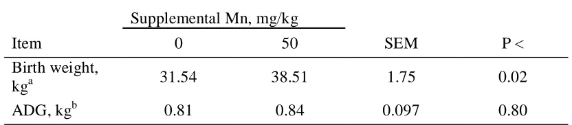
At the termination of the present study, 10 pregnant heifers from the control group and 10 pregnant heifers from the 50 mg supplemental Mn/kg DM group were selected to remain on treatments through gestation and early lactation. This study was designed to observe the effects of long term feeding of a low Mn diet on pregnant heifers and their
offspring. Calves born to control heifers were lighter at birth, and had lower whole blood Mn concentrations than those born to supplemented heifers (Hansen et al., 2005). Several calves born to control heifers exhibited signs linked to a deficiency of Mn, including dwarfism and superior brachygnathism. These results indicate that 15.8 mg Mn/kg DM is not sufficient for normal calf development in the gestating beef heifer.
42 concentrations in tissues, but has a number of important roles in biochemical systems in the body. The level of dietary Mn below which Mn stores and Mn-dependent processes in the body begin to be affected is unclear. Therefore, the establishment of criteria to evaluate the Mn status of cattle is necessary before current NRC (1996) recommendations for Mn in beef cattle can be further refined.
Implications
Manganese supplementation to a diet containing 15.8 mg Mn/kg DM did not affect performance characteristics. Numerically, the addition of 50 mg Mn/kg diet to the control diet increased the percent of heifers cycling and pregnancy rate of heifers, though results were not statistically significant. The results of this study indicate that a dietary Mn concentration of 15.8 mg Mn/kg DM is adequate for growth of beef heifers, but may be marginal for maximum fecundity. Further research is needed to determine the beef heifer requirement for Mn during her reproductive life cycle. Beneficial information could also come from investigation of the bioavailability of Mn in various beef cattle feedstuffs and forages.
Literature Cited
Bentley, O. G. and P. H. Phillips. 1951. The effect of low manganese rations upon dairy cattle. J. Dairy Sci. 34:396-403.
Curran, G. L., and D. L. Azarnoff. 1961. Effect of certain transition elements on cholesterol biosynthesis. Fed. Proc.20: 109-111.
43 DiCostanzo, A., J. C. Meiske, S. D. Plegge, D. L. Haggard, and K. M. Chaloner. 1986.
Influence of manganese, copper and zinc on reproductive performance of beef cows. Nutr. Rep. Int. 34: 287-292.
Engle, T. E, Spears, J. W. 2000. Effects of dietary copper concentration and source on performance and copper status of growing and finishing steers. J. Anim. Sci. 78:2446-2451.
Gengelbach, G. P., J. D. Ward, and J. W. Spears. 1994. Effect of dietary copper, iron, and molybdenum on growth and copper status of beef cows and calves. J. Anim. Sci. 72:2722-2727.
Gibbons, R. A., S. N. Dixon, K. Hallis, A. M. Russell, B. F. Sansom, and H. W. Symonds. 1976. Manganese metabolism in cows and goats. Biochem. Biophys. Acta. 444: 1-10.
Hansen, S. L., J. W. Spears, C. S. Whisnant, and K. E. Lloyd. 2005. Effects of dietary manganese concentration on performance and manganese status of pregnant beef heifers and their offspring. M. Sc. Thesis, North Carolina State University, Raleigh, NC.
Hidiroglou, M. 1975. 54Mn uptake by the ovaries and reproductive tract of cycling and anestrous ewes. Can. J. Phys. Pharm. 53: 969-972.
Hidiroglou, M., S. K. Ho, M. Ivan, and D. A. Shearer. 1978. Manganese status of pasturing ewes, of pregnant ewes and doe rabbits on low manganese diets and of dairy cows with cystic ovaries. Can. J. Comp. Med. 42: 100-107.
44 Ivan, M., and M. Hidiroglou. 1980. Effect of dietary manganese on growth and manganese
metabolism in sheep. J. Dairy Sci. 63: 385-390.
Kawano, J., D. M. Ney, C. L. Keen, and B. O. Schneeman. 1987. Altered high density lipoprotein composition in manganese-deficient Sprague-Dawley and Wistar rats. J. Nutr. 117: 902-906.
Lassiter, J. W., and J. D. Morton. 1968. Effects of a low manganese diet on certain ovine characteristics. J. Anim. Sci. 27:776-9.
Leach, R. M., Jr., and E. D. Harris. 1997. Manganese. In: Handbook of Nutritionally
Essential Mineral Elements. B. L. O’Dell and R. A. Sunde (eds.) p. 335-356. Marcel Dekker Inc., New York.
Masters, D. G., D. I. Paynter, J. Briegel, S. K. Baker, and D. B. Purser. 1988. Influence of manganese intake on body, wool and testicular growth of young rams and on the concentration of manganese and the activity of manganese enzymes in tissues. Aust. J. Agric. Res. 39: 517-524.
Mena, I. 1981. Manganese. In:Disorders of Mineral Metabolism. Bronner, F. & Coburn, J.
W., (eds.) p. 233-270, Academic Press, New York, NY.
NRC. 1996. Nutrient Requirements of Beef Cattle (7th Ed.). National Academy Press, Washington, DC.
45 Paynter, D. I. 1980. Changes in activity of the manganese superoxide dismutase enzyme in
tissues of the rat with changes in dietary manganese. J. Nutr. 110: 437-447.
Rojas, M. A., I. A. Dyer, and W. A. Cassatt. 1965. Manganese deficiency in the bovine. J. Anim. Sci. 24:664-667
Underwood, E. J, and N. F. Suttle. 1999. The Mineral Nutrition of Livestock. Commonwealth Agricultural Bureaux. Slough, England. Pp. 283-342.
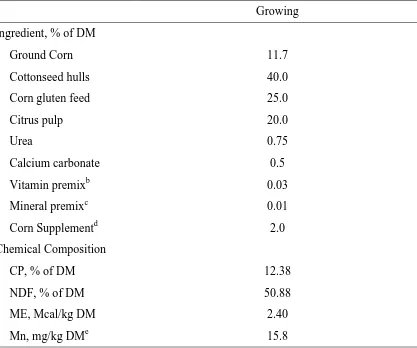
46 Table 1. Ingredient and calculated chemical composition of growing basal diet fed to beef heifersa
Growing Ingredient, % of DM
Ground Corn 11.7
Cottonseed hulls 40.0
Corn gluten feed 25.0
Citrus pulp 20.0
Urea 0.75
Calcium carbonate 0.5
Vitamin premixb 0.03
Mineral premixc 0.01
Corn Supplementd 2.0
Chemical Composition
CP, % of DM 12.38
NDF, % of DM 50.88
ME, Mcal/kg DM 2.40
Mn, mg/kg DMe 15.8
a
Diet contained Rumensin (Elanco Animal Health, Indianapolis, IN) at 33 mg/kg. b
Contained per kg of premix: 6,600,000 IU of vitamin A, 1,520,000 IU of vitamin D, and 6,600 IU of vitamin E.
c
Provided per kg of diet: 30 mg of Zn as ZnSO4; 10 mg of Cu as CuSO4; 0.5 mg of I as Ca(IO3)2(H2O); 0.2 mg of Se as Na2SeO3; and 0.1 mg of Co as CoCO3.
d
Provided supplemental Mn as MnSO4. e
47 Table 2. Effect of supplemental manganese on heifer performance
Supplemental Mn, mg/kg
Item 0 10 30 50 SEM
ADG, kg/d 0.98 0.97 0.98 1.01 0.02
DMI, kg/d 10.18 9.84 10.23 10.42 0.2
Gain:Feed 0.11 0.11 0.11 0.11 0.002
48 Table 3. Effects of manganese supplementation on plasma and liver manganese
concentrations and serum cholesterol concentrations of growing beef heifers
Supplemental Mn, mg/kg
0 10 30 50 SEM Significancea
Plasma Mnb 16.39 17.12 15.54 17.10 0.94 A**
d 0 9.85 13.55 9.83 13.23 1.58
d 98 19.71 20.15 20.47 17.90 1.65
d 168 19.61 17.67 16.31 20.15 1.63
Liver Mnc 9.08 9.43 9.69 10.25 0.29 A** B* C*
d 98 9.95 10.26 10.83 11.01 0.43 B† C*
d 196 8.19 8.57 8.69 9.40 0.32 B* C*
Serum Cholesterold 196.3 212.0 203.0 200.6 9.20 A**
†P < 0.10
*P < 0.05
**P < 0.01
a
A =sampling time effect; B =control vs. supplemental Mn; C =control vs. 30
and 50 mg supplemental Mn/kg DM.
b
Means across all sampling times expressed as ng/mL.
c
Means across all sampling times expressed as mg/kg DM.
d
49 Table 4. Effects of supplemental manganese on reproductive performance of heifers
Supplemental Mn, mg/kg
Item 0 10 30 50
Cyclinga, % 40 50 47 55
Response to Lutalyse doseb,% 40 40 47 50
Services to Conception 1.20 1.15 1.26 1.20
Conception Ratec,d, % 45 40 47 60
Pregnancy Rated, % 60 50 67 75
a
Based on serum progesterone of > 1.0 ng/mL.
b
Heifers that showed estrus in response to Lutalyse protocol.
c
At first service.
d
50
0
50
100
150
200
250
300
0
63
98
168
196
Day of study
C
h
o
le
s
te
ro
l, m
g
/d
L
Angus
Simmental
51 CHAPTER 2
Effects of dietary manganese concentration on performance and manganese status of pregnant beef heifers and their offspring1,2
S. L. Hansen, J. W. Spears3, C. S. Whisnant, and K. E. Lloyd
Department of Animal Science, North Carolina State University, Raleigh, NC 27695-7621
1
Use of trade names in this publication does not imply endorsement by the North Carolina Agric. Research Serv. or criticism of similar products not mentioned.
2
Appreciation is extended to Greg Shaeffer, Leon Legleiter, Heather Stahlhut, Joey Dickerson, Jay Woodlief, and Barbara Matthews for their assistance in sampling and animal care.
3