University of Windsor University of Windsor
Scholarship at UWindsor
Scholarship at UWindsor
Electronic Theses and Dissertations Theses, Dissertations, and Major Papers
10-19-2015
The Effects of Exercise Intensity and Relative Timing of Exercise
The Effects of Exercise Intensity and Relative Timing of Exercise
on Memory Performance
on Memory Performance
Alex Pennetti University of Windsor
Follow this and additional works at: https://scholar.uwindsor.ca/etd
Recommended Citation Recommended Citation
Pennetti, Alex, "The Effects of Exercise Intensity and Relative Timing of Exercise on Memory Performance" (2015). Electronic Theses and Dissertations. 5658.
https://scholar.uwindsor.ca/etd/5658
The Effects of Exercise Intensity and Relative Timing of Exercise on Memory
Performance
By
Alex Pennetti
A Thesis
Submitted to the Faculty of Graduate Studies through the Department of Kinesiology
in Partial Fulfillment of the Requirements for the Degree of Master of Human Kinetics
at the University of Windsor
Windsor, Ontario, Canada
2015
The Effects of Exercise Intensity and Relative Timing of Exercise on Memory
Performance
by
Alex Pennetti
APPROVED BY:
______________________________________________ Dr. Carlin Miller
Department of Psychology
______________________________________________ Dr. Kenji Kenno
Department of Kinesiology
______________________________________________ Dr. Kevin Milne, Advisor
Department of Kinesiology
AUTHOR’S DECLARATION OF ORIGINALITY
I hereby certify that I am the sole author of this thesis and that no part of this thesis
has been published or submitted for publication.
I certify that, to the best of my knowledge, my thesis does not infringe upon
anyone’s copyright nor violate any proprietary rights and that any ideas, techniques,
quotations, or any other material from the work of other people included in my thesis,
published or otherwise, are fully acknowledged in accordance with the standard
referencing practices. Furthermore, to the extent that I have included copyrighted material
that surpasses the bounds of fair dealing within the meaning of the Canada Copyright Act,
I certify that I have obtained a written permission from the copyright owner(s) to include
such material(s) in my thesis and have included copies of such copyright clearances to my
appendix.
I declare that this is a true copy of my thesis, including any final revisions, as
approved by my thesis committee and the Graduate Studies office, and that this thesis has
ABSTRACT
When external stimuli cause a physiological response associated with arousal
(increased adrenaline and cortisol), human memory is improved. Limited evidence
suggests that exercise, a potent physiological stress, can improve memory as well.
Consequently, this study aimed to further examine the exercise intensity-induced
enhancement in memory and the relative timing of stimulus presentation on this
phenomenon. 28 young adults were divided into 3 groups: viewing images before
exercise (TG1), viewing images immediately after exercise (TG2) and viewing images 30
minutes post exercise (TG3). Each participant completed either rest, low (40% of
VO2peak), moderate (60% of VO2peak), or high intensity (80% of VO2peak) cycle
ergometry on separate days as the exercise stress. Correctly recalled images 45min after
presentation were observed for memory. No significant differences were found between
exercise intensities or timing groups (p >0.05). However, further research is required to
DEDICATION
This thesis is dedicated to the greatest family this galaxy or any other has produced. First,
and foremost this thesis is dedicated to my parents, Mary Lou and Aristide Pennetti. This
thesis would not have been possible without their countless sacrifices over the course of
the past few decades. You have supported me in a variety of ways, and for that I am
eternally grateful. This dedication is a small token of my appreciation but it would be
impossible to return to you what you’ve provided me over the years. I’d also like to make
mention of the risk that all of 5 (yes, 5) of my grandparents took. All of my grandparents
made the epic journey migrating to Canada, fueled by hopes and dreams of a better life
they packed up and left the comforts of their home country of Italy, and arrived with next
to nothing but desire, courage and a massive reservoir of motivation. Unmistakably
without their massive undertakings I would not be where I am (and probably not alive
due to the fact my parents would never have met, but I digress). I also dedicate this thesis
to my sister Victoria, my aunts, uncles and cousins who have all helped me immensely
throughout my life in a variety of ways that would take up too many pieces of paper to
ACKNOWLEDGEMENTS
Firstly, I acknowledge my thesis committee, Dr. Carlin Miller, and Dr. Kenji
Kenno. Your time and effort to oversee this project is certainly much appreciated. You’ve
both made significant suggestions and contributions that have improved this project and I
again thank you helping me throughout this significant undertaking.
Secondly, I acknowledge and thank the lab volunteers who helped with this
project. I am grateful for all of your time, and energy you’ve given to ensure that the
project was run smoothly. My hope is that my passion for research has rubbed off on all
of you and that a drive for discovery is a part of your future endeavors. I wish all of you
the best in your academic and professional futures and I know that you will all be
successful no matter the path you choose.
Lastly and most importantly, I want to acknowledge and apologize to the Milnes.
Girls, I’m sorry for taking up so much of your dad’s time over the past couple of years
and adding to his collection of grey hair. I am leaving the nest now so when he is over
worked it’ll no longer be (entirely) my fault. Marcia, thanks for all of you’ve done as
well. You’ve always been so generous to me, and I am truly indebted to you and your
family. Finally, Kevin, I cannot do this acknowledgement justice without getting overly
mushy and sappy about it, so instead I’ll share this quote I saw one day on the internet
“Tell me and I forget, teach me and I may remember, involve me and I learn”. So, to the
TABLE OF CONTENTS
AUTHOR’S DECLARATION OF ORIGINALITY ... iii
ABSTRACT ... iv
DEDICATION ... v
ACKNOWLEDGEMENTS ... vi
List of Tables ... ix
List of Figures ...x
Abbreviations ... xi
Chapter 1: Literature Review ... 1
1.1 Memories connect our past with present ... 1
1.2 Adrenal stress hormones affect memory ... 7
1.3 Exercise is a physiological stressor ... 13
1.4 Acute and chronic exercise improves components of cognitive function ... 14
1.5 Effects of stress hormones during memory retrieval ... 15
1.6 Sex differences in memory consolidation ... 16
1.7 Sex differences in the stress response to exercise ... 16
1.8 The effects of the timing of stressor relative to information exposure and memory consolidation or retrieval ... 17
1.9 Aspects of the exercise and memory field requiring further investigation ... 18
1.10 References ... 19
Chapter 2: Manuscript ... 27
2.1 Introduction ... 27
2.2 Objectives ... 29
2.3 Hypotheses ... 29
2.4 Methods ... 30
Study Design ... 30
Recruitment ... 33
Procedures ... 35
Day 1/Baseline ... 35
Salivary Collection Procedure ... 35
Exercise/Image Days ... 42
Exercise Bouts ... 44
Salivary cortisol EIA ... 44
Recall scores ... 45
Statistical Analyses ... 46
2.5 Results ... 48
Responses to exercise... 48
Image Recall – Correct Images ... 54
Image Recall – Incorrect Images ... 57
Recall and Salivary Cortisol ... 57
Sleep, STAI, and Caloric Intake ... 60
2.6 Discussion ... 62
2.7 Further Research ... 65
2.8 Conclusion ... 66
Appendices ... 72
Appendix A: PAR-Q ... 72
Appendix B: RPE ... 73
Appendix C: Self Assessment Manikin ... 74
Appendix D: Image Day Questionnaire ... 75
Appendix E. STAI-Y1... 76
Appendix F: Ethics Approval ... 77
LIST OF TABLES
Table 2.1: Descriptive Statistics for Participants divided by Timing Group...34
Table 2.2: IAPS Standardized Emotional Ratings...38
Table 2.3: Mean correct image recalls by timing group and exercise intensity...55
Table 2.4: Mean correct image recalls by day...56
LIST OF FIGURES
Figure 1.1: General memory paradigm...5
Figure 1.2: Types of long-term memory...6
Figure 1.3: Modulation of memory consolidation by stress hormones...9
Figure 1.4: Inverted U pattern of recall based on salivary cortisol change in response to stressor...12
Figure 2.1: Study Design...32
Figure 2.2: Physiological Responses to Exercise Sessions...50
Figure 2.3: Salivary Cortisol Response to Exercise...51
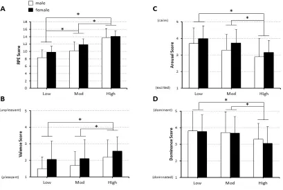
Figure 2.4: Subjective Ratings of Exercise Intensities...53
ABBREVIATIONS
CRH: Corticotropin Releasing Hormone
ACTH: Adrenocorticotropin Hormone
SAM: Self-Assessment Manikin
RPE: Rating of Perceived Exertion
PAR-Q: Physical Activity Readiness Questionnaire
IAPS: International Affective Picture System
STAI: State-Trait Anxiety Inventory
VO2: Volume of oxygen consumed
Chapter 1: Literature Review
1.1 Memories connect our past with present
How would you function without memories? You certainly wouldn’t be able to
comprehend this thesis if you couldn’t remember the previous words by the time you’ve
reached the end of this sentence. How could you create lasting bonds with family and
friends, without knowing whom you love or hate? Imagine the challenges of a single day
if you lost the capacity to store information? Our “selves” are nothing but a collection of
memories and our memories are what distinguish us from one another, memories connect
our past with our present and memories allow predictions of the future based on past
events. Within a memory, we can relive our most significant experiences, however joyful
or agonizing they may be. However, memory is still not fully understood. For example,
why do we “remember” some things and not others, especially those events that we
would rather “forget”? The difficulty in fully characterizing the memory paradigm is due
to its complexity. In fact, most theories on memory propose that there are many subtypes
and several steps in the formation, storage and retrieval of memories. This complexity is
apparent in the very first aspect of the presentation of new information. For example,
when presented with new information, there may be a very brief time where there is an
almost limitless amount of that new information that can be accessed (Cowan, 2001).
However, the ability to retrieve that information is gone in a very short period of time and
an individual becomes reliant on the capacities of short-term memory. A typical example
of short-term memory is noted when presented with a new phone number. The seven
auditory stimulus). Short term memory typically provides enough time to remember the
numbers long enough to write them down, but even within those few seconds, it is easy to
forget one or two numbers and they often must be repeated. Without rehearsal or some
other consolidating factor, the memory of the number may be lost in minutes and almost
certainly in the proceeding hours/days. Consequently, there are other factors responsible
for storing a memory in long-term memory. Moreover, it is often taught that in the theory
of short term memory, there is a general capacity limit of approximately seven
chunks/items of information, however, this limit may be too liberal or too conservative,
depending on environmental/personal factors (Cowan, 2001). Consequently, the ability to
shift information between initial presentation (or sensation), short-term memory and then
longer storage is still an actively studied area. In addition, the temporal distinctions
between sensory, short term and long term memory (discussed below) may very well be
not as mutually exclusive as the models would suggest. For example, a 45-minute delay
would certainly be considered long term memory, however, the mechanisms behind
sensory and short-term memory are crucial in the formation of long-term memory. Thus,
it is difficult to fully separate these memory subtypes in any investigation of memory.
Nonetheless, a well-accepted general proposal by Baddeley theorizes that when a
novel stimulus is presented, it is initially processed using the senses (e.g. sight, smell,
touch, etc.). However, attention must be given to any specific stimulus in order for
information associated with the stimulus to enter short-term memory for a brief period of
time (<30 seconds) (Baddeley, 2000; Baddeley and Hitch, 1974). However, the focus of
attention appears to be limited in capacity, thereby limiting the total amount of
Related to, but different, than short-term memory is the working memory or
temporary information storage that is used in cognitive tasks. Working memory has been
proposed to include executive processing of both auditory and visual information that is
needed for current tasks. The process of converting short-term memories into long-term
memories is termed memory consolidation or encoding, while the conversion of
long-term memories back to usable short-long-term memory or into working memory is long-termed
memory retrieval or recall (figure 1.1: Modified from (Baddeley, 2000 and Baddeley and
Hitch, 1974). Our current understanding of how humans store and retrieve memories is
still incomplete and a topic of great interest to many scientists.
Müller and Pilzecker (1900) first proposed the memory consolidation theory at
the turn of the 20th century. They observed that memories of newly learned information
could be interfered with by the introduction of additional information presented shortly
after the original information, suggesting that new memories are delicate and consolidate
into solidified long-term memories over time (Müller and Pilzecker,1900). This seminal
theory has guided the field of memory consolidation for over a century and continues to
guide research today.
Expanding upon the memory consolidation theory, researchers in the field of
memory and learning have tried to decipher the mechanisms of memory consolidation. In
the mid 1990’s, Cahill and McGaugh found that participants presented with an
emotionally arousing story were able to recall more details about the story than
participants presented with a similar yet emotionally neutral story (Cahill and McGaugh,
1995). Their findings preceded further evidence that via increases in glucocorticoid
human (Andreano and Cahill, 2006; Cahill and Alkire, 2003; van Ast et al., 2013; van
Stegeren, 2007), and animal (Roozendaal, 2002; Roozendaal, Okuda, et al., 2006)
memory.
It seems intuitive that emotionally stimulating memories are better remembered,
and this is supported through scientific evidence as well (McGaugh, 2003). We are better
able to remember the extraordinary, thrilling, or excruciating rather than the mundane,
dull, or monotonous. The most well supported hypothesis of why this is the case, is still
that of Müller and Pilzecker (Anderson, Wais, & Gabrieli, 2006; McGaugh, 2006), which
again suggests that new memories are delicate and consolidate into solidified long-term
memories over time, thus the memories that are optimally consolidated into long term
Figure 1.1: General memory paradigm
Figure 1.1: When a novel stimulus is presented it is initially processed using the senses
(e.g. sight, smell, touch, etc.). It then enters short-term memory for a brief period of time,
(<30 seconds). Often thought to be related, but different than short-term memory, is the
working memory or temporary information storage that is used in cognitive tasks.
Working memory has been proposed to include executive processing of both auditory and
visual information that is needed for current tasks. The process of converting short-term
memories into long-term memories is termed memory consolidation or encoding, while
the conversion of long-term memories back to short-term memory or into working
memory is termed memory retrieval or recall. Both consolidation and recall can be
modified by, for example, the physiological state of the individual or the use of different
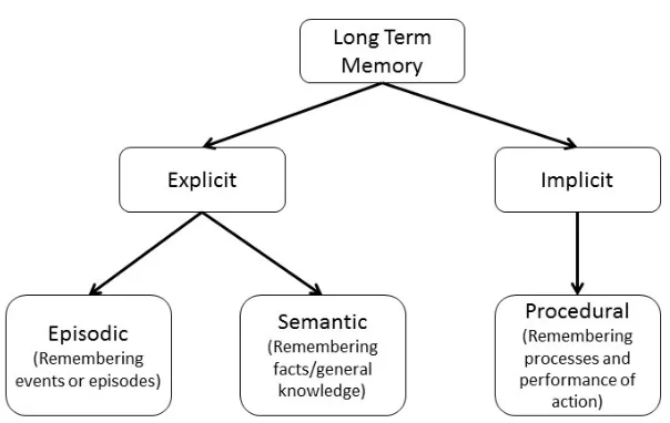
Figure 1.2: Types of long-term memory
Figure 1.2: Explicit long-term and implicit long-term memory differs in the degree that
conscious thought is required to access them. Implicit memories are typically accessed as
procedural memory (e.g. remembering how to walk). In contrast, explicit memories can
be further divided into semantic memory (e.g. remembering the 7 continents) and
episodic memory (e.g. remembering a significant life event such as the first day of a new
job, or a car accident). As such, the proposed study focused on episodic long-term
1.2 Adrenal stress hormones affect memory
The glucocorticoid, cortisol, and the catecholamines, epinephrine and
norepinephrine, are key biochemical regulators of the biological fight or flight response,
and consequently, they are significantly increased in the circulation during periods of real
and perceived stress (Miller et al., 2007). Cortisol is primarily synthesized and released
from steroidogenic cells in the zona fasciculata of the adrenal glands (Simpson and
Waterman, 1988). Typically initiated at the hypothalamus with the release of
corticotropin releasing hormone (CRH), the anterior pituitary responds to elevated CRH
by releasing adrenocorticotropin hormone (ACTH), which in turn stimulates the release
of cortisol from the adrenal gland (Dickerson and Kemeny, 2004). Cortisol circulates
both free and bound to corticosteroid binding globulin or albumin and has a half-life of
approximately one hour. Typically, cortisol signaling involving the glucocorticoid
receptor invokes changes in target cell gene expression through upregulated gene
transcription.
The catecholamines, specifically epinephrine and norepinephrine are secreted
from the sympathetic nervous system and the adrenal medulla. Typically, only secretion
from the adrenal medulla results in circulating concentrations of these hormones, but
spillover from adrenergic innervation of tissues may also occur during periods of high
activation (e.g. during stressful events). The half-life of circulating catecholamines is
much shorter than that of cortisol, typically a few minutes, and as such, their effects are
normally immediate and fast acting although the modulation of gene expression (i.e.
protein building) can occur resulting in slower effects. The catecholamines bind to
rate, increasing heart contractility and dilating bronchioles (Carrasco and Van de Kar,
2003)
Moreover, during an encountered stress, the catecholamines and cortisol act in
concert to increase blood glucose concentration, suppress immune function, induce
vasoconstriction, and increase heart rate, thus enhancing an animal’s chances of survival
(Dickerson and Kemeny, 2004). These homeostatic regulatory functions have no direct
impact on memory, however, when cortisol concentrations are high, cortisol can cross the
blood-brain-barrier and in conjunction with epinephrine and norepinephrine ultimately
influence crucial brain regions (McGaugh, 2004; McGaugh et al., 1996) involved in
memory consolidation such as the amygdala and hippocampus (Cahill and Alkire, 2003;
Kukolja et al., 2008; McMorris et al., 2011; McGaugh, 2004; McIntyre et al., 2003;
Revest et al., 2010; Roozendaal, Okuda et al., 2006; van Stegeren et al., 2007) (Figure
Figure 1.3: Modulation of memory consolidation by stress hormones
Figure 1.3: Modulation of memory consolidation by emotional arousal and the
subsequent release of adrenal stress hormones (cortisol and catecholamines). Emotional
arousal and stressful stimuli (e.g. exercise) activate the release of noradrenaline in the
basolateral amygdala and cause the adrenal gland to release stress hormones. In response
to the stress hormones, the amygdala increases noradrenergic signaling and modulates
From an evolutionary perspective, it is logical that animals that remember a
stressful environment or situation may be better able to cope with or avoid repeated
exposures to similar stressors in the future, thus making them more fit for survival. As
such, it is not surprising that an increase in the stress hormones, cortisol and the
catecholamines, would be a potential mechanism by which enhanced memory
consolidation would occur during periods of stress.
In fact, treatment with central adrenergic blockers in human (Cahill, Prins, Weber,
M., & McGaugh, 1994; van Stegeren, 1998; van Stegeren et al., 2007) and animal models
(Roozendaal Okuda et al., 2006) and glucocorticoid blockers in an animal model (Revest
et al., 2010) can impair memory performance. The evidence from these studies suggests
cortisol and the catecholamines have permissive and/or synergistic actions on memory
consolidation. For instance, van Stegeren et al. (2007) found that participants who
elicited an increase in endogenous cortisol levels in response to viewing images (ranging
from neutral to extremely negative) had increased amygdala activation when given a
placebo 90 minutes prior to image presentation, but no difference in amygdala activation
when given a beta-blocker 90 minutes prior to image presentation.
Moreover, animal (McGaugh, 2000; Roozendaal, 2000; Roozendaal, Hui, et al.,
2006; Roozendaal, Okuda et al., 2006) and human research (Buchanan and Lovallo,
2001; Cahill, Gorski, & Le, 2003; Felmingham et al., 2012; Kuhlmann and Wolf, 2006;
Lupien et al., 2005; van Stegeren et al., 1998) have demonstrated the beneficial effects of
increased circulating glucocorticoid and catecholamine concentrations on long-term
memory performance. The weight of the evidence demonstrates improvements in
demonstrated an inverted-U/quadratic dose response pattern of memory consolidation
(Andreano and Cahill, 2006; Lupien et al., 2005) suggesting that there is an optimal
concentration of cortisol and/or the catecholamines during new information exposure that
results in optimal memory consolidation, whereas with further increases in cortisol and/or
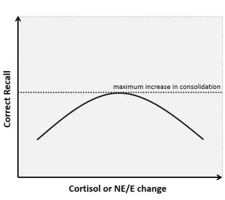
Figure 1.4. Inverted U pattern of recall based on salivary cortisol change in response to
stressor.
Figure 1.4. Inverted “U” relationship between cortisol or catecholamine change and
subsequent memory (number of items recalled correctly). (Adapted from Andreano and
At the present time, it is not currently known why this quadratic relationship may
exist. However, a potential reason may be due to high concentrations of cortisol
increasing the binding of low affinity glucocorticoid or other receptors, thus influencing
the consolidation processes by decreasing the amount of synaptic long-term potentiation
and increasing the amount of synaptic long-term depression (de Kloet, Oitzl, & Joëls,
1999). Long-term potentiation and long-term depression are the processes in which
neural synapses strengthen and weaken, respectively. Thus, the initial increases in
cortisol which lead to increased binding with high affinity receptors that, increase
long-term potentiation and decrease long-long-term depression could significantly increase the
likelihood that a given item or event is remembered sometime in the future while once
cortisol concentration is high enough to elicit significant low affinity receptor binding,
long-term depression is favoured (Malenka and Bear, 2004).
1.3 Exercise is a physiological stressor
Physical exercise elicits similar physiological changes as psychological stress.
During exercise, there is a need for increased blood flow to working muscle, increased
demand/release of metabolic fuels, increased blood acidity, increased temperature, and a
demand to clear potentially harmful metabolic by-products. These needs during exercise
pose challenges to several aspects of biological homeostasis (i.e. the body is under
stress). Exercise elicits significant increases in circulating cortisol (Fryer at el., 2014; Hill
et al., 2008; VanBruggen, Hackney, McMurray, & Ondrak, 2011) and catecholamine
(Weinberg et al., 2014) concentrations as global responses to many of those stressors.
above (i.e. increased cardiac output, glucose output, increased heart rate and blood
pressure) are clearly related to the need to engage in exercise (i.e. get away or fight for
survival, mating, food, etc.) during stress. Further, the exercise-induced increases in
stress hormones are positively correlated with exercise intensity (Crewther, Lowe,
Ingram, & Weatherby, 2010; Fryer et al., 2014; Hill et al., 2008; Hough et al., 2011;
Kindermann et al., 1982; VanBruggen et al., 2011; Winter et al., 2007). Consequently,
because of the aforementioned relationship between these hormones and memory, it has
been hypothesized that exercise can improve memory consolidation.
1.4 Acute and chronic exercise improves components of cognitive function
Animal (Berchtold, Castello, & Cotman, 2010; Speisman et al., 2013) and human
research (Labban and Etnier, 2011; Roig et al., 2013; Weinberg et al., 2014) provide
evidence that exercise may indeed impact memory consolidation, and this enhancement is
via the endogenous increase in circulating catecholamine and cortisol concentrations. For
example, Weinberg et al. (2014) found that in a randomized controlled trial, participants
in the “active” group who performed one leg knee extension/flexion exercise after
viewing images were better able to recall these images 48 hours later. Moreover,
improved overall physical fitness in children can enhance cognition, memory, and
academic achievement (Hillman et al., 2009; Hillman et al., 2014; Loprinzi and Kane,
2015; Pontifex et al., 2010), and physical activity interventions can improve a variety of
aspects related to cognition in older adults (Colcombe and Kramer, 2003; Smith et al.,
2010). While the exact mechanisms behind these improvements in cognitive function are
unknown, that they occur following exercise training suggests chronic neurological
acute bout of exercise or dependent on a chronic adaptation that requires repeated
exercise bouts has not been established and certain aspects of this phenomenon have yet
to be investigated. Potentially confounding these observations is that exercise is a broad
term defining many different frequencies, intensities, durations and types. In particular,
the investigation of different exercise intensities on memory consolidation in the same
study is rare.
1.5 Effects of stress hormones during memory retrieval
An item of interest within the stress-induced changes in memory is the potential
hindering effects of high concentrations of cortisol and catecholamines during memory
retrieval. Evidence suggests that high concentrations of these hormones can elicit poorer
performance when trying to recall stored memories (Buchanan and Tranel, 2008; de
Quervain et al., 2000; Domes et al., 2004; Domes, Rothfischer, Reichwald, & Hautzinger,
2005; Kuhlmann, Piel & Wolf, 2005; Roozendaal, 2002; Smeets et al., 2008; Smeets
2011). Moreover, the mechanism by which cortisol and the catecholamines improves
consolidation may be similar to the impairing effects on retrieval. For example, in a
study of 42 healthy volunteers, orally administered cortisone (exogenous cortisol)
impaired retrieval yet the impairment was attenuated by the concurrent administration of
propranolol (a beta adrenergic blocker that blocks the effects of catecholamines)(de
Quervain, Aerni, & Roozendaal, 2007).
Another potential similarity between the phenomena of cortisol and the
catecholamines improving consolidation and hindering retrieval is related to the dose
response action, as an inverted-U pattern may exist in the effects of retrieval hindrance as
cited studies of stress-induced impairment in memory retrieval used a form of verbal
memoryand all but one used a 24-hour delay between presentation and memory retrieval.
The present study examined episodic memory (which includes memories of events and
episodes and is a component of explicit long-term memory) using images as the material
to be remembered with a 45-minute delay between exposure and recall as a
representation of long-term memory performance.
1.6 Sex differences in memory consolidation
The examination of sex differences in the stress/memory response has produced
equivocal results. Evidence shows no difference between sexes (Weinberg et al., 2014),
some sex differences (Andreano and Cahill, 2006; Wolf, 2001) or in most cases, sex
differences are not explicitly investigated or reported. In the 2006 study by Andreano and
Cahill, it was found that a cold-pressor task (i.e. submersing the hand in ice water)
improved memory of a neutral story in men and not in women. It is possible that this sex
difference is specific to either the type of stressor, the type of memory or an interaction of
the two. Preliminary results from our lab, with a sample of 27 participants (14 male, 13
female) have shown no differences in memory (images recalled 1 week later) between
sexes using the same exercise intensities in this study (p= 0.525).
1.7 Sex differences in the stress response to exercise
As noted above, no sex differences in the stress response to exercise were evident
in the Weinberg et al., (2014) study. However, several studies cited above used all male
participants (Fryer at el., 2014; Hill et al., 2008; Van Bruggen et al, 2011). There is a gap
research study. In preliminary results from our lab, with a sample size of 27 (14 male, 13
female) no sex differences in the stress (cortisol) response to exercise were observed in
the response to the same exercise intensities that were used in the present study
(p=0.840). The gap in the literature stems from the exclusion of female participants and
the underreporting of sex as a variable.
1.8 The effects of the timing of stressor relative to information exposure and memory
consolidation or retrieval
It is not currently known how long the potential exercise-induced enhancement in
memory consolidation lasts or how exercise after the exposure of material modulates this
response. One human study used the relative timing of exercise compared to exposure of
memory material (paragraph recall) as a variable and it was found that exercise prior to
exposure improved memory in the intervention group versus a control group, but exercise
after exposure did not significantly differ from either the exercise before or the control
groups (Labban and Etnier, 2011). However, this study used only one exercise intensity
and did not use a time delay between the exercise and exposure in the pre-exposure
group. In another recent study, the relative timing effects of hydrocortisone
administration on long-term memory were assessed (van Ast et al., 2013). Participants
(64 men) given 10mg pills of hydrocortisone 210 minutes before encoding outperformed
participants that were administered 10mg pills of hydrocortisone 30 minutes before
encoding as well as those in the control group that were given placebo pills on a 24 hour
delay visual/verbal (words were presented with a corresponding picture in the
background) recall test. This may be due to slow acting effects of cortisol or could relate
had significantly lower salivary cortisol concentrations (van Ast et al., 2013).
Nonetheless, timing of stressor exposure is an important aspect of the modulation of
consolidation and retrieval processes and this requires further investigation.
1.9 Aspects of the exercise and memory field requiring further investigation
Given that the investigation of exercise-induced improvements on memory is a
relatively young field, many gaps exist in the literature. Specifically, few studies examine
more than one exercise intensity within the same cohort of participants, repeated
measures approaches are generally underutilized in memory studies due to challenges
related to learning effects, sex is an underreported variable, and the effects of the timing
of acute exercise relative to the exposure of the material to be remembered have not been
established. The present study attempted to address some aspects of the stress-induced
improvement in memory consolidation that have not yet been comprehensively
1.10 References
Anderson, A. K., Wais, P. E., & Gabrieli, J. D. E. (2006). Emotion enhances
remembrance of neutral events past. Proceedings of the National Academy of
Sciences of the United States of America, 103(5), 1599–1604.
Andreano, J. M., & Cahill, L. (2006). Glucocorticoid Release and Memory Consolidation
in Men and Women. Psychological Science, 17(6), 466–470.
Baddeley, A.D. (2000). The episodic buffer: a new component of working memory?
Trends in Cognitive Sciences, 4(11), 417-423.
Baddeley, A.D. and Hitch, G. (1974). Working Memory. Psychology of Learning and
Motivation, 8, 47-89.
Berchtold, N. C., Castello, N., & Cotman, C. W. (2010). Exercise and time-dependent
benefits to learning and memory. Neuroscience, 167(3), 588–597.
Buchanan, T. W., & Lovallo, W. R. (2001). Enhanced memory for emotional material
following stress-level cortisol treatment in humans. Psychoneuroendocrinology,
26(3), 307–317.
Buchanan, T. W., & Tranel, D. (2008). Stress and emotional memory retrieval: Effects of
sex and cortisol response. Neurobiology of Learning and Memory, 89(2), 134–141.
Cahill, L., & Alkire, M. T. (2003). Epinephrine enhancement of human memory
consolidation: Interaction with arousal at encoding. Neurobiology of Learning and
Memory, 79(2), 194–198.
Cahill, L., Gorski, L., & Le, K. (2003). Enhanced Human Memory Consolidation With
Post-Learning Stress: Interaction With the Degree of Arousal at Encoding. Learning
Cahill, L., & McGaugh, J. L. (1995). A Novel Demonstration of Enhanced Memory
Associated with Emotional Arousal. Consciousness and Cognition, 4(4), 410–421.
Cahill, L., Prins, B., Weber, M., & McGaugh, J. L. (1994). β-Adrenergic activation and
memory for emotional events. Nature, 371(6499), 702–704.
Carrasco, G. A., & Van de Kar, L. D. (2003). Neuroendocrine pharmacology of stress.
European Journal of Pharmacology, 463(1–3), 235–272.
Colcombe, S., & Kramer, A. F. (2003). Fitness Effects on the Cognitive Function of
Older Adults A Meta-Analytic Study. Psychological Science, 14(2), 125–130.
Cowan, N. (2001). The magical number 4 in short-term memory: A reconsideration of
mental storage capacity. Behavioral and Brain Sciences, 24(1), 87–114; discussion
114–85.
Crewther, B., Lowe, T., Ingram, J., & Weatherby, R. (2010). Validating the salivary
testosterone and cortisol concentration measures in response to short high-intensity
exercise. The Journal of Sports Medicine and Physical Fitness, 50(1), 85–92.
de Kloet, E. R., Oitzl, M. S., & Joëls, M. (1999). Stress and cognition: are corticosteroids
good or bad guys? Trends in Neurosciences, 22(10), 422–426.
de Quervain, D. J.-F., Roozendaal, B., Nitsch, R. M., McGaugh, J. L., & Hock, C. (2000).
Acute cortisone administration impairs retrieval of long-term declarative memory in
humans. Nature Neuroscience, 3(4), 313–314.
de Quervain, D. J.-F., Aerni, A, & Roozendaal, B. (2007). Preventive Effect of
β-Adrenoceptor Blockade on Glucocorticoid-Induced Memory Retrieval Deficits.
Dickerson, S. S., & Kemeny, M. E. (2004). Acute Stressors and Cortisol Responses: A
Theoretical Integration and Synthesis of Laboratory Research. Psychological
Bulletin, 130(3), 355–391.
Domes, G., Heinrichs, M., Rimmele, U., Reichwald, U., & Hautzinger, M. (2004). Acute
stress impairs recognition for positive words--association with stress-induced
cortisol secretion. Stress (Amsterdam, Netherlands), 7(3), 173–181.
Domes, G., Rothfischer, J., Reichwald, U., & Hautzinger, M. (2005). Inverted-U Function
Between Salivary Cortisol and Retrieval of Verbal Memory After Hydrocortisone
Treatment. Behavioral Neuroscience, 119(2), 512–517.
Felmingham, K. L., Tran, T. P., Fong, W. C., & Bryant, R. A. (2012). Sex differences in
emotional memory consolidation: The effect of stress-induced salivary
alpha-amylase and cortisol. Biological Psychology, 89(3), 539–544.
Fryer, S., Dickson, T., Hillier, S., Stoner, L., Scarrott, C., & Draper, N. (2014). A
Comparison of Capillary, Venous and Salivary Cortisol Sampling Following Intense
Exercise. International Journal of Sports Physiology and Performance.
Hill, E., Zack, E., Battaglini, C., Viru, M., Viru, V., & Hackney, A. (2008). Exercise and
circulating cortisol levels: the intensity threshold effect. Journal of Endocrinological
Investigation, 31(7), 587–591.
Hillman, C. H., Buck, S. M., Themanson, J. R., Pontifex, M. B., & Castelli, D. M. (2009).
Aerobic fitness and cognitive development: Event-related brain potential and task
performance indices of executive control in preadolescent children. Developmental
Hillman, C. H., Pontifex, M. B., Castelli, D. M., Khan, N. A., Raine, L. B., Scudder, M.
R., … Kamijo, K. (2014). Effects of the FITKids Randomized Controlled Trial on
Executive Control and Brain Function. Pediatrics, 134(4), e1063–e1071.
Hough, J. P., Papacosta, E., Wraith, E., & Gleeson, M. (2011). Plasma and Salivary
Steroid Hormone Responses of Men to High-Intensity Cycling and Resistance
Exercise: Journal of Strength and Conditioning Research, 25(1), 23–31.
Kindermann, W., Schnabel, A., Schmitt, W. M., Biro, G., Cassens, J., & Weber, F.
(1982). Catecholamines, growth hormone, cortisol, insulin, and sex hormones in
anaerobic and aerobic exercise. European Journal of Applied Physiology and
Occupational Physiology, 49(3), 389–399.
Kuhlmann, S., Piel, M., & Wolf, O. T. (2005). Impaired Memory Retrieval after
Psychosocial Stress in Healthy Young Men. The Journal of Neuroscience, 25(11),
2977–2982.
Kuhlmann, S., & Wolf, O. T. (2006). Arousal and cortisol interact in modulating memory
consolidation in healthy young men. Behavioral Neuroscience, 120(1), 217–223.
Kukolja, J., Schläpfer, T. E., Keysers, C., Klingmüller, D., Maier, W., Fink, G. R., &
Hurlemann, R. (2008). Modeling a Negative Response Bias in the Human Amygdala
by Noradrenergic–Glucocorticoid Interactions. The Journal of Neuroscience, 28(48),
12868–12876.
Labban, J. D., & Etnier, J. L. (2011). Effects of Acute Exercise on Long-Term Memory.
Lupien, S. J., Fiocco, A., Wan, N., Maheu, F., Lord, C., Schramek, T., & Tu, M. T.
(2005). Stress hormones and human memory function across the lifespan.
Psychoneuroendocrinology, 30(3), 225–242.
Malenka, R. C., & Bear, M. F. (2004). LTP and LTD: An Embarrassment of Riches.
Neuron, 44(1), 5–21.
McGaugh, J. L. (2000). Memory--a Century of Consolidation. Science, 287(5451), 248–
251.
McGaugh, J. L. (2003). Memory and Emotion: The Making of Lasting Memories.
Columbia University Press.
McGaugh, J. L. (2004). The Amygdala Modulates the Consolidation of Memories of
Emotionally Arousing Experiences. Annual Review of Neuroscience, 27(1), 1–28.
McGaugh, J. L. (2006). Make mild moments memorable: add a little arousal. Trends in
Cognitive Sciences, 10(8), 345–347.
McGaugh, J. L., Cahill, L., & Roozendaal, B. (1996). Involvement of the amygdala in
memory storage: Interaction with other brain systems. Proceedings of the National
Academy of Sciences, 93(24), 13508–13514.
McIntyre, C. K., Power, A. E., Roozendaal, B., & McGaugh, J. L. (2003). Role of the
Basolateral Amygdala in Memory Consolidation. Annals of the New York Academy
of Sciences, 985(1), 273–293.
McMorris, T., Sproule, J., Turner, A., & Hale, B. J. (2011). Acute, intermediate intensity
exercise, and speed and accuracy in working memory tasks: A meta-analytical
Miller, G. E., Chen, E., & Zhou, E. S. (2007). If it goes up, must it come down? Chronic
stress and the hypothalamic-pituitary-adrenocortical axis in humans. Psychological
Bulletin, 133(1), 25–45.
Müller, G., & Pilzecker, A. (1900). Experimental contributions to the theory of memory.
Z Angew Psychol, 1(1).
Pontifex, M. B., Raine, L. B., Johnson, C. R., Chaddock, L., Voss, M. W., Cohen, N. J.,
… Hillman, C. H. (2010). Cardiorespiratory Fitness and the Flexible Modulation of
Cognitive Control in Preadolescent Children. Journal of Cognitive Neuroscience,
23(6), 1332–1345.
Revest, J.-M., Kaouane, N., Mondin, M., Le Roux, A., Rougé-Pont, F., Vallée, M., …
Piazza, P. V. (2010). The enhancement of stress-related memory by glucocorticoids
depends on synapsin-Ia/Ib. Molecular Psychiatry, 15(12), 1140–1151.
Roig, M., Nordbrandt, S., Geertsen, S. S., & Nielsen, J. B. (2013). The effects of
cardiovascular exercise on human memory: A review with meta-analysis.
Neuroscience & Biobehavioral Reviews, 37(8), 1645–1666.
Roozendaal, B. (2000). Glucocorticoids and the regulation of memory consolidation.
Psychoneuroendocrinology, 25(3), 213–238.
Roozendaal, B. (2002). Stress and Memory: Opposing Effects of Glucocorticoids on
Memory Consolidation and Memory Retrieval. Neurobiology of Learning and
Memory, 78(3), 578–595.
Roozendaal, B., Hui, G. K., Hui, I. R., Berlau, D. J., McGaugh, J. L., & Weinberger, N.
corticosterone-induced enhancement of auditory fear conditioning. Neurobiology of Learning and
Memory, 86(3), 249–255.
Roozendaal, B., Okuda, S., Zee, E. A. V. der, & McGaugh, J. L. (2006). Glucocorticoid
enhancement of memory requires arousal-induced noradrenergic activation in the
basolateral amygdala. Proceedings of the National Academy of Sciences, 103(17),
6741–6746.
Simpson, E. R., & Waterman, M. R. (1988). Regulation of the Synthesis of Steroidogenic
Enzymes in Adrenal Cortical Cells by ACTH. Annual Review of Physiology, 50(1),
427–440.
Smeets, T. (2011). Acute stress impairs memory retrieval independent of time of day.
Psychoneuroendocrinology, 36(4), 495–501
Smeets, T., Otgaar, H., Candel, I., & Wolf, O. T. (2008). True or false? Memory is
differentially affected by stress-induced cortisol elevations and sympathetic activity
at consolidation and retrieval. Psychoneuroendocrinology, 33(10), 1378–1386.
Smith, P. J., Blumenthal, J. A., Hoffman, B. M., Cooper, H., Strauman, T. A.,
Welsh-Bohmer, K., … Sherwood, A. (2010). Aerobic Exercise and Neurocognitive
Performance: A Meta-Analytic Review of Randomized Controlled Trials.
Psychosomatic Medicine April 2010, 72(3), 239–252.
Speisman, R. B., Kumar, A., Rani, A., Foster, T. C., & Ormerod, B. K. (2013). Daily
exercise improves memory, stimulates hippocampal neurogenesis and modulates
immune and neuroimmune cytokines in aging rats. Brain, Behavior, and Immunity,
Van Ast, V. A., Cornelisse, S., Meeter, M., Joëls, M., & Kindt, M. (2013).
Time-Dependent Effects of Cortisol on the Contextualization of Emotional Memories.
Biological Psychiatry, 74(11), 809–816.
Van Stegeren, A. H., Everaerd, W., Cahill, L., McGaugh, J. L., & Gooren, L. J. G.
(1998). Memory for emotional events: differential effects of centrally versus
peripherally acting β-blocking agents. Psychopharmacology, 138(3-4), 305–310.
Van Stegeren, A. H., Wolf, O. T., Everaerd, W., Scheltens, P., Barkhof, F., & Rombouts,
S. A. R. B. (2007). Endogenous cortisol level interacts with noradrenergic activation
in the human amygdala. Neurobiology of Learning and Memory, 87(1), 57–66.
VanBruggen, M. D., Hackney, Anthony C., McMurray, R. G., & Ondrak, K. S. (2011).
The Relationship Between Serum and Salivary Cortisol Levels in Response to
Different Intensities of Exercise. International Journal of Sports Physiology &
Performance, 6(3), 396–407.
Weinberg, L., Hasni, A., Shinohara, M., & Duarte, A. (2014). A single bout of resistance
exercise can enhance episodic memory performance. Acta Psychologica, 153, 13–
19.
Winter, B., Breitenstein, C., Mooren, F. C., Voelker, K., Fobker, M., Lechtermann, A., …
Knecht, S. (2007). High impact running improves learning. Neurobiology of
Learning and Memory, 87(4), 597–609.
Wolf, O. T., Schommer, N. C., Hellhammer, D. H., McEwen, B. S., & Kirschbaum, C.
(2001). The relationship between stress induced cortisol levels and memory differs
Chapter 2: Manuscript
2.1 Introduction
Why do we remember the things we do, and forget the things that we forget? At
times we want this to be a conscious decision (e.g. remembering material for test, where
we left our keys, etc.), but often, it seems beyond our control (e.g. remembering the end
of a relationship). We don’t decide to remember or forget every piece of information the
moment that it is presented to us. Human memory is a complex multifaceted matter,
which is involved in virtually all of our daily tasks. Although human memory is not fully
understood there are several aspects that have been categorized.
The three major types of memory are sensory, short term and long-term memory.
The distinguishing factor between the three types is the length of time that a particular
memory is stored in each form. Sensory memory lasts for a very brief period of time,
generally not more than a few seconds. The brain processes senses such as sight, smell,
taste etc. and if enough significance/attention is given to a particular sensory memory it
can then be stored as short-term memory. Long-term memory includes the vast majority
of any individual’s memories, including names, places, facts, and events. Long-term
memories are created from short-term memories through a process called consolidation.
In order to be useful in the future, however, long-term memories must be converted back
into short-term memories through a process called memory retrieval or recall. Thus, both
the process of consolidation and recall are vital to any practical measurement of memory
(Baddeley, 2000; Baddeley and Hitch, 1974).
As humans, we remember the exciting, unique and thrilling as opposed to the
amount of research (Cahill and McGaugh, 1995; McGaugh, 2003; McGaugh, Cahill &
Roozendaal, 1996). Likely, life threatening events were more critical to survival, thus
remembering these arousing events improved an animal’s chances of survival and this
trait could be passed on to its progeny. Since it has been repeatedly shown that arousing
events and items are more likely remembered, it followed that researchers examined
whether external stressors could influence memory. To date, a variety of stressors have
been shown to improve memory in humans (Andreano and Cahill, 2006; Smeets et al.,
2008; Weinberg, Hasni, Shinohara, & Duarte, 2014) Moreover, the arousal-induced
improvement in memory has been linked to the adrenal stress hormones epinephrine,
norepinephrine and cortisol in human (Buchanan and Lovallo, 2001; Cahill, Prins,
Weber, M., & McGaugh, 1994; Cahill, L., Gorski, L., & Le, K. 2003; Felmingham et al.,
2012; Kuhlmann and Wolf, 2006; Lupien et al., 2005; van Stegeren, 1998; van Stegeren
et al., 2007) and animal models (McGaugh, 2000; Revest et al., 2010; Roozendaal, 2000;
Roozendaal, Hui, et al., 2006; Roozendaal, Okuda et al., 2006). The pattern of the
relationship between the stress hormones and memory performance has in some cases
been in the shape of an inverted U (Andreano and Cahill, 2006; Lupien et al., 2005;),
however, this is still not conclusive. Recently, researchers have tested exercise, one of the
most significant stressors on the human body, to examine its effects on memory.
Exercise elicits a variety of physiological responses including the release of the
previously mentioned adrenal stress hormones (Fryer at el., 2014; Hill et al., 2008;
VanBruggen, Hackney, McMurray, & Ondrak, 2011; Weinberg, Hasni, Shinohara, &
Duarte, 2014). Thus, it is logical to suggest that exercise and the concomitant release of
investigation of exercise as a method to enhance cognition in general is a relatively young
field, but findings suggest that exercise can enhance memory (Berchtold, Castello, &
Cotman, 2010; Labban and Etnier, 2011; Roig et al., 2013; Speisman et al., 2013;
Weinberg et al., 2014) and other cognitive factors (Loprinzi and Kane, 2015; Hillman et
al., 2009; Hillman et al., 2014; Pontifex et al., 2010). The current study aimed to utilize
this potential enhancing effect of exercise on memory.
In order to investigate further the effects of exercise on memory, exercise of
different intensities were used in a dose response manner between exercise and memory.
Multiple exercise intensities have not been typically examined in previous research.
Moreover, the timing of exercise relative to the presentation of new information was
manipulated in order to establish whether exercise prior to image presentation or exercise
after image presentation was more advantageous. Additionally, another timing group
involved a 30 minute delay between the completion of exercise and the presentation of
images to examine if the memory enhancing effects of exercise were transient or if they
could last long enough in order to have an appreciable effect.
2.2 Objectives
The objectives of this study were to a) further investigate the exercise-induced
enhancement in memory, and establish a dose response relationship with exercise
intensity, b) investigate if the relative timing of stimulus presentation affects this
enhancement, and c) attempt to expand the knowledge base of this phenomenon to more
applied scenarios.
2.3 Hypotheses
i) There would be a dose response effect with respect to exercise and memory
consolidation such that higher intensity exercise would result in enhanced
recall.
ii) Viewing images immediately after exercise (timing group 2; see below) would
result in the greatest recall.
iii) Enhanced recall would be positively correlated with salivary cortisol change
prior to image viewing.
2.4 Methods
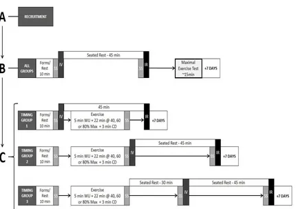
Study Design
This study utilized a repeated measures design (exercise intensity) with a between
group factor (timing of image presentation relative to exercise session; Figure 2.1). 28
young adults (10 males and 18 females) were randomly divided into one of three timing
of exercise groups consisting of: i) images presented immediately prior to exercise
(timing group 1); ii) images presented immediately after exercise (timing group 2); iii)
images presented 30 minutes after exercise (timing group 3).
Each participant underwent his or her timing group specific protocol on three of
the four image days. The first image day for each participant was a rest/baseline day. On
this control day they did not exercise and instead rested for 45 minutes between image
viewing and image recall. The order of exercise intensity for the three subsequent image
days was randomized for each participant. The three exercise intensities used in this study
were low (aimed to elicit 40% of peak oxygen consumption, VO2 peak), moderate (aimed
to elicit 60% VO2 peak) and high (aimed to elicit 80% VO2 peak). Image recall occurred
on all days at baseline, immediately prior to image recall, immediately prior to and post
exercise, and prior to image recall. Note that some saliva samples served as dual samples.
For example, the post exercise sample for those in timing group 2 was also the pre
image-viewing sample. On the rest day and for timing group 1 the baseline saliva sample also
served as the pre image-viewing sample. Additionally, for those in timing group 1, the
post exercise sample served as the pre image recall sample. For timing group 2, the post
exercise sample also served as the pre image-viewing sample. Thus, on exercise days for
timing group 1 and on the rest/control day, 2 samples were collected, on exercise days for
timing group 2, 3 samples were collected per day and 4 saliva collections were made on
exercise days for timing group 3. Thus a total of 8 salivary samples were collected from
those in timing group 1, 11 salivary samples from participants in timing group 2 and 14
samples from participants in timing group 3. Salivary samples were used to measure
cortisol due to the reduced invasiveness compared to blood collection and the potential of
blood or injection phobias that could have invoked a significant stress response in some
participants (Ritz, Meuret, & Ayala, 2010). In addition, salivary cortisol levels reliably
reflect serum cortisol concentrations (Dorn et al., 2007). In addition to saliva collection at
Figure 2.1. Study Design
Recruitment
Healthy (answer of “yes” to all questions on the physical activity readiness
questionnaire, see below), young adult (18-30 years of age) participants were recruited
from the University of Windsor using email, and by word of mouth. Using these
recruitment methods, 47 people expressed interest in participating in the study. After
initial contact, 32 individuals subsequently volunteered to participate in the study and
attended the Physical Activity and Cardiovascular Research (PACR) Lab in the Faculty
of Human Kinetics for baseline assessment. Three participants did not return to the lab
after the baseline assessment, and one participant did not return after completing both the
baseline assessment and one exercise day. Consequently, 28 individuals (18 females, 10
males; 20.07 ± 2.28 years of age) fully completed the study and were used in final
analyses. This resulted in an 87.5% retention and completion rate. Participant
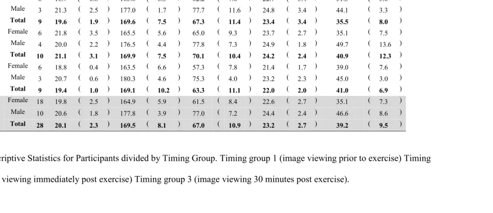
Table 2.1: Descriptive Statistics for Participants divided by Timing Group
Table 2.1: Descriptive Statistics for Participants divided by Timing Group. Timing group 1 (image viewing prior to exercise) Timing
group 2 (image viewing immediately post exercise) Timing group 3 (image viewing 30 minutes post exercise). Group Sex n
Age
(y) SD
Height
(cm) SD
Weight
(kg) SD BMI SD
VO2 Peak
(ml/kg/min) SD
Timing Group
1
Female 6 18.7 ( 0.8 ) 165.8 ( 6.3 ) 62.2 ( 7.6 ) 22.7 ( 3.4 ) 31.3 ( 5.6 )
Male 3 21.3 ( 2.5 ) 177.0 ( 1.7 ) 77.7 ( 11.6 ) 24.8 ( 3.4 ) 44.1 ( 3.3 )
Total 9 19.6 ( 1.9 ) 169.6 ( 7.5 ) 67.3 ( 11.4 ) 23.4 ( 3.4 ) 35.5 ( 8.0 )
Timing Group
2
Female 6 21.8 ( 3.5 ) 165.5 ( 5.6 ) 65.0 ( 9.3 ) 23.7 ( 2.7 ) 35.1 ( 7.5 )
Male 4 20.0 ( 2.2 ) 176.5 ( 4.4 ) 77.8 ( 7.3 ) 24.9 ( 1.8 ) 49.7 ( 13.6 )
Total 10 21.1 ( 3.1 ) 169.9 ( 7.5 ) 70.1 ( 10.4 ) 24.2 ( 2.4 ) 40.9 ( 12.3 )
Timing Group
3
Female 6 18.8 ( 0.4 ) 163.5 ( 6.6 ) 57.3 ( 7.8 ) 21.4 ( 1.7 ) 39.0 ( 7.6 )
Male 3 20.7 ( 0.6 ) 180.3 ( 4.6 ) 75.3 ( 4.0 ) 23.2 ( 2.3 ) 45.0 ( 3.0 )
Total 9 19.4 ( 1.0 ) 169.1 ( 10.2 ) 63.3 ( 11.1 ) 22.0 ( 2.0 ) 41.0 ( 6.9 )
Total
Female 18 19.8 ( 2.5 ) 164.9 ( 5.9 ) 61.5 ( 8.4 ) 22.6 ( 2.7 ) 35.1 ( 7.3 )
Male 10 20.6 ( 1.8 ) 177.8 ( 3.9 ) 77.0 ( 7.2 ) 24.4 ( 2.4 ) 46.6 ( 8.6 )
Total 28 20.1 ( 2.3 ) 169.5 ( 8.1 ) 67.0 ( 10.9 ) 23.2 ( 2.7 ) 39.2 ( 9.5 )
3
Procedures
Participants who responded to the call for participants were asked to complete the
Physical Activity Readiness Questionnaire (PAR-Q, Appendix A). The PAR-Q is a
standard pre-exercise questionnaire recommended by the Canadian Society of Exercise
Physiology. Subsequently, participants were asked to report to the Physical Activity and
Cardiovascular Research (PACR) Lab in the Human Kinetics building in appropriate
exercise attire (e.g. shorts, gym shoes, t-shirt, etc.). The intent and procedures of the
project were verbally explained to each participant, and subsequently, he/she read and
signed a written informed consent form. As part of this procedure, participants were then
asked to read, complete and sign the PAR-Q in the presence of the investigator to ensure
that they understand and were indeed able to partake in the exercise study. Once enrolled
in the study, participants were given a random identification code to ensure
confidentiality of any personal information recorded.
Day 1/Baseline
On each participant’s first visit, after signing an informed consent form, and
completing the PAR-Q questionnaire (Appendix A) a memory performance test was
conducted. A salivary sample was collected both prior to image viewing and image recall
based on the collection procedure for all samples as follows:
Salivary Collection Procedure
Participants were asked to rinse their mouths with a cup of water and a salivary
sample was obtained using a commercially available saliva collection swab (Salimetrics,
participant’s mouth, it was placed in a collection tube and centrifuged for 15 min at
1500rpm. Typically, approximately 250 – 2000 μl was collected in this fashion.
Collected saliva was then stored in the collection tube in an ultra-low temperature (-70oC)
freezer for future analysis (see cortisol analysis below).
On the baseline-testing day, immediately after saliva collection, image viewing
took place. In all image-viewing instances, participants were escorted to a designated
image viewing area within 1 minute of other events. Once in the image viewing area,
participants were asked to sit comfortably in an adjustable cushioned office chair in front
of a desk and standard computer widescreen monitor positioned at approximately eye
level. Prior to the presentation of images, all participants were read the following script:
“You are about to be shown 15 images. You will likely find some of the images
pleasant, some unpleasant and some neutral. Each image will be presented to
you for 10 seconds. When I cue you by saying, “go ahead” you should name the
slide (orally) using a word or short phrase. For example, if there is a woman
holding a tennis racquet, you might say “tennis, or tennis player” when cued.
After 10 seconds of viewing a black screen, the images will be presented one
after another. Your image viewing session will begin shortly.”
Subsequently, a set of 15 images rated for valence, arousal, and dominance by the
International Affective Picture System (IAPS) were presented in semi-random order (i.e.
first and last images remained the same to account for order effects) on the computer
monitor. The participant was separated from the experimenter by an office wall, but
On all days, 45 minutes after image viewing was completed, participants were
given a pen and booklet of paper and asked to recall as many images as possible in as
much time as needed, and were told that the order in which they wrote the images did not
matter. Once participants indicated that they could not recall any further images, the
booklet was returned to the experimenter.
The sets of images that were shown on each day were different, but standardized
based on ratings of valence, arousal and dominance. Moreover, using redundant images
was avoided (i.e. presenting a picture of a dog on more than one image day). Image IDs,
Table 2.2: IAPS Standardized Emotional Ratings
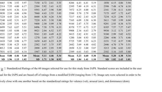
Table 2.2. Standardized Ratings of the 60 images selected for use for this study from IAPS. Standard scores are included in the user
manual for the IAPS and are based off of ratings from a modified SAM (ranging from 1-9). Image sets were selected in order to be
Day 1 Day 2 Day 3 Day 4
Image
ID Val Aro Dom Image ID Val Aro Dom Image ID Val Aro Dom Image ID Val Aro Dom
8465 5.96 3.93 5.97 7150 4.72 2.61 5.55 8206 6.43 6.41 5.19 1850 6.15 4.06 5.94 2314 7.55 4.00 6.17 2384 5.92 3.41 6.32 2395 7.49 4.19 6.31 2398 7.48 4.74 6.18 5300 6.91 4.36 4.14 5593 6.47 3.98 5.89 7472 6.25 4.00 6.31 2341 7.38 4.11 6.44 9830 2.54 4.86 4.96 7060 4.43 2.55 5.85 2580 5.70 2.79 5.88 7175 4.87 1.72 6.47 7224 4.45 2.81 6.26 8600 6.38 4.26 5.54 7217 4.82 2.43 6.25 7234 4.23 2.96 5.73 7240 6.02 5.51 6.37 7220 6.91 5.30 5.80 7165 6.09 3.50 6.30 5611 7.05 3.99 6.04 8230 2.95 5.91 4.05 9400 2.50 5.99 3.78 9184 2.47 5.75 3.86 9291 2.93 4.38 4.75 8499 7.63 6.07 5.51 8496 7.58 5.79 6.33 8370 7.77 6.73 5.37 8185 7.57 7.27 5.47 9405 1.83 6.08 3.40 9921 2.04 6.52 3.57 9908 2.34 6.63 2.79 9930 3.12 5.71 2.97 9331 2.87 3.85 4.72 9341 2.85 4.49 4.22 9415 2.82 4.91 4.22 9180 2.99 5.02 4.52 9445 3.87 4.49 4.51 1270 3.68 4.77 5.25 1280 3.66 4.93 5.05 2206 4.06 3.71 4.46 1610 7.69 3.98 6.77 1650 6.65 6.23 4.29 5725 7.09 3.55 6.23 2151 7.32 4.37 5.90 1903 5.50 4.25 6.01 2382 5.67 3.75 5.97 2682 3.69 4.48 4.02 2446 4.70 3.79 5.51 3005.2 5.98 4.84 5.97 2890 4.95 2.95 5.99 2850 5.22 3.00 5.87 3213 2.96 6.82 3.92 7031 4.52 2.03 6.14 1947 5.85 4.35 5.77 2389 6.61 5.63 5.90 2102 5.16 3.03 5.80
Mean 5.08 4.46 5.40 Mean 5.11 4.46 5.34 Mean 5.23 4.60 5.30 Mean 5.20 4.38 5.34 SD 1.96 1.15 1.02 SD 1.71 1.30 0.92 SD 1.84 1.41 1.10 SD 1.82 1.43 1.01
3
Immediately after image viewing on each day, participants completed the Image
Day Questionnaire (Appendix D), which asked the participant to recall in writing:
a) The approximate total time of sleep on the night immediately preceding the
current visit
b) Whether the participant had exercised in the 48 hours prior to the current visit
c) Food and drink consumption since waking on the morning of the current visit
d) Whether the participant had consumed any stimulants in the 24 hours prior to the
current visit.
The questionnaire was used solely as a method to justify exclusion of potential outlying
data. Since no outliers were observed, no participant had data excluded.
On the baseline testing day after completing the Image Day Questionnaire,
participants underwent maximal exercise testing.Prior to beginning the test, participants
were fitted with a telemetric heart rate monitor (Cosmed, Italy) and asked to mount an
electronically braked cycle ergometer (Ergoline, Germany). The cycle seat height was
adjusted for comfort and proper positioning of the participant. A head strap was used to
secure a mouthpiece for gas (expired oxygen and carbon dioxide) analyses. Prior to
exercise, it was explained to participants that the RPE is a scale from 6-20 with a rating
of 6 being equivalent to no effort exerted and a rating of 20 equated with maximal
exertion. Further, participants were told that the Self-Assessment Mannequin
(SAM-Appendix C) requires participants to point to one of 5 pictures in each row (3 total rows)
to assess their emotional state at a given time. The first row rated the emotion of valence,
the second arousal, and the third dominance. The valence rating ranges from pleasant to