Mir-434-5p mediates skin whitening and lightening
David TS Wu Jack S Chen Donald C Chang Shi-Lung Lin
Institute of Mello Biotechnology, Taipei, Taiwan, ROC
Correspondence: Shi-Lung Lin Institute of Mello Biotechnology, Taipei, Taiwan, ROC
Tel +2 1 323 442 1856 Fax +2 1 626 294 9950 Email shilungl@sbcglobal.net or lins@usc.edu
Abstract: Utilization of gene silencing effectors, such as microRNA (miRNA) and small hairpin RNA (shRNA), provides a powerful new strategy for human skin care in vivo, particularly for hyperpigmentation treatment and aging prevention. In this study, tyrosinase (Tyr), the rate-limiting enzyme of melanin (black pigment) biosynthesis, was served as a target for treatment of hyperpigmentation in mouse and human skins. There are over 54 native microRNA capable of silencing human tyrosinase for skin whitening and lightening. To this, we have designed a mir-434-5p homologue and used it to successfully demonstrate the feasibility of miRNA-mediated skin whitening and lightening in vitro and in vivo. Under the same experimental condition in the trials, Pol-II-directed intronic mir-434-5p expression did not cause any detectable sign of cytotoxicity, whereas siRNAs targeting the same sequence often induced certain nonspecifi c mRNA degradation as previously reported. Because the intronic miRNA-mediated gene silencing pathway is tightly regulated by multiple intracellular surveillance systems, including Pol-II transcription, RNA splic-ing, exosomal digestion and nonsense-mediated RNA decay (NMD), the current fi ndings underscore the fact that intronic miRNA agents, such as manually re-designed mir-434-5p homologues, are effective, target-specifi c and safe to be used for skin whitening without any detectable cytotoxic effect. Given that the human skins also express a variety of other native miRNAs, we may re-design these miRNAs based on their individual functions for skin care, which may provide signifi cant insights into areas of opportunity for new cosmetic and/or therapeutical applications.
Keywords:microRNA, miRNA, mir-434, intron, gene silencing, RNAi, tyrosinase, melanin, cosmetics, pigmentation, skin whitening
Introduction
Prevention of hyperpigmentation (ie, sun-burn) and aging is the key means for having healthy skins. However, many of the skin pigmentation and aging processes are asso-ciated with personal gene activities. For example, tyrosinase (Tyr), a melanocytic membrane-bound glycoprotein, is the rate-limiting enzyme critical for melanin (black pigment) biosynthesis in skins and hairs, while hyaluronidase (Hyal) often causes skin wrinkle by degrading subcutaneous hyaluronan (HA), the major antiaging extracel-lular matrix in skins. Therefore, a good skin care can be achieved by suppressing the activities of these enzymes.
With the advance of recent RNA interference (RNAi) technologies, novel small RNA agents have been found to mediate more potent effects in targeted gene suppression. These include double-stranded short interfering RNA (eg, dsRNA/siRNA) and doxyribonucleotidylated-RNA interfering molecules (eg, D-RNAi) (Fire et al 1998; Elbashir et al 2001; Lin and Ying 2001). Conceivably, these small RNA agents may be used to develop new cosmetic designs and products for skin care. In principle, the RNAi mechanism elicits a post-transcriptional gene silencing (PTGS) phenom-enon capable of inhibiting specifi c gene function with high potency at a few nanomolar dosage, which has been proven to be effective longer and much less toxic than conventional gene-knockout methods using antisense oligonucleotides or small molecule chemical inhibitors (Lin and Ying 2001). As reported in many previous studies, the siRNA-induced gene silencing effects may last over one week, while the D-RNAi effects can even sustain up to one month after one treatment (Grant 1999; Elbashir et al 2001; Lin and Ying 2001; Lin and Ying 2004). These siRNA/D-RNAi agents evoke a series of intracellular sequence-specifi c mRNA degradation and/or translational suppression processes, affecting all highly homologous gene transcripts, namely co-suppression. It has been observed that such co-suppression results from the generation of small RNA products (21–25 nucleotide bases) by the enzymatic activities of RNaseIII endoribonucleases (Dicer) and/or RNA-directed RNA polymerases (RdRp) on aberrant RNA templates, which are usually the derivatives of foreign transgenes or viral genomes (Grant 1999; Elbashir et al 2001; Lin and Ying 2001).
Limitations of siRNA/shRNA-based
gene silencing agents
Although the modern RNAi technologies may offer a new avenue for suppressing unwanted gene function in skins, the applications thereof have not been demonstrated to work consistently and safely in higher vertebrates, including fi sh, avian, mammal and human. For example, almost all of the current siRNA agents are based on a double-stranded RNA (dsRNA) conformation, which has been shown to cause inter-feron-mediated nonspecifi c RNA degradation in vertebrates (Stark et al 1998; Elbashir et al 2001). Such an interferon-mediated cytotoxic response reduces the target specifi city of siRNA-induced gene silencing effects and often results in global RNA degradation in vertebrate cells. Particularly in mammalian cells, it has been noted that the RNAi effects are disturbed when the siRNA/dsRNA size is longer than 25 basepairs (bp) (Elbashir et al 2001). Transfection of
siRNA or small hairpin RNA (shRNA) sized less than 25 bp may not completely overcome such a problem, because both Sledz and colleagues (2003) and Lin and Ying (2004) have reported that the high dosage of siRNAs and shRNAs (such as ⬎250 nM in human T cells) is able to cause strong cytotoxic effects similar to those of long dsRNAs. This toxicity is due to their double-stranded RNA conformation, which activates the interferon-mediated nonspecifi c RNA degradation and programmed cell death through the activation of cellular PKR and 2-5A signaling pathways. It has been well established that interferon-activated protein kinase PKR can trigger cell apoptosis, while the activation of interferon-induced 2’,5’-oligoadenylate synthetase (2-5A) system leads to extensive cleavage of single-stranded RNAs, such as mRNAs (Stark 1998). Both PKR and 2-5A systems contain dsRNA-binding motifs, which possess high affi nity to the double-stranded RNA conformation. Further, the most diffi cult problem is that these small siRNA/shRNA agents are not stable enough to be maintained at an optimal dose in vivo due to the abundant RNase activities in higher vertebrates (Brantl 2002).
As the RNAi effects are naturally caused by the produc-tion of small RNA products (21–25 nucleotide bases) from a transcriptional template derived from foreign transgenes or viral genomes (Grant 1999; Lin and Ying 2001), the recent utilization of Pol-III-directed siRNA/shRNA expression vectors has been shown to offer relatively stable RNAi effects
These disadvantages discourage the use of Pol-III-based RNAi vector systems in health care. In order to improve the delivery stability, targeting specifi city and safety aspects of modern RNAi technologies for healthy skin care, a better transduction and maintenance strategy is highly desired.
Intronic microRNA-mediated
RNAi mechanism
Research based on gene transcript (eg, mRNA), an assembly of protein-coding exons, is fully described throughout the literature, taking the fate of spliced noncoding introns to be completely digested for granted (Nott et al 2003). Is it true that the intronic portion of a gene is destined to be a genetic waste without function or is there an yet undiscovered func-tion for it? Recently, this misconcepfunc-tion was corrected by the discovery of intronic microRNA (miRNA) (Lin et al 2003; Ying and Lin 2004; Ying and Lin 2005). Intronic miRNA is a new class of small single-stranded regulatory RNAs derived from the introns, which are spliced out of the precur-sor messenger RNA (pre-mRNA) of the encoding gene and further processed into small mature miRNAs (Lin et al 2003). A miRNA is usually about 18–27 nucleotides (nt) in length and is capable of either directly degrading its messenger RNA (mRNA) target or suppressing the protein translation of its targeted mRNA, depending on the complementarity between the miRNA and its target. In this way, the intronic miRNA is
functionally similar to previously described siRNA/shRNA, but differs from them in the requirement of intracellular type II RNA polymerase (Pol-II) transcription and RNA splicing processes for its biogenesis (Lin et al 2003). In addition, since introns naturally contain multiple translational stop codons for recognition by the intracellular nonsense-mediated decay (NMD) system (Zhang et al 1994; Danin-Kreiselman et al 2003), most of the unstructured intron sequences can be quickly degraded after RNA splicing to prevent excessive accumulation, which is toxic to the cells. It has been deter-mined that approximately 10%–30% of a spliced intron is preserved after the exosome and NMD digestion in cytoplasm with a relatively long half-life, indicating the cellular origin of native intronic miRNAs (Clement et al 1999).
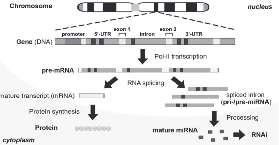
Natural intronic miRNA biogenesis relies on the coupled interaction between nascent Pol-II-mediated pre-mRNA transcription and intron splicing/excision (Figure 1), which take place within certain nuclear regions proximal to genomic perichromatin (Ghosh and Garcia-Blanco 2000; Lin and Ying 2004). In eukaryotic cells, gene transcription is mediated by Pol-II, and the resulting precursor mRNA (pre-mRNA) consists of four major components: 5’-untranslated region (UTR), protein-coding exon, noncoding intron and 3’-UTR. Broadly speaking, both 5’- and 3’-UTR can be seen as a kind of intron extension. Introns occupy the largest proportion of the noncoding sequences in the pre-mRNA. Each intron can
range up to 30 kilobases and is excised out of the pre-mRNA during RNA splicing executed by intracellular spliceosomes. Subsequently, some of the intron-derived RNA fragments are further processed to form microRNA (miRNA) derivative molecules, which can effectively silence their respec-tive targeted genes through an RNA interference (RNAi)-like mechanism. Exons, on the other hand, are ligated together to form a mature mRNA for protein synthesis.
Differences between miRNA
and siRNA biogenesis pathways
We have demonstrated that effective mature miRNAs can be generated from the introns of vertebrate genes, of which the biogenetic process is different from those of siRNA and intergenic miRNA (Lin et al 2003; Lin et al 2005). Figure 2 compares the native biogenesis and RNAi mechanisms among siRNA, intergenic (exonic) miRNA
and intronic miRNA. Presumably, an siRNA is formed by two perfectly complementary RNAs transcribed by two reversely positioned promoters from one DNA template; these complementary RNAs are then hybridized and further processed into 20–25-bp duplexes by RNaseIII endoribonucleases, namely Dicer. Different from this siRNA model, the biogenesis of intergenic miRNA, eg,
lin-4 and let-7, involves a long noncoding precursor RNA transcript (pri-miRNA), which is directly transcribed from a Pol-II or Pol-III RNA promoter, whereas intronic miRNA is co-transcribed with its encoding gene by only Pol-II and released after RNA splicing as a spliced intron. The spliced intron then serves as a pri-miRNA for processing into an intronic precursor miRNA (pre-miRNA) or a multi-pre-miRNA cluster. In the cell nucleus, the pri-miRNA is further excised by either Drosha-like RNases (for intergenic miRNA) or spliceosomal components (for intronic miRNA)
to form a hairpin-like stem-loop precursor or a cluster of multiple stem-loop structures, termed pre-miRNA, and then exported to cytoplasm for fi nal processing into mature miRNA by a miRNA-associated Dicer (Dicer*) (Lee et al 2003). Subsequently, all three small regulatory RNAs are incorporated into a RNA-induced silencing complex (RISC), which contains either strand of siRNA or the mature strand of miRNA. The Dicers and RISCs for siRNA and miRNA pathways are known to be different (Tang 2005). For example, some enzymes of the nonsense-mediated decay (NMD) system may play the role of Dicer* in the mechanism of intronic miRNA maturation. As a result, the effect of miRNA is generally more specifi c and less adverse than that of siRNA because only one strand is involved. On the other hand, siRNAs primarily trigger mRNA degradation, whereas miRNAs can induce either mRNA degradation or suppression of protein synthesis, or both, depending on the sequence complementarity to their targeted gene transcripts. Because the intronic miRNA pathway is tightly regulated by multiple intracellular surveillance systems, such as Pol-II transcription, RNA splicing, exosomal digestion and NMD processing, the gene silencing effects of intronic miRNA are considered to be more effective, specifi c and safer than those of siRNA and intergenic miRNA (Lin et al 2008).
Development of miRNA-based
gene silencing agents
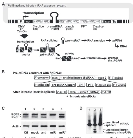
Based on the intronic RNA splicing and processing mecha-nisms (Figures 3A and 3B), we have designed and developed a Pol-II-mediated recombinant gene expression system con-taining at least a splicing-competent intron, namely SpRNAi, capable of inhibiting the EGFP gene with high complemen-tarity to the intron sequence. The SpRNAi is co-transcribed with the precursor mRNA (pre-mRNA) of the recombinant gene by Pol-II RNA polymerases (P) and cleaved out of the pre-mRNA by RNA splicing. Subsequently, the spliced
SpRNAi is further processed into mature gene silencing agents, such as shRNA and miRNA, capable of triggering RNAi-related gene silencing. After intron removal, the exons of the recombinant gene transcript are linked together to form a mature mRNA molecule for translational synthesis of a marker or functional protein.
As shown in Figure 3A, the essential components of the
SpRNAi intron include several consensus nucleotide ele-ments, consisting of a 5’-splice site, a branch-point motif (BrP), a poly-pyrimidine tract (PPT), and a 3’-splice site. In addition, a hairpin RNA-like pre-miRNA sequence is inserted
inside the SpRNAi intron located between the 5’-splice site and the branch-point motif (BrP). This portion of the intron normally forms a lariat structure during RNA splicing and processing. We have observed that spliceosomal U2 and U6 snRNPs, both helicases, are involved in the unwinding and excision of the lariat RNA fragment into pre-miRNA; however, the detailed processing remains to be elucidated. Further, the 3’-end of the SpRNAi construct contains a mul-tiple translational stop codon region (T codon) in order to increase the accuracy of intronic RNA splicing and NMD processing. When presented in a cytoplasmic mRNA, this T codon will signal the activation of the nonsense-mediated decay (NMD) pathway to degrade any unstructured RNA accumulation in the cell. However, the highly secondary structured hairpin RNA and pre-miRNA insert will be preserved for further Dicer cleavage, so as to form mature siRNA and miRNA, respectively. Moreover, for intracellular expression, we manually incorporate the SpRNAi construct in the DraII restriction site of a red fl uorescent protein (RGFP) gene isolated from mutated chromoproteins of the coral reef
Heteractis crispa, so as to form a recombinant SpRNAi-RGFP
gene. The cleavage of RGFP at its 208th nucleotide site by the restriction enzyme DraII generates an AG–GN nucleotide break with three recessing nucleotides in each end, which will form 5’- and 3’-splice sites respectively after the SpRNAi
insertion. Because this intronic insertion disrupts the struc-ture of a functional RGFP protein, which can be recovered by intron splicing, we can determine the release of intronic shRNA/miRNA and RGFP-mRNA maturation through the appearance of red RGFP around the affected cells. The RGFP
gene also provides multiple exonic splicing enhancers (ESEs) to increase RNA splicing accuracy and effi ciency.
and then triggers a desired gene silencing effect on specifi c gene transcripts with high complementarity to the inserted RNA sequence, while the exons of the recombinant gene transcript are linked together to form mature mRNA for expression of a desirable gene function, such as translation of a reporter or marker protein selected from the group of red/green fl uorescent protein (RGFP/EGFP), luciferase, lac-Z, and their derivative homologues. The presence of the reporter/marker protein is useful for signaling the produc-tion of the inserted shRNA/miRNA molecules in treated
cells, thus facilitating the identifi cation of the desired gene silencing/RNAi effects.
gene knockout or RNA interference (RNAi) effects, which are useful for inhibiting targeted gene function. The intron-derived gene silencing molecules so obtained may include antisense RNA, ribozyme, short temporary RNA (stRNA), double-stranded RNA (dsRNA), small interfering RNA (siRNA), tiny noncoding RNA (tncRNA), short hairpin RNA (shRNA), microRNA (miRNA), and RNAi-associated pre-cursor RNA constructs (pri-/pre-miRNA). The use of these intronic RNA-derived gene silencing agents is a powerful tool for targeting and silencing unwanted genes selected from the group consisting of pathogenic transgenes, viral genes, mutant genes, oncogenes, disease-related small RNA genes and any other types of protein-coding as well as noncoding genes.
Using this novel Pol-II-mediated SpRNAi-RGFP expres-sion system, we have successfully generated mature shRNA and miRNA molecules with full gene silencing capacity in vitro in human prostate cancer (LNCaP), human cervical cancer (HeLa) and rat neuronal stem (HCN-A94-2) cells as well as in vivo in zebrafi sh, chicken and mouse (Lin and Ying 2006; Lin et al 2006). We have tested different pre-miRNA insert constructs targeting against green EGFP and other cellular gene expression in zebrafi sh and various human cell lines, and have learned that effective gene silencing miRNAs are derived from the 5’-proximity of the intron sequence between the 5’-splice site and the branching point. As shown in Figure 3C, a strong gene silencing effect was observed only in the transfection of anti-EGFP pre-miRNA insert (lane 4), whereas no effect could be detected in those of other inserts indicated by lanes from left to right: 1, blank vector control (Ctl); 2, pre-miRNA insert targeting HIV-p24
(mock); 3, antisense EGFP insert without the hairpin loop structure (anti); and 5, reverse pre-miRNA sequence which was completely complementary to the anti-EGFP pre-miRNA (miR*). No effect was detected on off-target genes, such as marker RGFP and housekeeping β-actin, suggest-ing that such an intronic miRNA-mediated gene silencsuggest-ing effect is highly target-specifi c. To confi rm the role of RNA splicing in this intronic RNAi effect, we further tested three different SpRNAi-RGFP expression systems as shown in Figure 3D: 1, vector expressing intron-free RGFP without any pre-miRNA insert; 2, vector expressing RGFP with an intronic anti-EGFP pre-miRNA insert; and 3, vector similar to the 2 construct but with a defective 5’-splice site in the
SpRNAi intron. As a result, northern blotting revealed that mature miRNAs were released only from the spliced intron of the vector 2 construct, which was exactly identical to the
SpRNAi vector construct with the anti-EGFP pre-miRNA
insert in Figure 3C. Thus, RNA splicing is required for intronic miRNA biogenesis.
Evaluation of natural antityrosinase
miRNA agents
We have adopted the proof-of-principle design of the
SpRNAi-RGFP expression system and use it for developing novel cosmetic products for skin care. In this new approach, we apply skins a nonnaturally occurring intron capable of being processed into hairpin-like precursor microRNA (pre-miRNA) molecules by the skin cells and thus inducing specifi c gene silencing effects on epidermal pigment-related genes and/or aging-causing genes. In this case, the RNA splicing- and processing-generated gene silencing molecule is the hairpin-like pre-miRNA insert located within the intron area of the recombinant gene and was capable of silencing a targeted gene, such as tyrosinase (Tyr), hyaluronidase (Hyal), hyaluronan receptors CD44 and CD168, and other pigmentation-related and/or aging-related genes and onco-genes. Alternatively, such a pre-miRNA insert can also be artifi cially incorporated into the intron region of a cellular gene in the skin. In general, this kind of intronic insertion technology includes plasmid-like transgene transfection, homologous recombination, transposon delivery, jumping gene integration and retroviral infection.
In the present design, the recombinant SpRNAi-RGFP
a desired hairpin-like intronic RNA and 90%∼100% for a linear intronic RNA molecule.
In addition, the 5’-end of the nonnaturally occurring intron contains a donor splice site homologous to 5’-GTAAGAGK-3’ motifs, while its 3’-end is an acceptor splice site that is homologous to 5’-GWKSCYRCAG-3’ motifs. Moreover, a branch point sequence is located between the 5’- and 3’-splice sites, containing homology to 5’-TACTWAY-3’ motifs. The adenosine “A” nucleo-tide of the branch-point sequence forms a part of (2’–5’)-linked lariat intron RNA by cellular (2’–5’)-oligoadenylate synthetases and spliceosomes in almost all spliceosomal introns. Furthermore, a poly-pyrimidine tract is closely located between the branch-point and 3’-splice site, containing a high T or C content oligonucleotide sequence homologous to either 5’-(TY)m(C/−)(T)nS(C/−)-3’ or 5’-(TC)nNCTAG(G/−)-3’ motifs. The symbols of “m” and “n” indicate multiple repeats ⱖ1; most preferably, the m number is equal to 1∼3 and the n number is equal to 7∼12. The symbol “–” refers to an empty nucleotide in the sequence. There are also some linker nucleotide sequences for the con-nection of all these intron components. By defi nition, the symbol W refers to an adenine (A) or thymine (T)/uracil (U), the symbol K refers to a guanine (G) or thymine (T)/uracil (U), the symbol S refers to a cytosine (C) or guanine (G), the symbol Y refers to a cytosine (C) or thymine (T)/uracil (U), the symbol R refers to an adenine (A) or guanine (G), and the symbol N refers to an adenine (A), cytosine (C), guanine (G) or thymine (T)/uracil (U).”
Based on the above design, we have tested an optimized
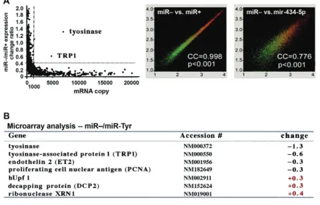
SpRNAi-RGFP gene construct expressing either anti-Tyr or anti-Hyal pre-miRNA directed against the pigmentation-related gene Tyr or aging-related gene Hyal in mouse skins. These pre-miRNAs target a highly conserved region (⬎98% homology) in both human Tyr and mouse Hyal genes. In nature, there are 54 native miRNAs capable of targeting human tyrosinase (Tyr; 2082 bp) for pigmentation gene silencing, including 1, 15a, 16, 31, mir-101, mir-129, mir-137, mir-143, mir-154, mir-194, mir-195, mir-196b, mir-200b, mir-200c, mir-206, mir-208, mir-214, 221, 222, 292-3p, 299-3p, 326, mir-328, mir-381, mir-409-5p, mir-434-5p, mir-450, mir-451, mir-452, mir-464, mir-466, mir-488, mir-490, mir-501, 522, 552, 553, 570, 571, 582, mir-600, mir-619, mir-624, mir-625, mir-633, mir-634, mir-690, mir-697, mir-704, mir-714, mir-759, mir-761, mir-768-5p, and mir-804. According to the miRNA-target database of the miRBase::Sequences program (http://microrna.sanger.ac.uk),
all these anti-Tyr miRNAs are directed against a region within the fi rst 300 nucleotides of the Tyr gene transcript (NCBI accession number NM000372). On the other hand, there are 9 native miRNAs capable of targeting hyaluronidase (Hyal; 2518 bp; NCBI accession number NM007312) for aging gene silencing, including mir-197, mir-349, mir-434-5p, mir-549, mir-605, mir-618, mir-647, mir-680, mir-702, and mir-763. Among these native miRNAs, mir-434-5p is the only one that targets both Tyr and Hyal genes in human and it is also one of the most effi cient miRNAs targeting the least off-target genes other than Tyr and Hyal. However, because almost all native miRNAs target several to over fi fty cellular genes and they tend to bind with some of the target genes more strongly than others, the use of these native miRNAs is likely not specifi c and safe enough for the purpose of skin care.
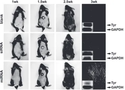
To test the feasibility of miRNA-mediated skin whit-ening, we utilized an SpRNAi-RGFP expression vector to express native pre-mir-434-5p in mouse skin. As shown in Figure 4, patched albino (white) skins of melanin-knockdown mice (W-9 black) were observed after a daily intra-cutaneous (i.c.) injection of the pre-mir-434-5p expression vector (50 μg) for four times (total 200 μg). Tyr, a type-I mem-brane protein and copper-containing enzyme, catalyzes the critical and rate-limiting step of tyrosine hydroxylation in the biosynthesis of melanin (black pigment) in skins and hairs; thus, the silencing of Tyr expression will result in a great loss of skin/hair pigments. Approximately two weeks after the fi rst i.c. injection, we could clearly observe that the skin and hair pigments were signifi cantly lost only in mice transfected with the pre-mir-434-5p. In contrast, the blank control and the transfection of a Pol-III (U6)-transcribed siRNA agent against the same Tyr sequence presented no sig-nifi cant effects. Northern blot analyses using mRNAs isolated from the hair follicles of the pre-mir-434-5p-transfected mice showed a markedly reduction of the Tyr expression (76.1% ± 5.3%) two days post-transfection, whereas mild, nonspecifi c degradation of random gene transcripts was detected in the siRNA-transfected skins (seen from the smearing patterns of both house-keeping control GAPDH
LYPLA2 and Hyal, its off-target gene silencing effects remain to be determined.
Re-design of mir-434-5p for skin
whitening use in human
In order to improve the target-specifi city and safety of
anti-Tyr miRNA agents, we have re-designed the seed sequence of the mir-434-5p to form a highly matched region binding to either Tyr nucleotides 3–25 (miR-Tyr) or Hyal nucleotides 459–482 (miR-Hyal). The pre-miRNA insert sequence for pre-miR-Tyr is 5’-GTCCGATCGT CGCCCTACTC TATTGCCTAA GCCGCTAAGC CAGGCGGCTT AGGCAATAGA GTAGGGCCGA CGCGTCAT-3’, which forms a hairpin-like RNA after splicing and can be further processed into a mature miR-Tyr microRNA (miRNA) sequence containing or homologous to 5’-GCCCTACTCT ATTGCCTAAG CC-3’. Alternatively, the sequence for pre-miR-Hyal is 5’-GTCCGATCGT CAGCTAGACA GTCAGGGTTT GAAGCTAAGC CAGGCTTCAA ACCCTGACTG TCTAGCTCGA CGCGTCAT-3’, which
forms a different kind of hairpin-like RNA after splicing and is further processed into a mature miR-Hyal miRNA sequence containing or homologous to 5’-AGCTAGACAG TCAGGGTTTG AA-3’. Although both pre-miR-Tyr and pre-miR-Hyal constructs were re-designed based on the same mir-434-5p backbone and mir-302 stem-loop, the mature miR-Tyr and miR-Hyal were shown to be totally different from each other. As shown in Figure 5, the transfective expressions of miR-Tyr and miR-Hyal in mouse skins spe-cifi cally knocked down the targeted Tyr (reduction ⬎90%) and Hyal genes (reduction ⬎67%), respectively, without any crossover off-target effect. The expression levels of mature miR-Tyr and miR-Hyal microRNAs were directly measured by northern blot analyses, while the knockdown rates of the targeted Tyr and Hyal proteins were assessed by western blot analyses.
After understanding the optimized gene silencing effects of the re-designed miR-Tyr and miR-Hyal miRNAs, we continued to test their effi cacy, target specifi city and safety in human skins. For effi cient vector transfection into the Figure 4 Effects of miRNA-mediated tyrosinase (Tyr) gene silencing on the depigmentation of mouse skins and hairs. Targeted gene silencing in epidermal tissues was shown to be feasible using the ectopic transfection of a recombinant SpRNAi-RGFP gene vector expressing the native mir-434 pre-miRNA. Subcutaneous transfection of mir-434-5p induced a strong and specifi c gene silencing effect on Tyr but not house-keeping GAPDH expression, whereas transfection of a U6 promoter-driven siRNA against the same
human epidermal cell layers, a 1 μg/ml SpRNAi-RGFP
vector solution was prepared by mixing 100 μg of the purifi ed
SpRNAi-RGFP vector in 1 ml of autoclaved ddH2O with 99 ml of 100% free glycerin (or glycerol). DNase-free glycerin was used to encapsulate miR-Tyr for deep skin delivery and cell membrane penetration. This formed the major ingredient base for skin whitening and lightening products. Based on this, more other cosmetic ingredients could be added to increase the color, fragrance, effectiveness and/or stability of the fi nal cosmetic products. As shown in Figure 6A, Asian male arm skins were treated with 2 ml of this major ingredient base expressing either miR-Tyr (right) or empty SpRNAi-RGFP vector alone (glycerin control, left). The whitening effect of the miR-Tyr treatment on human skins (loss of the black pigment–melanin) was clearly observed within three days after two treatments per day.
Next, a primary skin cell culture was obtained by trypsin-dissociated skin explants from the tested donor with personal consent. The SpRNAi-RGFP vector transfection (fi nal 6 μg/ml) in the primary skin culture was performed using FuGene liposomal reagent (Roche Biochemicals,
Indianapolis, IN), as described previously (Lin et al 2003; Lin et al 2006). Figure 6B shows that western blot analyses of the loss of the targeted tyrosinase proteins and its substrate melanin were statistically signifi cant (p⬎ 0.001). The reduc-tion levels of Tyr and its substrate melanin in skins were proportional to the treated concentrations of the miR-Tyr expression vector, suggesting a positive correlation between the increase of the miR-Tyr treatment and the loss of the targeted Tyr proteins and its substrate melanin. No effect was observed in other treatments, such as SpRNAi-RGFP
vector alone (glycerin control) and an SpRNAi-RGFP vector expressing an anti-EGFP pre-miRNA (miR-gfp). At 1 μg/ml of the miR-Tyr expression vector transfection, the optimal gene silencing rates were approximately 55%–60% for Tyr
and 30%–45% for melanin, while the expression of nontarget house-keeping control ß-actin was not affected by miR-Tyr treatment. Taken together, these results demonstrated the high target-specifi city of our re-designed manmade miRNA agents.
Figure 6C further showed that the skin melanin levels were signifi cantly reduced as shown in bright-fi eld (BF) Figure 5 Effects of Tyr and Hyal gene silencing on skin pigmentation in mice. The specifi city of tyrosinase (Tyr) gene silencing was markedly improved, using a man-made anti-Tyr
photographs of the primary skin cell culture (upper panels), while melanin (black dots around the cell nuclei) is highly expressed in normal skin cells without miR-Tyr treatment (ie, blank and glycerin only). The miR-Tyr-treated skin cells showed very limited melanin accumulation, demonstrating an effective skin-whitening effect in vivo. In regard to this loss of skin melanin, the targeted tyrosinase expression was concurrently reduced in the miR-Tyr-treated skin cells, as determined by immunocytochemical (ICC) staining analy-sis (Figure 6C, lower panels). According to these results, the re-designed miR-Tyr microRNA effectively knocked down the expression of Tyr proteins and successfully blocked melanin production in human skins in vivo.
Microarray analyses of target
speci
fi
city and safety
After the effi cacy of miR-Tyr-mediated gene silencing was established in human skins, we applied human genome microarray analyses (Human GeneChip U133A&B arrays, Affymetrix, Santa Clara, CA) to assess the changes of approximately 32,668 human gene expression patterns in miR-Tyr-transfected and control primary skin cell cul-tures. As shown in Figure 7A (left), microarray analyses in nontreated (miR–) versus miR-Tyr-transfected (miR+)
for increasing the safety and effi ciency of these miRNAs used in cosmetic applications.
Materials and methods
Construction of SpRNAi-containing
recombinant transgene (SpRNAi-RGFP)
Synthetic nucleic acid sequences used for generation of three different SpRNAi introns containing either sense-, antisense- or hairpin-EGFP insert were listed as follows: N1-sense,
GCGCTCCTGA-3’; N3-antisense, 5’-pCGCGTCAGGA GCGCACCATC TTCTTCAAGT TAACTTGAAG AAGATGGTGC GCTCCTGCGA TCGGATCCTC TTAC-3’; N4-sense, 5’-pCGCGTTACTA ACTGGTACCT C T T C T T T T T T T T T T T G A T A T C C T G C A G - 3 ’ ; N4-antisense, 5’-pGTCCTGCAGG ATATCAAAAA AAAAAGAAGA GGTACCAGTT AGTAA-3’. In addition, two exon fragments were generated by DraII restriction enzyme cleavage of a red fl uorescent RGFP gene at its 208th nucleotide (nt) site and the 5’ fragment was further blunt-ended by T4 DNA polymerase. RGFP refers to a new red-shifted fl uorescent chromoprotein gene generated by insertion of an additional aspartate at the 69th amino acid (a.a.) site of HcRed1 chromoproteins from Heteractis crispa. (BD Biosciences, CA), developing less aggregate and almost twice intense far-red fluorescent emission at 570-nm. We also cleaved a pHcRed1-N1/1 plasmid (BD Biosciences, CA) with XhoI and XbaI restriction enzymes and purifi ed a full 769-bp RGFP gene fragment and a 3,934-bp empty plasmid separately isolated from 2% agarose gel electrophoresis.
Hybridization of N1-sense to N1-antisense, N2-sense to N2-antisense, N3-sense to N3-antisense and N4-sense to N4-antisense was separately performed by heating each complementary mixture of sense and antisense (1:1) sequences to 94 °C for 2 min and then 70 °C for 10 min in 1x PCR buffer (eg, 50 mM Tris-HCl, pH 9.2 at 25 °C, 16 mM (NH4)2SO4, 1.75 mM MgCl2). Continuously, sequen-tial ligation of either N1, N2 or N3 hybrid to the N4 hybrid was performed by gradually cooling the mixture of N1-N4, N2-N4 or N3-N4 (1:1) hybrids respectively from 50 °C to 10 °C over a period of 1 hr, and then T4 ligase and relative buffer (Roche, IN) were mixed with the mixture for 12 hr at 12 °C, so as to obtain introns for insertion into exons with proper ends. After the RGFP exon fragments were added into the reaction (1:1:1), T4 ligase and buffer were adjusted accordingly to reiterate ligation for another 12 hr at 12 °C. To clone the properly sized recombinant RGFP gene, 10 ng of the ligated nucleotide sequences were amplifi ed by PCR with a pair of RGFP-specifi c primers 5’-CTCGAGCATG GTGAGCGGCC TGCTGAA-3’ and 5’-TCTAGAAGTT GGCCTTCTCG GGCAGGT-3’ at 94 °C, 1 min, 52 °C, 1 min and then 68 °C, 2 min for 30 cycles. The resulting PCR products were fractionated on a 2% agarose gel, and a ∼900-bp nucleotide sequences was extracted and purifi ed by a Gel Extraction kit (Qiagen, CA). The composition of this ∼900 bp SpRNAi-containing RGFP gene was further confi rmed by sequencing.
Since the recombinant SpRNAi-RGFP gene possesses an XhoI and an XbaI restriction site at its 5’- and 3’-end, respectively, it can be easily cloned into a vector with cohe-sive ends to the XhoI and XbaI cloning sites. The vector must be a skin-compatible, expressing-competent organism or suborganism selected from the group consisted of plasmids, cosmids, jumping genes, transposons, adeno-associated viral and retroviral vectors. Moreover, since the insert within the intron is also fl anked with a PvuI and an MluI restriction site at its 5’- and 3’-end, respectively, we can remove and replace the insert with another different insert sequence pos-sessing cohesive ends to the PvuI and MluI cloning sites. The inserted sequence is preferably a hairpin-like gene silencing effector containing high complementarity to a gene target selected from the group consisted of fl uorescent protein (GFP) genes, luciferase genes, lac-Z genes, viral genes, bacterial genes, plant genes, animal genes and human genes. The complementarity and/or homology rate between the gene-silencing effector insert and its targeted gene is ranged from about 30%–100%, more preferably 35%–49% for a hairpin-shRNA insert and 90%–100% for a sense-stRNA or antisense-siRNA insert.
Cloning of the SpRNAi-containing transgene
into an expression-competent vector
We mixed the SpRNAi-RGFP gene with the linearized 3,934-bp empty pHcRed1-N1/1 plasmid at 1:16 (w/w) ratio, cooled the mixture from 65 °C to 15 °C over a period of 50 min, and then added T4 ligase and relative buffer accord-ingly into the mixture for ligation at 12 °C for 12 hrs. This forms the SpRNAi-RGFP expression vector, which can be propagated in an E.coli DH5α LB culture containing 50 μg/ml kanamycin (Sigma Chemical, St. Louis, MO). A positive clone was confi rmed by PCR with the RGFP -specifi c primers at 94 °C, 1 min and then 68 °C, 2 min for 30 cycles, and for further sequencing. For cloning into viral vec-tors, the same ligation procedure was repeated except using an
XhoI/XbaI-linearized pLNCX2 retroviral vector (BD). Since the insert within the SpRNAi intron was fl anked with a PvuI
and a MluI restriction site at its 5’- and 3’-end, respectively, we could remove and replace the anti-EGFP shRNA insert with the miR-Tyr or miR-Hyal insert sequences possessing cohensive ends to the PvuI and MluI cloning sites.
GTAGGGCCGA CGCGTCAT-3’; miR-Tyr-antisense, 5’-ATGACGCGTC GGCCCTACTC TATTGCCTAA GCCGCCTGGC TTAGCGGCTT AGGCAATAGA GTAGGGCGAC GATCGGAC-3’; and miR-Hyal-sense, 5’-GTCCGATCGT CAGCTAGACA GTCAGGGTTT GAAGCTAAGC CAGGCTTCAA ACCCTGACTG TCTAGCTCGA CGCGTCAT-3’; miR-Hyal-antisense, 5’-ATGACGCGTC GAGCTAGACA GTCAGGGTTT GAAGCCTGGC TTAGCTTCAA ACCCTGACTG TCTAGCTGAC GATCGGAC-3’. These inserts were formed via hybridization of sense to miR-Tyr-antisense and miR-Hyal-sense to miR-Hyal-miR-Tyr-antisense, respectively. These miR-Tyr- and miR-Hyal-expressing vectors could be propagated in E.coli DH5α LB-culture containing either 50 μg/ml kanamycin (for pHcRed1-N1/1
plasmid-based vector) or 100 μg/ml ampicillin (for pLNCX2
viral vector). The propagated SpRNAi-RGFP vectors could be isolated and purifi ed using a Mini-prep or Maxi-prep Plasmid Extraction kit (Qiagen, CA), following the manufacturer’s suggestion. For pLNCX2 vectors, we also used a packaging cell line PT67 (BD) for producing infectious but replication-incompetent virus. The transfected PT67 cells were grown in 1x DMEM medium supplemented with 10% fetal bovine serum with 4 mM L-glutamine, 1 mM sodium pyruvate, 100 μg/ml streptomycin sulfate and 50 μg/ml neomycin (Sigma Chemical, MO) at 37 °C under 5% CO2. The titer of virus produced by PT67 cells was determined to be at least 106 cfu/ml before transfection.
Vector transfection
For vector transfection into cultivated cells or mouse skins, we fi rst mixed the SpRNAi-RGFP expression plasmid vectors containing either anti-EGFP, miR-Tyr or miR-Hyal pre-miRNA insert using FuGene reagent (Roche, IN), following the manufacturer’s suggestion. Thereafter, the mixtures were directly applied to the cultivated cells (ie, primary human skin culture) or mouse skins, respectively. Vectors containing an insert-free RGFP gene and an SpRNAi-RGFP gene with a pre-miRNA insert against the HIV gag-p24 gene were used as negative controls. Tissue or cell morphology and fl uorescence imaging was photographed at 0-, 24-, 48-, and 72-hr time points after the fi rst transfection. For transfec-tion to the human skins in vivo, a pre-made SpRNAi-RGFP
vector solution was formed by mixing certain amounts (ie, 1–1000 μg) of the purifi ed SpRNAi-RGFP vector with or without either anti-EGFP, miR-Tyr or miR-Hyal pre-miRNA insert in 1 ml of autoclaved ddH2O with 99 ml of 100% DNase-free glycerin (or glycerol). This solution
was then directly applied to the skins with gentle massage for 3 min.
Northern blot analysis
RNA (20 μg total RNA or 2 μg poly[A+] RNA) was fractionated on 1% formaldehyde-agarose gels and transferred onto nylon membranes (Schleicher and Schuell, Keene, NH). Synthetic probes complementary to either the 75-bp junction sequence fl anking between the RGFP
5’-exon and the anti-EGFP pre-miRNA insert or miR-Tyr, or miR-Hyal, were labeled with the Prime-It II kit (Stratagene, La Jolla, CA) by random primer extension in the presence of [32P]-dATP (⬎3000 Ci/mM, Amersham International,
Arlington Heights, IL), and purifi ed with 10 bp-cutoff Micro Bio-Spin chromatography columns (Bio-Rad, Hercules, CA). Hybridization was carried out in the mixture of 50% freshly deionized formamide (pH 7.0), 5x Denhardt’s solu-tion, 0.5% SDS, 4x SSPE and 250 mg/mL denatured salmon sperm DNA fragments (18 hr, 42 °C). Membranes were sequentially washed twice in 2x SSC, 0.1% SDS (15 min, 25 °C), and once in 0.2x SSC, 0.1% SDS (45 min, 37 °C) before autoradiography.
SDS-PAGE electrophoresis
and Western blot analysis
detected using primary antibodies directed against EGFP (1:5,000; JL-8, BD), RGFP (1:10,000; BD), Tyr (1:2,000; Santa Crutz), or Hyal (1:2,000; Santa Crutz) incubated overnight at 4 °C. The protein blots were then rinsed 3 times with TBS-T and incubated with goat antimouse secondary antibody conjugated with Alexa Fluor 680 reactive dye (1:2,000; Molecular Probes) for 1 hr at room temperature. After three more TBS-T rinses, scanning and image analysis were completed with Li-Cor Odyssey Infrared Imager and Odyssey Software v.10 (Li-Cor).
Intronic mir-434-5p and miR-Tyr-mediated
gene silencing in mouse skins
in vivo
Patched albino (white) skins of melanin-knockdown mice (W-9 black strain) were created either by a succession of intra-cutaneous (i.c.) injection of an isolated SpRNAi-RGFP
gene expression vector with the native mir-434-5p pre-miRNA insert for 4 days (total 200 μg) or by a direct skin infusion of a liposome-encapsulated SpRNAi-RGFP gene expression vector with the designed miR-Tyr pre-miRNA insert for two times per day for six days (total 240 μg). To generate the SpRNAi-RGFP gene expression vector with the native mir-434-5p pre-miRNA insert, we followed the same procedure as aforementioned, except using a synthetic mir-434-5p pre-miRNA for intronic insertion (ie, 5’-GTCCGATCGT CUCGACUCUG GGUUUGAACC AAAGCUCGAC UCAUGGUUUG AACCAUUACU UAAUUCGUGG UUUGAACCAU CACUCGACUC CUGGUUCGAA CCAUCCGACG CGTCAT-3’). For effi cient delivery into target tissues, the construct was mixed with a FuGene liposomal transfection reagent (Roche, IN).
Intronic miR-Tyr-mediated gene
silencing in human skins
For effi cient vector transfection into the human multiple skin cell layers, a 1 μg/ml SpRNAi-RGFP vector solution is made by mixing 100 μg of the purifi ed SpRNAi-RGFP vector in 1 ml of autoclaved ddH2O with 99 ml of 100% DNase-free glycerin (or called glycerol). DNase-free glycerin is used to encapsulate miR-Tyr for deep skin delivery and cell mem-brane penetration. This forms the major ingredient base for our present skin whitening and lightening inventions. There-after, one (left arm) of the author’s arm skins was directly treated with one 2 ml of the major ingredient base solution containing SpRNAi-RGFP vector with (right side) or without (left side) miR-Tyr. The effect of miR-Tyr treatment on skin
whitening (loss of the black pigment–melanin) was assessed three days after two single treatments per day.
Immunocytochemical (ICC) staining assay
An immunochemical staining kit was obtained from Imgenex (San Diego, CA) and used according to the manufacturer’s suggestion. Specimens were fi rst rinsed in PBS three times and incubated with Zeller’s solution (10 mM Tris, 100 mM MgCl2, 5% fetal calf serum, 1% BSA and 0.5% Tween-20, pH 7.4) for 30 min. They were then incubated with primary antibody (diluted in Zeller’s solution) overnight in a humidi-fi ed chamber at 4 °C. Thereafter, the specimens were washed with TBST three times and incubated with secondary anti-body for 2 hr, using biotinylated goat antirabbit or horse antimouse antibody as the secondary antibody (Chemicon, Temecula, CA). Specimens were then washed once with TBST and incubated with streptavidin-HRP as the tertiary antibody for 2 hrs and followed by PBT washings. The bound antibody was subsequently detected with DAB substrates and observed under a 100x microscope with whole fi eld scanning and recorded at 100x and 400x magnifi cation (TE2000 inverted microscopic quantitation system).
Microarray analysis
To prepare labeled probes for microarray hybridization, total RNAs (2 μg) were converted into double-stranded cDNAs, using a Superscript Choice system kit (Gibco/BRL, Gaithersburg, MD) with a modifi ed oligo(dT)24-T7 promoter primer, such as 5’-GGCCAGTGAA TTGTAATACG ACTCACTATA GGGAGGCGG-(dT)24-3’, following the manufacturer’s protocol. Double-stranded cDNAs were purifi ed by phenol/chloroform extractions, precipitated with ethanol, and resuspended at a concentration of 0.5 μg/μl in diethyl pyrocarbonate (DEPC)-treated ddH2O. Phase-Lock Gel (5’Prime →3’Prime, Inc., Boulder, CO) was used for all organic extractions to increase recovery. In-vitro
transcription was performed with T7 RNA polymerase and with 1 μg of cDNA, 7.5 mM unlabeled ATP and GTP, 5 mM unlabeled UTP and CTP, and 2 mM biotin-labeled CTP and UTP (biotin-11-CTP, biotin-16-UTP, Enzo Diagnostics). Reactions were carried out for 4 hr at 37 °C, and cRNA was purifi ed by RNeasy spin columns (Qiagen, CA). A sample was separated on a 1% agarose gel to check the size range, and then μg of cRNA was fragmented randomly to an average size of 50 bases by heating at 94 °C for 35 min in 40 mM Tris-acetate, pH 8.0, 100 mM KOAc/30 mM MgOAc.
total 32,668 genes were used for hybridization. Hybridizations were completed in 200 μl of AFFY buffer (Affymetrix) at 40 °C for 16 hr with constant mixing. After hybridization, arrays were rinsed three times with 200 μl of 6x SSPE-T buffer (1 × 0.25 M sodium chloride/15 mM sodium phos-phate, pH 7.6/1 mM EDTA/0.005% Triton) and then washed with 200 μl of 6x SSPE-T for 1 hr at 50 °C. The arrays were rinsed twice with 0.5x SSPE-T and washed with 0.5x SSPE-T at 50 °C for 15 min. Staining was done with 2 μg/ml streptavidin-phycoerythrin (Molecular Probes) and 1 mg/ml acetylated BSA (Sigma) in 6x SSPE-T (pH 7.6). The arrays were read at 7.5 μm with a confocal scanner (Molecular Dynamics) and analyzed with Affymetrix Microarray Suite version 4.0 software. The samples were normalized by using the total average difference between perfectly matched probe and the mismatched probe. The differential signals that were induced greater than 2-fold are collected.
Statistical analysis
Results were presented as mean ±SE. Statistical analysis of data was performed by one-way ANOVA. When main effects were signifi cant, the Dunnett’s post-hoc test was used to identify the groups that differed signifi cantly from the controls. For pairwise comparison between two treatment groups, the two-tailed student t test was used. For experi-ments involving more than two treatment groups, ANOVA was performed followed by a post-hoc multiple range test. Probability values of p⬍ 0.05 were considered signifi cant. All p values were determined from two-tailed tests.
Conclusion
In summary, the utilization of intronic hairpin-like miRNA expression provides a potentially powerful and innovative strategy for human skin care, particularly for hyperpigmen-tation and aging prevention. Under the same treatment in animal trials, Pol-II-directed intronic miRNA expression did not cause any detectable cytotoxicity, whereas Pol-III-directed siRNAs induced nonspecifi c mRNA degradation as previously reported (Elbashir 2001; Lin and Ying 2001; Sledz et al 2003; Lin and Ying 2004). This underscores the fact that the intronic miRNA agent is effective, target-specifi c and safe for use in skin care. Because the intronic miRNA pathway is regulated by multiple intracellular surveillance systems, such as Pol-II transcription, RNA splicing, exosomal digestion and NMD processing, the gene silencing effect of intronic miRNA is considered to be most effective, specifi c and safest among all three currently known RNAi pathways. By utilizing the intronic miRNA expression strategy,
many cosmetic applications can be designed and developed for skin care, offering more long-term effectiveness, better target-specifi city and higher safety in skin gene manipulation, which prevents the unspecifi c off-target cytotoxicity as commonly seen in the conventional siRNA methods.
Disclosure
The authors report no confl icts of interest in this work.
References
Brantl S. 2002. Antisense-RNA regulation and RNA interference.
Biochimica et Biophysica Acta, 1575:15–25.
Clement JQ, Qian L, Kaplinsky N, et al. 1999. The stability and fate of a spliced intron from vertebrate cells. RNA, 5:206–20.
Danin-Kreiselman M, Lee CY, Chanfreau G. 2003. RNAse III-mediated degradation of unspliced pre-mRNA and lariat introns. Mol Cell, 11:1279–89.
Elbashir SM, Harborth J, Lendeckel W, et al. 2001. Duplexes of 21-nucleotide RNAs mediate RNA interference in cultured mammalian cells. Nature, 411:494–8.
Fire A, Xu S, Montgomery MK, et al. 1998. Potent and specifi c genetic interference by double-stranded RNA in Caenorhabditis elegans.
Nature, 391:806–11.
Ghosh S, Garcia-Blanco MA. 2000. Coupled in vitro synthesis and splicing of RNA polymerase II transcripts. RNA, 6:1325–34.
Grant SR. 1999. Dissecting the mechanisms of posttranscriptional gene silencing: divide and conquer. Cell, 96:303–6.
Grimm D, Streetz KL, Jopling CL, et al. 2006. Fatality in mice due to oversaturation of cellular microRNA/short hairpin RNA pathways.
Nature, 441:537–41.
Gunnery S, Ma Y, Mathews MB. 1999. Termination sequence require-ments vary among genes transcribed by RNA polymerase III. J Mol Biol, 286:745–57.
Lee NS, Dohjima T, Bauer G, et al. 2002. Expression of small interfering RNAs targeted against HIV-1 rev transcripts in human cells. Nat Biotechnol, 20:500–5.
Lee Y, Ahn C, Han J, et al. 2003. The nuclear RNase III Drosha initiates microRNA processing. Nature, 425:415–9.
Lin SL, Chang D, Wu DY, et al. 2003. A novel RNA splicing-mediated gene silencing mechanism potential for genome evolution. Biochem Biophys Res Commun, 310:754–60.
Lin SL, Chang D, Ying SY. 2005. Asymmetry of intronic pre-microRNA structures in functional RISC assembly. Gene, 356:32–8.
Lin SL, Chang SJE, Ying SY. 2006. Transgene-like animal model using intronic microRNAs. Methods Mol Biol, 342:321–34.
Lin SL, Kim H, Ying SY. 2008. Intron-mediated RNA interference and microRNA (miRNA). Front Biosci, 2008; 13:2216–30.
Lin SL, Ying SY. 2001. D-RNAi (messenger RNA-antisense DNA interference) as a novel defense system against cancer and viral infections. Current Cancer Drug Targets, 1:241–7.
Lin SL, Ying SY. 2004. Combinational therapy for HIV-1 eradication and vaccination. Int J Oncol, 24:81–8.
Lin SL, Ying SY. 2004. Novel RNAi therapy – Intron-derived microRNA drugs. Drug Design Reviews, 1:247–55.
Lin SL, Ying SY. 2006. Gene silencing in vitro and in vivo using intronic microRNAs. Methods Mol Biol, 342:295–312.
Miyagishi M, Taira K. 2002. U6 promoter-driven siRNAs with four uridine 3’ overhangs effi ciently suppress targeted gene expression in mammalian cells. Nat Biotechnol, 20:497–500.
Nott A, Meislin SH, Moore MJ. 2003. A quantitative analysis of intron effects on mammalian gene expression. RNA, 9:607–17.
Schramm L, Hernandez N. 2002. Recruitment of RNA polymerase III to its target promoters. Genes Dev, 16:2593–620.
Sledz CA, Holko M, de Veer MJ, et al. 2003. Activation of the interferon system by short-interfering RNAs. Nat Cell Biol, 5:834–9.
Solano F, Briganti S, Picardo M, et al. 2006. Hypopigmenting agents: an updated review on biological, chemical and clinical aspects. Pigment Cell Res, 19:550–71.
Stark GR, Kerr IM, Williams BR, et al. 1998. How cells respond to inter-ferons. Annu Rev Biochem, 67:227–64.
Tang G. 2005. siRNA and miRNA: an insight into RISCs. Trends Biochem Sci, 30:106–14.
Tuschl T. 2002. Expanding small RNA interference. Nat Biotechnol, 20:446–8.
Ying SY, Lin SL. 2005. Intronic microRNAs. Biochem Biophys Res Commun, 326:515–20.
Ying SY, Lin, SL. 2004. Intron-derived microRNAs–fi ne tuning of gene functions. Gene, 342:25–8.