Association of miR-497-5p and miR-4417 Expression in Oral Squamous Cell Carcinoma and Clinical Parameters
Author list and affiliations:
Cintia Micaela Chamorro Petronacci1*, Mario Pérez-Sayans García2, María Elena Padín Iruegas3, José M Suárez Peñaranda4, Alejandro Ismael Lorenzo Pouso5, Andrés Blanco Carrión6, Abel García García7.
1*PhD, DDS; Oral Medicine, Oral Surgery and Implantology Unit; Faculty of Medicine and Dentistry; Instituto de Investigación Sanitaria de Santiago (IDIS); Santiago de Compostela; Spain. E-mail: cintiamica.chamo@yahoo.es
2PhD, DDS; Oral Medicine, Oral Surgery and Implantology Unit; Faculty of Medicine and Dentistry; Instituto de Investigación Sanitaria de Santiago (IDIS); Santiago de Compostela; Spain. E-mail: perezsayans@gmail.com.
3MD, PhD; Human Anatomy and Embryology Area; Faculty of Physiotherapy; Department of Functional Biology and Health Sciences; Pontevedra; Vigo University; Spain. E-mail:
mepadin@uvigo.es
4MD, PhD; Pathological Anatomy Service; Hospital Clinico Universitario de Santiago (CHUS); Santiago de Compostela; Spain. E-mail: jm.suarez.penaranda@gmail.com
5DDS, MSc; Oral Medicine, Oral Surgery and Implantology Unit; Faculty of Medicine and Dentistry; Instituto de Investigación Sanitaria de Santiago (IDIS); Santiago de Compostela; Spain. E-mail: alexlopo@hotmail.com
6MD, PhD; Oral Medicine, Oral Surgery and Implantology Unit; Faculty of Medicine and Dentistry. Email: Andres.blanco@usc.es
7MD, PhD; Oral Medicine, Oral Surgery and Implantology Unit; Faculty of Medicine and Dentistry; Instituto de Investigación Sanitaria de Santiago (IDIS); Santiago de Compostela, Spain. E-mail: abel.garcia@usc.es
*Corresponding author: Cintia Micaela Chamorro Petronacci. Address: Entrerríos s/n, Santiago de Compostela C.P. 15782 Spain. TLF: 0034651011815, Fax: 0034986295424.
Abstract
MicroRNAs are small non-coding RNAs that are implicated in several physiological processes such as cell development, proliferation, differentiation, apoptosis, immune response and angiogenesis. In the last couple of decades, several studies on miRNA profiling in OSCC have tried to associate miRNAs with clinical characteristics such as relapse, metastasis, and survival, however, the results have been found to vary considerably, sometimes even being contradictory. The main objective of our study was to analyse and verify miRNA expression in oral squamous cell carcinoma in a Spanish population. Second, we attempted to associate the identified deregulated miRNAs with molecular pathways. 8 Oral Squamous Cell Carcinoma patients and 8 healthy control samples were analysed by a microarray Affymetrix® miRNA 4.1 array plate and validated with 8 more cases using RT-qPCR. Deregulated miRNAs were studied using Diana Tools miRPath 3.0 to associate miRNA targets with molecular pathways. Microarray analysis identified 80 deregulated miRNAs (35 over-expressed and 45 under-expressed). Only miR-497-5p and miR-4417 maintained its deregulated expression after validation with qPCR. Among the molecular pathways in which deregulated miRNAs could be implicated, the most statistically significant pathway was ‘proteoglycans in cancer’. No relationship was found between miR-497-5p or miR-4417 expression and clinical or pathological parameters except of nodular affectation (high miR-4417 expression in patients with nodular affectation, p = 0.035) and radiotherapy (diminished miR-497-5p expression in patients who needed radiotherapy, p = 0.05). We have verified the altered expression of miR-497-5p and miR-4417 in Oral Squamous Cell Carcinoma samples and related the deregulated miRNAs with the ‘proteoglycans in cancer’ pathway.
Keywords: microRNA; mouth neoplasms; expression profile; microarray analysis.
INTRODUCTION
Oral squamous cell carcinoma (OSCC) is one of the most frequent cancers worldwide. The five-year survival rate of OSCC patients is only about 50% because most patients are diagnosed in advanced cancer stages (1). Despite biological and technological advances, its prognosis has not improved in the last few decades, and its incidence has increased. Owing to the severity and complexity of OSCC, improvements in diagnostic and prognostic tools in the clinical setting are needed. The determination of affected molecular pathways permits the identification of biomarkers that might be used to make an early diagnosis or anticipate tumour progression.
MicroRNAs (miRNAs) are small, noncoding RNAs (18–25 nucleotides long) that regulate protein expression at the post-transcriptional level. MiRNAs participate in different physiological processes such as cell development, proliferation, differentiation, apoptosis, immune response and angiogenesis (2, 3). The deregulation of specific miRNAs can lead to cancer initiation and progression (4).
MiRNA expression profiles studies demonstrate differences among populations (11). The majority of the miRNA profiling reports on OSCC have been based on Asian samples. Given that an epigenetic mechanism underlies the origin of many cancers, several authors agree on the necessity of studying miRNA profiles from different populations to improve the quality of evidence (12). Also, the ongoing discovery of new miRNAs force investigators to develop new studies with updated technologies to identify such miRNAs.
Hence, our main objective was to analyse miRNA expression in frozen samples of OSCC and healthy control samples from a Spanish population using microarrays. Second, we validated some of our results by qRT-PCR. Finally, we aimed to identify the miRNA targets and their molecular pathways through an in silico enrichment analysis.
MATERIALS AND METHODS
Patients and samples
OSCC samples were obtained from 16 patients treated by the Maxillofacial Surgery Unit of Complejo Hospitalario de Santiago de Compostela (Santiago Teaching Hospital) from 1994 to 2015. All samples were preserved at −80°C at the hospital’s biobank (SERGAS). The
keratinized gingiva surrounding a third molar, which was extracted, was extracted as a control sample in 8 healthy patients by the same unit in 2015.
The approval of the Clinical Research Ethics Committee of Galicia Ref. 2015/132 (CEIC), currently known as the Comité Autonómica da Ética da Investigación (CAEI), was obtained for this study. All procedures were carried out with the adequate understanding and written consent of the subjects in accordance with the Declaration of Helsinki.
Criteria and data
Inclusion criteria OSCC patients: all patients treated by Maxillofacial Surgery Unit with no history of cancer, radiotherapy or chemotherapy, adults (older than 18 years old), with access to their clinical data.
Exclusion criteria OSCC patients: patients younger than 18 years old, with previous history of cancer, radiotherapy and/ or chemotherapy.
Inclusion criteria of healthy patients: adults, with the necessity of third molar extraction.
Exclusion criteria of healthy patients: previous history of cancer, radiotherapy and / or chemotherapy.
Clinical parameters noted from all patients were as follows: sex, age smoking (smoker, non-smoker, ex-smoker). Additionally, from OSCC patients following parameters were also noted: tumour localization, differentiation, stage, TNM classification, posterior necessity of radiotherapy and / or chemotherapy, relapse and vital situation (death or not).
RNA isolation
MicroRNA profiling
Once total RNA was extracted, samples were labelled with the Affymetrix® FlashTagTM Biotin HSR RNA Labeling Kit. The Affymetrix® miRNA 4.1 array plate was used for the microarray, since it contains enough probes to identify 2,578 human miRNAs. The analysis of the differential expression was developed using the software Transcriptome Analysis Computer 4.0 by Affymetrix (free access).
Enrichment analysis
Enrichment analysis for molecular pathways was performed with miRNAs that were downregulated or upregulated in OSCC with a statistically significant difference (p < 0.001). Kyoto Encyclopedia of Gene and Genomes (KEGG) analysis was carried out through Diana Tools mirPath v3.0 (free access). Results were determined following the ‘pathway-union’ criteria; p values were calculated by Diana software.
MiRNA quantitative RT-PCR
Targets of those miRNAs with higher significance level in microarray analysis were searched in Diana Tools miRPath, and four miRNAs with OSCC known targets (497-5p, 617, miR-4417, and miR-6825-5p) were selected to corroborate the results with RT-qPCR. RNU6B was used as the endogenous control as previously described (14). Retrotranscription was performed with the TaqMan MicroRNA Reverse Transcription Kit (Applied Biosystems) with the following thermal cycling profile: 16°C for 30 min, 42°C for 30 min, 85°C for 5 min, and finally, 4°C.
TaqMan® microRNA assay (Thermo Fisher) was used for RT-qPCR with samples from 16 patients and 4 control individuals, with the following thermal cycling conditions: 40 cycles of 95°C for 10 min, 95°C for 15 s, and 60°C for 1 min, following the manufacturer’s instructions. Each sample was spotted thrice. Ct values were normalised, and miRNA expression levels were calculated using Ct comparative level: CtSAMPLE − Ct MEANNORMALIZERS.
Statistical analysis
For the first analysis, a standard descriptive analysis was performed, which included mean, standard deviation, frequency, and percentage.
For array data analysis, the Transcriptome Analysis Console 4.0 (TAC) developed a univariate ANOVA analysis that selected genes with ±2-fold change and p-values <0.05. Diana Tools miRPath 3.0 used Fisher’s exact test to relate microRNA targets and their molecular pathways. For the RT-qPCR statistical analysis, we first normalised the data. Next, we performed Pearson’s chi-square test, to check statistical differences between cases and controls. The relationship between clinical and pathological parameters and the expression of deregulated miRNAs were calculated using non-parametric statistics with Mann−Whitney’s U test and the parametric chi-square test, depending on the application conditions. Kaplan−Meier analysis and the log-rank test were performed to identify survival differences in OSCC patients.
RESULTS
Descriptive analysis
A total of 24 patients participated in this study (16 cases and 8 controls); the clinical and demographic parameters are summarized in table 1. The TNM staging system could not be obtained for one case.
CASES CONTROLS
Characteristic N % N %
Exitus
Yes
No
8
8
50
50
Mean Age (SD) (min-max) 60,46 (11,64) (38-75) 37,75 (19,20) (18-75)
Table 1. Descriptive analysis. *TNM stage was not determined in one of 16 cases.
Profiling of differentially expressed miRNAs
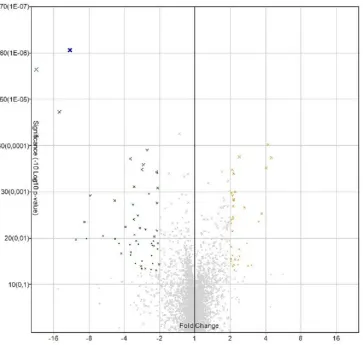
80 miRNAs exhibited statistically significant different expression levels in OSCC samples compared with healthy controls (Figure 1); 35 and 45 miRNAs were upregulated and downregulated, respectively. In table 2, deregulated miRNAs are summarized.
Figure 1. Volcano Plot. Deregulated miRNA in microarray analysis.
Deregulation MiRNA
Down-regulated
miR-29c-3p, miR-486-3p, miR-6762-5p, miR-6838-5p, miR-99a-3p, miR-7641, miR-1224-5p, miR-6868-5p, miR486-5p, 3659, 451a, 19a-3p, miR-138-5p, miR-139, miR-1295a, miR874-5p, miR-514b-5p, miR-758-3p, miR-497-miR-514b-5p, miR-30c-1-3p, 204-5p, 99a-204-5p, 6756-204-5p, 139-204-5p, 6086, 101-3p, 494-3p, 27b-3p, 7162-3p, 664a-3p, 509-5p, 3911,
miR-7977, miR-23b-5p.
Up-regulated
6877-3p, 1908-5p, 3780b-5p, 3180, 6738-5p, 1237-5p, 4443, 6870-5p, 3180-3p, 4442, 4788, miR-548w, miR-6126, miR-4495, miR-6805-5p, miR6765-5p, miR-4706, miR-149-3p, miR6754-3p, miR7846-3p,
miR-8072, miR-146b-5p, miR-135a-3p, miR-5585-3p, miR-5196-5p, miR-6753-3p, miR-885-3p, miR-3175, 3937, miR3934-5p, 663b, 1290,
miR-7111-5p, miR6825-5p, miR-4417.
Table 2. Up regulated and down regulated miRNAs in microarray analysis.
MiRNA expression analysis by RT-qPCR
Following the screening criteria of a ±2.5-fold change and p-values of <0.01, and after in silico
analysis of possible targets, four deregulated miRNAs were chosen for subsequent PCR analysis (miR-617, miR-497-5p, miR-4417, and miR-6825-5p). Only miR-497-5p maintained its diminished expression in all cases compared with controls (p = 0.001), while miR-4417 expression increased (p = 0.007).
As a complementary validation method, we performed a correlation study (Spearman’s rho) between miRNAs and the different platforms used, which yielded rho values of 0.657 and 0.840 for miR-497-5p CC (p = 0.020) and miR-4417 CC = (p < 0.001), respectively.
Clinical characteristics and miRNA expression association
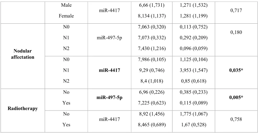
No significant association between patient sex and miR-497-5p or miR-4417 expression was found by the PCR analysis (p = 0.208 for miR-497-5p, p = 0.717 for miR-4417) or microarray analysis (p = 0.299 for miR-497-5p, p = 0.114 for miR-4417).
We detected significant differences between miR-4417 expression and nodular affectation (N1, 3.95; SD = 0.10 vs. N0, 1.13; SD = 1.55; p = 0.035). We also found significantly diminished expression of miR-497-5p in patients who needed radiotherapy (p = 0.05).
The expression level of neither miRNA showed significant differences according to tumour localisation (miR-497-5p, p = 0.475 for PCR and p = 0.111 for microarray; miR-4417, p = 0.212 for PCR and p = 0.164 for microarray). Expression distribution of both miRNAs was the same for differentiation, tumour size, tumour stage, metastasis, chemotherapy, relapse, or survival categories (Table 3).
Variable MicroRNA Microarray Mean (SD) qPCR Mean (SD) PCR (p value)
Sex Male
Female miR-497-5p
8,047 (0,79)
7,581 (1,025)
0,252 (0,361)
Male
Female miR-4417
6,66 (1,731)
8,134 (1,137)
1,271 (1,532)
1,281 (1,199) 0,717
Nodular affectation N0 N1 N2 miR-497-5p 7,063 (0,320) 7,073 (0,332) 7,430 (1,216) 0,113 (0,752) 0,292 (0,209) 0,096 (0,059) 0,180 N0 N1 N2 miR-4417 7,986 (0,105) 9,29 (0,746) 8,4 (1,018) 1,125 (0,104) 3,953 (1,547) 0,85 (0,618) 0,035* Radiotherapy No
Yes miR-497-5p
6,96 (0,226)
7,225 (0,623)
0,385 (0,233)
0,115 (0,089) 0,005*
No
Yes miR-4417
8,92 (1,456)
8,465 (0,689)
1,775 (1,067)
1,67 (0,528) 0,758
Table 3. Association between miRNA expression and clinical parameters.
Survival analysis
The mean follow-up time for the patients was 52.71 months (SD = 71.13), with a range from 7.69 to 243.97 months. The average time of patient survival was 30.24 months (SD = 19.66), with a range from 11.51 to 73.46 months. To study the relationship between miRNA expression and survival, confirmed by PCR, we determined as a cut-off the mean expression level of both miRNAs. The mean expression values for miR-497-5p and miR-4417 were 0.149 and 1.71, respectively.
Cox regression analysis confirmed that patients with miR-497-5p expression levels below the mean value lived for an estimated 24.31 months (IC: 13.93–34.69) compared to those with higher expression levels (40.13 months; IC: 7.38–72.88), although these differences were not statistically significant (p = 0.538). Patients with miR-4417 expression levels above the mean value had a shorter survival (7.48 months; IC: 2.10–12.8) compared to patients with expression levels less than the mean value (12.86 months; IC: 3.35–22.38). However, these differences were not statistically significant either (p = 0.449).
KEGG pathway enrichment analysis
Figure 2. Molecular pathways. Diana Tools results.
We normalised the p-values and confirmed that the most statistically significant was ‘proteoglycans in cancer’ (Figure 3).
Figure 3. Proteoglycans molecular pathway and miRNAs deregulated in our study.
DISCUSSION
In the past few decades, several studies have associated miRNA expression and clinical parameters in OSCC with recurrence, metastasis, and survival. However, to apply those results to clinical practice, more studies are needed. Lin et al. carried out a meta-analysis to evaluate the potential of miRNAs as biomarkers to detect OSCC. They selected eight profiling studies and found different and contradictory results. They ascribed the heterogeneity among studies to differences in study design, sample size, sample type, and origin or population (15). Other recent meta-analyses of OSCC and miRNAs have been conducted by Zahra et al., Zeljic et al., and Troiano et al., which showed some correspondence and some differences in results (7, 12, 16). Authors conclude that a possible bias in meta-analysis is the origin of the samples. Most profiling studies of OSCC and miRNAs have been carried out in Asia. The meta-analysis by Troiano et al. was the only one to include a study in Europe (Denmark).
OSCC prognosis can differ among ethnic groups, as carcinogenic patterns are different, for example, the mutation rates of TP53 are significantly lower in Asian patients than in Caucasian patients (12).
The need to perform more profiling studies of miRNA in OSCC in different parts of the world is justified by the lack of evidence to facilitate expression patterns on the basis of which clinicians can use miRNAs as biomarkers or as possible OSCC therapies.
miRNAs are already in the clinical trial phase for treating other cancers as MRX34 for hepatic carcinoma (17), but owing to variations in tobacco consumption habits, environment, and microbiome influences in miRNA expression, we still do not have enough evidence to apply the same findings to OSCC (8, 18). The statistical analysis in our study revealed a relationship between low miR-497-5p expression (lower than the mean level) and radiotherapy requirement. Moreover, statistically significant over-expression of miR-4417 was found in patients with nodular affectation. These results suggest that the under-expression of miR-497-5p might be associated with more aggressive tumours that need more than only resection.
Our results of differential expression agree with some miRNA results of other profiling studies, but corresponded better with those of Setién-Olarra et al., who used a population similar to the one we used herein. They determined 92 deregulated microRNAs in OSCC patients, and 10 of them coincided with ours (19).
In the designing of our study, the mirVanaTM miRNA isolation kit was chosen as the RNA isolation method because it permits the analysis of all RNAs of a sample, including those with <200 nucleotides. Although other methods exist, the mirVanaTM miRNA isolation kit remains the most used (20). Furthermore, RNU6B was also used as an internal control for RT-qPCR as it has already been applied in other studies (21, 22). Keratinized gingiva tissue was used as the control sample, because sometimes OSCC can also proceed from the gingiva, is exposed to risk factors in a manner similar to the other parts of the oral cavity, and taking it during the extraction of the third molar is a less invasive manner to obtain control tissue from healthy patients. In future studies, it would be better to use healthy samples of the same type of tissue (buccal mucosa, soft palate, retromolar region, tongue, and floor of the mouth), thus matching the tissue origin of the OSCC samples.
of CD44 in saliva has been proposed as an OSCC biomarker, and CD44 concentration is reconsidered a likely risk factor (15, 23). Insulin growth factor 1 receptor (IGF1R), another miR-497-5p target, is a transmembrane receptor activated by IGF1 and IGF2, and its over-expression has also been associated with OSCC (9, 23). Other miR-497-5p targets are the MYC oncogene (24) and MMP9 (25), the high expression of which has long been studied in oral cancer (26). However, profiling studies from Asian patients have found miR-497-5p expression in OSCC patients as being upregulated (27, 28).
Most deregulated miRNAs in this study have been identified only recently; therefore, there is still not enough evidence about their targets or the effects that can produce their altered expression in tissues. Among the 18 screened miRNAs with more significant values (p ≤ 0.001), only six, all of them downregulated, had known targets. In silico enrichment analysis revealed different molecular pathways, like proteoglycans in cancer; protein processing in the endoplasmic reticulum; adherens junctions; focal adhesion; and the Hippo, thyroid hormone, TGF-beta, mTOR, AMPK, p53, insulin, FoxO, HIF-1, and sphingolipid signalling pathways. Most of them are known pathways implicated in tumour development (30). This fact confirms the association between under-expressed miRNAs and oncogenes.
Proteoglycans are the main component in connective tissue that can be linked to the plasma membrane or extracellular matrix, and permit motility and cellular migration and filter the substances that can be transported inside the cell. Proteoglycans in the tumour microenvironment have been demonstrated to participate in proliferation, angiogenesis, or metastasis. For example, Perlecan, a heparan sulphate or sulphatase 2, is over-expressed in cancerous and precancerous oral lesions (31, 32).
Our study demonstrates that the expression of miRNAs in OSCC can be used as a prognostic biomarker, but the differences in expression levels among different populations is a field of research that requires further investigation. Possibly, the use of big data is necessary to be able to take advantage of these small RNAs as a tool for OSCC diagnosis and prognosis, and this study contributes to make possible this analysis in the future.
CONCLUSIONS
We have verified the altered expression of miR-497-5p and miR-4417 in Oral Squamous Cell Carcinoma samples and related the deregulated miRNAs with the ‘proteoglycans in cancer’ pathway.
Acknowledgments
We wish to thank our colleagues from the Maxillofacial Surgery Unit, the Pathological
Anatomy Unit and the biobank of SERGAS in the Santiago de Compostela Teaching Hospital.
Author contributions
Conflict of Interest Statement No conflict of interest.
REFERENCES
1. Malik UU, Zarina S, Pennington SR. Oral squamous cell carcinoma: Key clinical questions, biomarker discovery, and the role of proteomics. Arch Oral Biol. 2016 Mar;63:53-65.
2. Perez-Sayans M, Pilar GD, Barros-Angueira F, Suarez-Penaranda JM, Fernandez AC, Gandara-Rey JM, et al. Current trends in miRNAs and their relationship with oral squamous = cell carcinoma. J Oral Pathol Med. 2012 Jul;41(6):433-43.
3. Park EJ, Shimaoka M, Kiyono H. MicroRNA-mediated dynamic control of mucosal immunity. Int Immunol. 2017 Apr 1;29(4):157-63.
4. Momen-Heravi F, Bala S. miRNA regulation of innate immunity. J Leukoc Biol. 2018 Apr 14.
5. Gorenchtein M, Poh CF, Saini R, Garnis C. MicroRNAs in an oral cancer context - from basic biology to clinical utility. J Dent Res. 2012 May;91(5):440-6.
6. Manikandan M, Deva Magendhra Rao, A. K., Munirajan AK. Altered levels of 21, miR-125b-2*, miR-134, miR-155, miR-184, and miR-205 in oral squamous cell carcinoma and association with clinicopathological characteristics. J Oral Pathol Med. 2014 Dec 8.
7. Zeljic K, Jovanovic I, Jovanovic J, Magic Z, Stankovic A, Supic G. MicroRNA meta-signature of oral cancer: evidence from a meta-analysis. Ups J Med Sci. 2018 Mar;123(1):43-9.
8. Janiszewska J, Szaumkessel M, Szyfter K. microRNAs are important players in head and neck carcinoma: A review. Crit Rev Oncol Hematol. 2013 Aug 12.
9. Chen Z, Jin Y, Yu D, Wang A, Mahjabeen I, Wang C, et al. Down-regulation of the microRNA-99 family members in head and neck squamous cell carcinoma. Oral Oncol. 2012 Aug;48(8):686-91.
10. Min A, Zhu C, Peng S, Rajthala S, Costea DE, Sapkota D. MicroRNAs as Important Players and Biomarkers in Oral Carcinogenesis. Biomed Res Int. 2015;2015:186904.
11. Troiano G, Mastrangelo F, Caponio VCA, Laino L, Cirillo N, Lo Muzio L. Predictive Prognostic Value of Tissue-Based MicroRNA Expression in Oral Squamous Cell Carcinoma: A Systematic Review and Meta-analysis. J Dent Res. 2018 Mar 1:22034518762090.
13. Sauer E, Madea B, Courts C. An evidence based strategy for normalization of quantitative PCR data from miRNA expression analysis in forensically relevant body fluids. Forensic Sci Int Genet. 2014 Jul;11:174-81.
14. Lin N, Lin Y, Fu X, Wu C, Xu J, Cui Z, et al. MicroRNAs as a Novel Class of Diagnostic Biomarkers in Detection of Oral Carcinoma: a Meta-Analysis Study. Clin Lab. 2016;62(3):451-61.
15. Zahra A, Rubab I, Malik S, Khan A, Khan MJ, Fatmi MQ. Meta-Analysis of miRNAs and Their Involvement as Biomarkers in Oral Cancers. Biomed Res Int. 2018 Jan 4;2018:8439820.
16. Tiwari A, Shivananda S, Gopinath KS, Kumar A. MicroRNA-125a Reduces Proliferation and Invasion of Oral Squamous Cell Carcinoma Cells by Targeting Estrogen-related Receptor alpha: IMPLICATIONS FOR CANCER THERAPEUTICS. J Biol Chem. 2014 Nov
14;289(46):32276-90.
17. Van Roosbroeck K, Calin GA. MicroRNAs in chronic lymphocytic leukemia: miRacle or miRage for prognosis and targeted therapies? Semin Oncol. 2016 Apr;43(2):209-14.
18. Setien-Olarra A, Bediaga NG, Acha-Sagredo A, Marichalar-Mendia X, de Pancorbo MM, Aguirre-Urizar JM. Genomewide miRNA profiling of oral lichenoid disorders and oral squamous cell carcinoma. Oral Dis. 2016 Nov;22(8):754-60.
19. Mraz M, Malinova K, Mayer J, Pospisilova S. MicroRNA isolation and stability in stored RNA samples. Biochem Biophys Res Commun. 2009 Dec 4;390(1):1-4.
20. Sun L, Liu L, Fu H, Wang Q, Shi Y. Association of Decreased Expression of Serum miR-9 with Poor Prognosis of Oral Squamous Cell Carcinoma Patients. Med Sci Monit. 2016 Jan 27;22:289-94.
21. Hung PS, Tu HF, Kao SY, Yang CC, Liu CJ, Huang TY, et al. miR-31 is upregulated in oral premalignant epithelium and contributes to the immortalization of normal oral keratinocytes. Carcinogenesis. 2014 May;35(5):1162-71.
22. Yen YC, Shiah SG, Chu HC, Hsu YM, Hsiao JR, Chang JY, et al. Reciprocal regulation of microRNA-99a and insulin-like growth factor I receptor signaling in oral squamous cell carcinoma cells. Mol Cancer. 2014 Jan 10;13:6.
23. Sasahira T, Kirita T, Kuniyasu H. Update of molecular pathobiology in oral cancer: a review. Int J Clin Oncol. 2014 Jun;19(3):431-6.
24. Lu L, Xue X, Lan J, Gao Y, Xiong Z, Zhang H, et al. MicroRNA-29a upregulates MMP2 in oral squamous cell carcinoma to promote cancer invasion and anti-apoptosis. Biomed
Pharmacother. 2014 Feb;68(1):13-9.
25. Huang WC, Chan SH, Jang TH, Chang JW, Ko YC, Yen TC, et al. miRNA-491-5p and GIT1 serve as modulators and biomarkers for oral squamous cell carcinoma invasion and metastasis. Cancer Res. 2014 Feb 1;74(3):751-64.
27. Wong N, Khwaja SS, Baker CM, Gay HA, Thorstad WL, Daly MD, et al. Prognostic microRNA signatures derived from The Cancer Genome Atlas for head and neck squamous cell carcinomas. Cancer Med. 2016 Apr 25.
28. Languino LR, Singh A, Prisco M, Inman GJ, Luginbuhl A, Curry JM, et al. Exosome-mediated transfer from the tumor microenvironment increases TGFbeta signaling in squamous cell carcinoma. Am J Transl Res. 2016 May 15;8(5):2432-7.