R E V I E W
Alveolar epithelial and endothelial cell apoptosis
in emphysema: What we know and what we need
to know
Mathieu C Morissette Julie Parent
Julie Milot
Centre de Recherche de l’Hôpital Laval, Institut Universitaire de Cardiologie et de Pneumologie de l’Université Laval, Québec, Canada
Correspondence: Julie Milot 2725, chemin Ste-Foy, Québec (Qc), Canada, G1V 4G5
Tel +1 418 656 4747 Fax +1 418 656 4762 Email julie.milot@crhl.ulaval.ca
Abstract: Emphysema is mainly caused by cigarette smoking and is characterized by the loss of alveolar integrity and an enlargement of the alveolar space. However, mechanisms involved in its development are not fully understood. Alveolar cell apoptosis has been previously investigated in the lung of emphysematous subjects as a potential contributor to the loss of alveolar cell and has been found abnormally elevated. Though, mechanisms involved in the increased alveolar apoptosis that occurs in emphysema have now become a prolifi c fi eld of research. Those mechanisms are reviewed here with special focus on how they affect cell viability and how they may be implicated in emphysema. Moreover, we sug-gest a model that integrates all those mechanisms to explain the increased alveolar apoptosis observed in emphysema. This review also includes some refl ections and suggestions on the research to come.
Keywords: emphysema, apoptosis, proteases, VEGF, oxidative stress, TRAIL, autoimmunity
Introduction
According to the World Health Organization, chronic obstructive pulmonary disease (COPD) is believed to become the third cause of death by 2030 with cigarette
smok-ing as the main cause.1 COPD features two main phenotypes: chronic bronchitis and
emphysema with different physiopathology and symptoms. Emphysema has retained attention for its micro- and macroscopical manifestations: the loss of alveolar
integ-rity and an enlargement of the alveolar space.2 This leads to a poor gas exchange
at the alveolar level and to the retention of air caused by airways collapse due to
the loss of elastic recoil (hyperinfl ation).2 But what is literally destructing the lung
parenchyma of emphysematous patients? Proteases have been blamed very early, as
defi ciency in α1-antitrypsin (A1AT), the most important antiprotease of the lung,
causes noxious gas-independent emphysema at an early age.3 Emphysema was fi rst
considered a neutrophilic disease, neutrophils being the major protease generator
of all immune cells.4 However, as research progressed, emphysema was revealed to
be a much more complex disease also involving alveolar macrophages (AM)5 and
cytotoxic CD8+ T lymphocytes (CTL).6 Therefore, emphysema is now considered
a complex infl ammatory disease with only partly understood physiopathology. But the question remains: why and how alveolar structure is disappearing? Recently, new hypothesis rose trying to answer this question, among those was “apoptosis”, a complex and well-regulated process that leads to cell death. In this review, we will focus on the recent developments made on the involvement of apoptosis in emphysema and on the possible molecular mechanisms involved in the initiation and progression of the disease.
International Journal of Chronic Obstructive Pulmonary Disease downloaded from https://www.dovepress.com/ by 118.70.13.36 on 22-Aug-2020
Morissette et al
What we know
Apoptosis
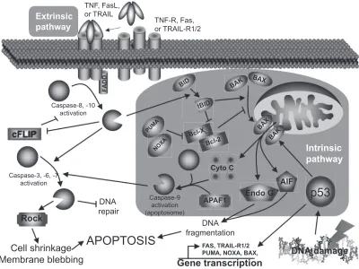
Mechanisms involved in the programmed cell death are very complex but quite well described. There are many reasons why a cell would undergo apoptosis, among them: growth factor deprivation, mitotic aberrations, loss of contact with the extracellular matrix, direct induction by immune cells, activation of death receptors by soluble death ligands, and heavy damages caused by various stresses. All these “stimuli” will lead to cell apoptosis but will trigger different intracel-lular pathways to achieve their goal. The two main apoptotic pathways are the extrinsic and the intrinsic pathways, both leading to DNA fragmentation and cell death.
The extrinsic pathway
The extrinsic pathway (Figure 1) is triggered mostly by death ligands such as tumor necrosis factor (TNF), Fas ligand (FasL), and TNF-related apoptosis-inducing ligand (TRAIL)
through their respective receptors.7 Briefl y, the intracellular
death domains of those receptors will, after autophosphoryla-tion, recruit various adaptor molecules such as Fas-associated
death domain (FADD). If all recruiting steps are encountered, procaspase-8/10 will be cleaved into their active form, caspase-8/10. Caspases (cystein-aspartase) are cystein pro-teases found in their pro-form (inactive form) in living cells. Following specifi c cleavage, they will acquire their protease activity and will start to cleave specifi c substrates and activate other caspases. Once cleaved, those substrates will transmit the apoptotic signal to the nucleus or mitochondria (caspase-3, 6, 7) or interfere with the anti-apoptotic protection (cellular FLICE-like inhibitory protein [cFLIP]). In addition, cleavage of the BH3-interacting domain death agonist (Bid) to trun-cated Bib (tBid) will activate the intrinsic pathway.
The intrinsic pathway
The intrinsic pathway (Figure 1) is mainly triggered by cellular stresses that cause DNA damages like oxidative
stress or UV light.8 Briefl y, DNA damages will lead to the
tumor suppressor p53 activation, stabilization, and to the acquisition of its transcription factor activity. Among its targets, p53 will either induce cell-cycle associated genes
(eg, p21, 14-3-3σ) or apoptosis-related genes (eg, Bax
TNF, FasL, or TRAIL
Caspase-8, -10 activation
cFLIP
Rock
Cell shrinkage Membrane blebbing
APOPTOSIS
DNA repair
DNA fragmentation
FAS, TRAIL-R1/2 PUMA, NOXA, BAX,
Gene transcription
NOXA
BID
BAX BAX
tBID
BAK
BAK
APAF1
Intrinsic pathway
Cyto C PUMA
Bcl-XL
Bcl-2
AIF
Caspase-9 activation (apoptosome) Caspase-3, -6, -7
activation
TNF-R, Fas, or TRAIL-R1/2 Extrinsic
pathway
p53
Endo GFigure 1 Apoptosis pathways.
Abbreviations: cFLIP,cellular FLICE-like inhibitory protein; FADD, Fas-associated death domain; FasL, Fas ligand; TNF, tumor necrosis factor; TRAIL, TNF-related apoptosis-inducing ligand; TRAIL-R, TRAIL receptor; Rock, Rho kinase; AIF, apoptosis-inducing factor; PUMA, p53 upregulated modulator of apoptosis; NOXA, damage.
International Journal of Chronic Obstructive Pulmonary Disease downloaded from https://www.dovepress.com/ by 118.70.13.36 on 22-Aug-2020
Epithelial and endothelial apoptosis in emphysema
[Bcl-2–associated X protein], NOXA [Latin for damage], PUMA [p53 upregulated modulator of apoptosis], Fas, TRAIL-R1/2) transcription, depending on the DNA dam-age severity. If damdam-ages are too severe, p53 will lead to apoptosis: Bax, NOXA, and PUMA will massively trans-locate to the mitochondria outer-membrane and facilitate the formation of channels that will allow pro-apoptotic factors to move to cytoplasm (DNAse, cytochrome c, and anti-apoptotic factors inhibitors). Cytochrome c will join Apaf-1 and caspase-9 to form the so-called apoptosome (activated caspase-9) that will activate caspase-3. As mentioned earlier, the extrinsic pathway can also activate the intrinsic pathway through Bid cleavage (tBid) by caspase-8/10, and help cytosolic Bax to translocate to the mitochondria and induce the release of the mitochondria-contained apoptosis factors.
Finalities
No matter how apoptosis is induced or which pathway
is involved, the fi nalities will be very similar.9 The most
obvious observation is the dynamic membrane blebbing that precedes apoptotic bodies formation. Moreover, the membrane lipid phosphatidyl serine (PS) normally localized on the cytoplasmic side of the cell membrane will be exter-nalized and used by scavenger cells to recognize apoptotic cells, leading to effi cient cell clearance. DNA will also be affected, with features like chromatid condensation, its fragmentation into 180–200 bp fragments (endonuclease G [Endo G], apoptosis-inducing factor [AIF]), and redis-tribution of the genetic material into the forming apoptotic bodies (Rho kinase [ROCK]). All those changes will lead to a clean cell death without intracellular material loss and to the phagocytosis of apoptotic bodies by immune or adjacent structural cells.
Apoptosis and emphysema: cellular mechanisms
Presence of higher apoptosis level in the human emphysema-tous lung has been reported numerous times in previous years (Table 1) and has been associated to decreased alveolar
sur-face area,10 but also to increased alveolar cell proliferation,11
suggesting that regeneration processes of the emphysematous lung might be overwhelmed by its destruction or impared by unknown mechanisms. To explain this phenomenon, mechanisms involved in the regulation of cell survival and
cell death have been studied in vivo (animal models, see
Table 2), ex vivo, and in vitro. Here, we are summarizing the
main mechanisms and their impacts on alveolar apoptosis that occurs in emphysema.
Protease-induced apoptosis
What are proteases?
Proteases are proteins able to cleave specifi c amino-acid sequences and are implicated in many biological processes. In the lung, as in other tissues, proteases activity is closely controlled by anti-proteases. Though, protease/anti-protease balance is an extremely important factor for tissue homeostasis.
What is known about proteases in apoptosis?
In addition to their role in protein turnover, cell migration, and lung immunity, it has been shown that proteases can also induce lung epithelial and endothelial cell apoptosis
(Figure 2).12–14 In fact, Suzuki and colleagues14 showed
that leukocyte elastase (LE), mainly released by activated polymorphonuclear leucocytes, can induce small airway and alveolar epithelial cell apoptosis through the intrinsic pathway and by decreasing AKT phosphorylation (anti-apoptotic factor) following proteinase-activated receptor 1
(PAR-1) activation. Yang and colleagues12 also demonstrated
a similar effect of neutrophil elastase (NE) and proteinase 3 (PR3), mainly secreted by activated neutrophils, on primary bovine arterial endothelial cells. On the other side, it has
been demonstrated that the protease inhibitor α1-antitrypsin
(A1AT) may act as a survival factor by preventing lung
endothelial cell death induced by staurosporine15 and
ciga-rette smoke16 through active caspase inhibition. Moreover,
increasing evidences suggest that “tissues inhibitor of metalloproteinases-1” (TIMP-1) may act as a survival factor through CD63-mediated extracellular signal-regulated kinase
(ERK) and AKT activation.17 Taken together, these data
suggest that protease/anti-protease imbalance can promote apoptosis both through direct activation of PAR-1 signaling and through the reduction of antiproteases’ ability to inhibit apoptotic processes.
What is known about proteases in promoting apoptosis in emphysema?
Discovering that A1AT defi ciency, the main inhibitor of
NE, confers susceptibility to emphysema development18 and
that emphysema-like changes can be induced by elastolytic enzymes instillation in animal models (elastase
emphy-sema)19 were at the fundaments of the protease hypothesis
of emphysema development. In fact, proteases such as NE,20
cathepsins L,21 and matrix metalloproteinases (MMP)-1,22,23
-2,22,24 -8,22 -9,22,24 and -1225 are increased in the
emphysema-tous lung mainly due to a higher number of activated neu-trophils and macrophages. The lung protease hyperactivity
International Journal of Chronic Obstructive Pulmonary Disease downloaded from https://www.dovepress.com/ by 118.70.13.36 on 22-Aug-2020
Morissette et al
is believed to affect alveolar integrity by degrading elastic fi bres responsible for the maintenance of alveolar structure and lung stretch. However, it seems that elastase-induced emphysema may not be attributable only to elastic fi bre degradation. In fact, elastase activity is detectable for only 45 to 50 minutes after elastase instillation while emphysema
phenotype and higher alveolar apoptosis remain for weeks,26
probably due to the installation of an infl ammatory state.
Supporting that hypothesis, mice knockout for IL-1β and
TNF receptors gene and submitted to elastase instillation showed fewer emphysematous lesions as well as alveolar
cells undergoing apoptosis,27 suggesting that elastase may
promote disease progression and alveolar apoptosis through
IL-1β and TNF-dependent mechanism. Moreover, in a mouse
model of emphysema induced by local lung overexpression
of interferon-γ, cathepsin S blockade, a protease involved
Table 1 Characteristics of studies reporting elevated apoptosis in the emphysematous lung
Authors Studied group Control group Apoptosis detection
technique
Apoptotic cell type
Segura-Valdez et al 200022 Smokers with emphysema Smokers and nonsmokers without COPD
In situ end labeling Mainly endothelial cells
Kasahara et al 200040 Smokers with emphysema Smokers and nonsmokers without emphysema
DNA fragmentation (TUNEL and LM-PCR) Single DNA detection (IHC)
Epithelial and endothelial cells
Yokohori et al 200411 Subjects with emphysema (smoking state not specifi ed)
Smokers and nonsmokers without emphysema
DNA fragmentation (TUNEL) Epithelial cells (mainly type II alveolar cells) Calabrese et al 200597 Subjects with AAT-defi ciency
emphysema
Young subjects with unspecifi ed smoking history
DNA fragmentation (TUNEL and agarose gel separation)
Alveolar wall cells
Imai et al 200510 Ex-smokers (⬎6 months) with emphysema
Nonsmokers with unspecifi ed lung functions
cleaved caspase-3 and PARP (Western blot) and DNA fragmentation (agarose gel separation)
Not mentionned
Abbreviations: COPD, chronic obstructive pulmonary disease; IHC, immunohistochemistry; LM-PCR, ligation-mediated polymerase chain reaction; PARP, poly(ADP-ribose) polymerase; TUNEL, terminal deoxynucleotidyl transferase biotin-dUTP nick end labeling.
Table 2 Animal models used to study apoptosis in emphysema
Authors Model of emphysema Apoptosis detection technique Apoptotic cell type
Kasahara et al 200040 Subcutaneaous injection of a VEGF receptor blocker (Sprague-Dawley rats)
DNA fragmentation (TUNEL and LM-PCR) Active caspase-3 (IHC)
Alveolar epithelial and endothelial cells Lucey et al 200227 Intratracheal instillation of porcine
pancreatic elastase (B6129SF2/J mice)
DNA fragmentation (TUNEL) Alveolar epithelial cells
Aoshiba et al 200398 Single intratracheal delivery of caspase-3 (C57BL/6 mice)
DNA fragmentation (TUNEL) and ssDNA detection (IHC)
Mainly alveolar epithelial cells
Bartalesi et al 200599 Whole-body exposure to cigarette smoke (C57BL/6 and DBA/2 mice)
DNA fragmentation (TUNEL) General increase but unspecifi ed cell type Kuo et al 2005100 Whole-body exposure to cigarette
smoke (Wistar rats)
DNA fragmentation (TUNEL) General increase but unspecifi ed cell type Zheng et al 200528 Transgenic lung
IFN-γ overexpression (CC10-rtTA-IFN-γ mice)
DNA fragmentation (TUNEL) Mainly alveolar Type I and Type II cells Petrache et al 2005101 Single intratracheal delivery of
C12ceramide (C57BL/6 mice)
DNA fragmentation (TUNEL) Mainly endothelial cells but also Type II alveolar epithelial cells Taraseviciene-Stewart
et al 200585
Anti-endothelial cells immunization that triggered autoimmune response (Sprague-Dawley rats)
DNA fragmentation (TUNEL) and active caspase-3 (IHC)
Alveolar epithelial cells
Brass et al 2008102 Exposure to lipopolysaccharide (C57BL/6 mice)
DNA fragmentation (TUNEL) and active caspase-3 (Western blot)
Alveolar epithelial cells
Abbreviations: IFN, interferon; IHC, immunohistochemistry; LM-PCR, ligation-mediated polymerase chain reaction; TUNEL, terminal deoxynucleotidyl transferase biotin-dUTP nick end labeling; VEGF, vascular endothelial growth factor.
International Journal of Chronic Obstructive Pulmonary Disease downloaded from https://www.dovepress.com/ by 118.70.13.36 on 22-Aug-2020
Epithelial and endothelial apoptosis in emphysema
in the antigen presentation process, limited the increase of
apoptosis,28 linking proteases once more with apoptosis in
emphysema.
Decreased VEGF signaling
What is VEGF?
Vascular endothelial growth factor (VEGF) is a powerful angiogenic molecule with two receptors (VEGFR1 and 2) triggering opposite cell responses. VEGFR1 is mainly respon-sible for the inhibition of endothelial cell migration and proliferation, when VEGFR2 tends to promote those processes. VEGF is found in great amount in the lung and
act as a key factor in the endothelial maintenance.29
What is known about VEGF in apoptosis?
In regard to apoptosis and cell survival, it is known that VEGF, by itself, can inhibit serum deprivation-induced
endothelial cell apoptosis in vitro.30,31 VEGF increases cell
survival through phosphoinositide-3 kinase (PI3K)/Akt
pathway activation32 leading to caspase-9 and Bad
inac-tivation33,34 and also by increasing the expression of the
anti-apoptotic molecules Bcl-2, A1,30 survivin, and XIAP.35
VEGF is also able to prevent ceramide-induced HMEC (human microvascular endothelial cells) apoptosis through MAPK/ERK pathway activation and SAPK/JNK pathway
inhibition.31
What is known about decreased VEGF signaling in promoting apoptosis in emphysema?
The role of VEGF signaling in the emphysema
pathophysiol-ogy has raised signifi cant interest in the past years.36,37 It has
been shown that VEGF and VEGFR2 levels were decreased in the lung of emphysematous subjects compared to healthy
controls38 and were related to increased alveolar cell
apop-tosis.39 Cigarette smoke-exposed Sprague-Dawley rats also
show decreased lung VEGF and VEGFR2.38 Moreover, rats
treated with the VEGF receptor blocker SU5416 developed emphysema-like phenotype, condition that may be prevented
by the administration of the caspase inhibitor Z-Asp-CH2
-DCB,40 suggesting that decreased VEGF signaling may
induce apoptotic process leading to emphysema. In addition, SU5416 treated rats presented evidences of alveolar oxida-tive stress (higher 8-hydroxyguanine and 4-hydroxynonenal
staining).41 In this model, oxidative stress and emphysema
may be prevented by the administration of the superoxyde
dismutase mimic M40419,41 by N-acetylcystein (NAC),42
and, interestingly, by the caspase inhibitor Z-Asp-CH2
-DCB,41 revealing that the apparition of oxidative stress is
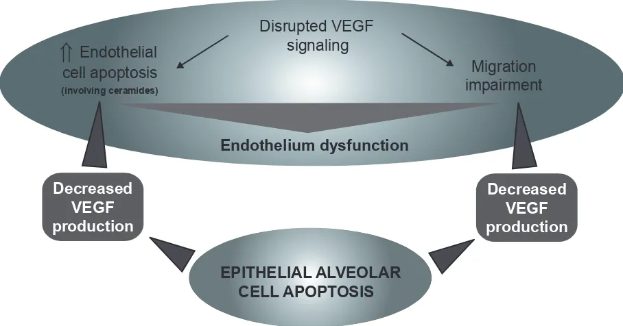
caspase-dependent. Taken together, these data suggest that decreased VEGF signaling may be responsible for endothelial cell apoptosis. However, the capillary bed appears essential
for the growth and maintenance of alveolar septa.43 Moreover,
as demonstrated in human fetal lung44,45 and in adult lung,46
VEGF is released principally by respiratory epithelial cells. Therefore, it may lead to a potential vicious circle that will cause endothelial and epithelial cell apoptosis (Figure 3).
Oxidative stress
What is oxidative stress?
Basically, oxidative stress happens when a cell is exposed to molecules with important oxidative power such as free radicals. If the oxidative defenses are strong enough, cel-lular damages will be negligible. However, if the oxidative aggression is too strong or if it persists for too long, cellular damages may then be very important.
What is known about oxidative stress in apoptosis?
It is well known that oxidative stress can trigger numerous cell responses, including the signaling cascade that will lead to apoptosis (through the intrinsic pathway), and many studies
are available on the subject.47In vitro, exogenous hydrogen
peroxide (H2O2) or compounds that promote endogenous
production of reactive oxygen species (ROS) (eg, arsenic
tri-oxide,48 anthracyclines,49 bleomycin,50N-(4-hydroxyphenyl)
retinamide51) are usually used to create oxidative stress.
A1AT
APOPTOSIS
ActiveCaspase-3
Intrinsic
pathway Erk
Akt A1AT
PA
R
-1
TIMP-1
CD63
TIMP-1 MMPs
NE PR3
LE
PA
R
-1
Figure 2 Mechanisms by which leucocyte elastase (LE), neutrophil elastase (NE), and proteinase 3 (PR3) may induce alveolar epithelial and endothelial cell apoptosis. α-1 antitrypsin (A1AT), proteinase activated receptor 1 (PAR-1), matrix metalloproteinase (MMP), tissue inhibitor of matrix metalloproteinase (TIMP), extracellular-signal regulated kinase (ERK).
International Journal of Chronic Obstructive Pulmonary Disease downloaded from https://www.dovepress.com/ by 118.70.13.36 on 22-Aug-2020
Morissette et al
In the majority of cell types, those treatments will trigger apoptosis through the intrinsic pathway as previously
discussed (Figure 1).47 Another feature of oxidative
stress-induced apoptosis is the activation of the mitogen-activated protein kinase (MAPK) pathways ERK 1/2, jun N-terminal kinase (JNK), and p38 MAPK). The JNK pathway seems to be pro-apoptotic as its inhibition promotes survival in primary
rat alveolar epithelial cells treated with H2O2.52 On the other
side, ERK 1/2 and p38 MAPK tend to have important
anti-apoptotic effects as their inhibition impairs cell survival.52
However, activation and cellular effects of MAPK pathways are known to be different according to cell types studied.
Main defenses against oxidative stress are antioxidant molecules and enzymes. The major antioxidants found in the lung are superoxide dismutase (superoxide anion scavenger),
catalase (converts H2O2 to water), and glutathione peroxidase
(converts organic hydroperoxides to organic hydroxides).53
What is known about oxidative stress in promoting apoptosis in emphysema?
In the case of emphysema, oxidant molecules have two main origins. The fi rst is of course cigarette smoke (active smoker), which contains free radicals with tremendous
oxi-dative power such as superoxide (O2•−), hydroxyl radical
(•OH), and hydrogen peroxide (H2O2).54 It is important to
note that nonemphysematous smokers are also exposed to the oxidative agents contained in cigarette smoke, suggesting that smokers who have developed emphysema (15%–20%) reacted differently or were more susceptible to this stress. The second source, and probably the most sustained, comes from the chronic inflammation inherent to emphysema
that persists even after smoking cessation.55 Activated
macrophages and neutrophils, present in higher number
in the emphysematous lung,56–59 are powerful producers of
reactive oxygen species (ROS).60,61 It is well known that the
lung antioxidant defenses of subjects with emphysema are overwhelmed, leading to important oxidative damages. In fact, compared to those of normal smokers and ex-smokers, the emphysematous lung presents more damaged proteinsand
more peroxidated lipids.62,63 All those oxidative damages
may then lead to the increased alveolar apoptosis observed in emphysema (Figure 4).
TRAIL-mediated apoptosis
What is TRAIL?
Tumor necrosis factor-related apoptosis-inducing ligand (TRAIL) is a member of the death ligand TNF family that also includes FasL. TRAIL has four receptors: TRAIL-R1
Cigarette smoke
(Oxidative stress)
Disrupted VEGF
signaling
Migration
impairment
Endothelium dysfunction
EPITHELIAL ALVEOLAR
CELL APOPTOSIS
Decreased
VEGF
production
Decreased
VEGF
production
⇑
Endothelial
cell apoptosis
(involving ceramides)
Figure 3 Cigarette smoke (oxidative stress)-mediated VEGF signaling disruption leading to endothelial cell death, migration impairment, and general endothelium dysfunction causing epithelial cells apoptosis.
Abbreviation: VEGF,vascular endothelial growth factor.
International Journal of Chronic Obstructive Pulmonary Disease downloaded from https://www.dovepress.com/ by 118.70.13.36 on 22-Aug-2020
Epithelial and endothelial apoptosis in emphysema
and R2 which contain functional death domains, TRAIL-R3 which contains a truncated death domain and acts as a decoy, and TRAIL-R4 which is unable to induce apoptotic signals but, as well as TRAIL-R1 and R2, can activate kinases that
will lead to NF-κB activation.64
What is known about TRAIL in apoptosis?
First interest on TRAIL has been brought by its ability to induce transformed/cancer cell apoptosis while having no
such effects on normal cells.65 Further research has shown
that not all transformed/cancer cells were sensitive to its
apoptotic signal.66 Interestingly, DNA damage and ROS
generating agents are able to suppress cellular resistance to TRAIL-mediated apoptosis in many cell lines, including
the lung adenocarcinoma cell line A549.67–69 Moreover, in a
mouse model of Alzheimer’s disease, a disease characterized by a progressive lost of neuronal cells, blockade of
TRAIL-R2 prevents β-amyloid-induced neuronal cell apoptosis.70
The main cells releasing TRAIL are activated macrophages,
NK cells, and T lymphocytes.71–73
What is known about TRAIL in promoting apoptosis in emphysema?
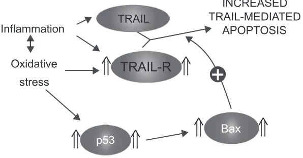
TRAIL receptors 1, 2, and 3 have been shown to be upregu-lated in the alveolar epithelial cells of emphysematous
smokers and ex-smokers.74 Moreover, their expression was
closely related to the levels of the tumor suppressor p53 in the
emphysematous lung parenchyma.74 Interestingly, A549 cells
(lung adenocarcinoma cell line) exposed in vitro to oxidative
stress (H2O2) and TNF had higher expression of TRAIL-R1,
2, and 3 but also higher levels of p53,74 suggesting that the
modulation of the TRAIL system observed in the emphyse-matous lung may be attributable to oxidative stress and/or infl ammatory cytokines. As TRAIL is released by activated infl ammatory cells, oxidative stress and infl ammation present in the emphysematous lung may sensitize alveolar cells to its apoptotic effects (Figure 5).
Killer cells and autoimmunity
What are killer cells and autoimmunity?
Killer cells are immune cells specialized in the act of killing infected or unwanted cells. There are mainly two types of
killer cells: cytotoxic CD8+ T lymphocytes (CTL), members
of the adaptive immunity, and natural killer (NK) cells, mem-bers of the innate immunity. The main difference between these two cell types is the way they recognize cells that have to be killed. When a given cell is infected with intracellular pathogens, major histocompatibility complex (MHC) class I will present pathogen peptides at the cell surface allowing CTL to recognize it with their T cell receptor (TCR) and kill
the infected cell.75 On its side, with no TCR, NK cell cannot
use by itself antigen-based recognition to identify cells to be killed. However, specifi c antigen-mediated cytotoxicity can be indirectly induced through antibody-dependent cellular
Figure 4 Increased alveolar apoptosis mediated through oxidative stress-induced cellular damages.
International Journal of Chronic Obstructive Pulmonary Disease downloaded from https://www.dovepress.com/ by 118.70.13.36 on 22-Aug-2020
Morissette et al
cytotoxicity (ADCC) via the binding of FcγRIII (CD16) to
the Fc portion of IgG bound to specifi c target cell membrane
antigen.76
Autoimmunity is an inappropriate immune response against antigens of the host (self-antigen) and can be
trig-gered by a variety of mechanisms;77 we describe here only
those that may be relevant in emphysema. Autoimmune response can be triggered by modified self-antigen, as exposure of cells to agents that may affect protein integrity (eg, oxidants, proteases) can create molecules unknown to the immune system and cause autoimmune response. Moreover, molecules normally hidden from the immune system (located into the nucleus or the cytoplasm) may trigger autoimmune response if released. Failed clearance of apoptotic cells or high levels of necrosis may expose molecules normally sequestered to the cytoplasm or to the nucleus, as it is
observed in systemic lupus erythematosus.78
What is known about killer cells and autoimmunity in apoptosis?
When an immune response is initiated against a self-antigen, processes involved in the targeting and recognition by antibodies or killer cells are very similar to those raised against pathogens. First, the humoral response is involved: subjects with autoimmune diseases like systemic lupus erythematosus and rheumatoid arthritis are presenting
autoantibodies in their serum.79 Those antibodies secreted
by activated B cells will, after recognition of their specifi c antigens, trigger local infl ammatory response to eliminate
the cells that bears autoantigen/antoantibody complexes.80
As mentioned earlier, NK cell can recognize these cells and induce their apoptosis. In fact, following recognition, NK cell will induce target cell’s apoptosis mainly trough the release of
granules that contain cytotoxic molecules like “pore forming protein” (perforin), granzymes, and granulisin. Death ligands such as FasL and TRAIL also play an important role in the
NK-mediated cell apoptosis.81 In addition to the
antibody-mediated immune response, a specifi c cellular response can
be mediated by CD4+ and CD8+ T lymphocytes. Once CD4+
T lymphocytes have encountered their antigen presented by an antigen-presenting cell (APC) like dendritic cells, they will proliferate (oligoclonal expansion) and be involved in the activation of B cells and alveolar macrophages through
cell/cell contact or the release of infl ammatory mediators.82
On their side, activated CD8+ T lymphocytes will directly
target cells that express the specifi c autoantigen and, with mechanisms similar to those used by NK cell, induce target
cell apoptosis.83 All these processes will lead to the deletion
of a specifi c autoantigen-bearing cell population.
What is known about killer cells and autoimmunity in promoting apoptosis in emphysema?
There are evidences that emphysema may have an autoim-mune component. Autoreactive antibodies with avidity for pulmonary epithelial cells were found in the serum of about 70% of subjects with emphysema when only 10% of normal
smokers or nonsmokers were positive.84 Moreover, in vitro
peripheral blood mononuclear cells cytotoxicity against pul-monary epithelial cells was stronger in presence of plasma
from emphysematous subjects than from normal subjects,84
suggesting that autoreactive antibodies may initiate immune cell-mediated apoptosis. Moreover, a model of autoimmune emphysema (Table 2) revealed that immune response against lung endothelial cells can lead to alveolar endothelial and
epithelial cell apoptosis and cause emphysema.85 Antibodies
against elastin protein have also been found in the serum
Inflammation
INCREASED
APOPTOSIS TRAIL-MEDIATED
Oxidative stress
TRAIL
TRAIL-R
p53 Bax
Figure 5 Sensitization to TRAIL-mediated apoptosis.
Abbreviation: TRAIL, tumor necrosis factor-related apoptosis-inducing ligand; TRAIL-R, TRAIL receptors.
International Journal of Chronic Obstructive Pulmonary Disease downloaded from https://www.dovepress.com/ by 118.70.13.36 on 22-Aug-2020
Epithelial and endothelial apoptosis in emphysema
of emphysematous subject86 as well as a higher number
of B cells secreting anti-elastin antibodies in the
emphy-sematous lung.86 In the same study, they also found that
peripheral blood CD4+ T lymphocytes from emphysematous
subjects were releasing more gamma interferon (IFN-γ) and
interleukin-10 (IL-10) than those from normal subjects upon
in vitro elastin exposure.86 Oligoclonal CD4+ T lymphocytes
able to proliferate in vitro upon interleukin-2 stimulation87,88
as well as oligoclonal CD8+ T lymphocytes89 were found
in the lung of subjects with emphysema. Moreover, CD8+
T lymphocytes from the lung of COPD subjects expressed higher levels of perforin and cytotoxic activity than those
from controls,90.91 phenomenon that is not observed in the
peripheral blood.92 All these data suggest the existence
of a local antigen-driven cellular immunity in the lung of emphysematous subjects (Figure 6).
What we need to know
Which cell type is dying?
There are three major structural cell types in the alveolar tissue: Type I alveolar cells, Type II alveolar cells, and endothelial cells. Type I alveolar is the most abundant cell type found in the alveolar tissue. They are responsible for the maintenance of the alveolar structure and gas diffusion toward alveolar capillaries. Type II alveolar cells are the major source of surfactant proteins and are the progenitor of Type I alveolar cells. Endothelial cells are in charge of lung
perfusion and, with Type I alveolar cells, of gas diffusion between alveoli and blood. As all these cell types are impor-tant to maintain alveolar tissue integrity, the accelerated loss of one or another will have direct effect on the whole alveolar tissue. The technique of choice to detect apoptotic cells in the emphysematous lung is terminal deoxynucleotidyl transfer-ase biotin-dUTP nick end labeling (TUNEL) analysis. Unlike tissue lysat-based analyses that only give a general view of the tissue apoptosis level (eg, Ligation-mediated polymerase chain reaction [LM-PCR], caspase-3 activity, poly(ADP-ribose) polymerase [PARP]) cleavage), TUNEL allows a precise identifi cation of the cells that undergo apoptosis. However, despite comparative TUNEL analyses of normal and emphysematous lung (Table 1), no real consensus has been established on which cell type seems to die predomi-nantly from apoptosis in the emphysematous lung.
According to the different hypothesis that attempt to explain the increased alveolar apoptosis that occurs in emphy-sema, different cell types are targeted as the earliest cell type touched by apoptosis. The VEGF hypothesis points mainly
at endothelial cells40 when the proteases imbalance,14,27
oxidative stress,62 and TRAIL hypothesis74 tend to point at
epithelial cells. Autoimmunity84,85 seems to be touching both
endothelial and epithelial cells. If we consider the confor-mation of the alveoli, epithelial alveolar cells are exposed directly to cigarette smoke and may be acting as a “cellular shield” for endothelial cells. It is then logic to assume that
CD8+
act CD8+
act CD4+ APC
Protease/anti-protease imbalance
Oxidative stress
CD4+
B Proliferation
ALVEOLAR CELL
NK
APOPTOSIS
Figure 6 Induction of an autoimmune response against immunogenic self-antigens after protease and oxidative stress-induced modifi cations. Antigen-presenting cell (APC). Self-antigen (), modifi ed self-antigen (¤), antibody against modifi ed self-antigen (Y).
International Journal of Chronic Obstructive Pulmonary Disease downloaded from https://www.dovepress.com/ by 118.70.13.36 on 22-Aug-2020
Morissette et al
most of the damages induced by cigarette smoke will be fi rst done to epithelial rather than endothelial cells. There is a lot of in vitro data on the effects of cigarette smoke on signal-ing pathways of alveolar epithelial and endothelial cells. However, no data are currently available on the crosstalk of alveolar epithelial cells exposed to cigarette smoke with endothelial cells. This would be extremely relevant to under-stand how damaged alveolar epithelial cells may affect the functions and the faith of endothelial cells, and would give important information on which cell type may be susceptible to undergo apoptosis fi rst.
When does it die?
Until now, publications on apoptosis in emphysema com-pared the apoptosis levels in the lung of emphysematous subjects with well-developed disease with those of nonem-physematous subjects. From this, we can say that apoptosis levels are higher in emphysematous subjects despite smoking cessation and we can suppose that apoptosis is involved in the progression of the disease. We also know that alveolar epithelial cells’ proliferation is increased, but does not seem to compensate for cell loss (apoptosis/proliferation imbal-ance). However, we do not know if the apoptotic mecha-nisms that have been described in this review are involved in the initiation of the disease or only in the progression once the disease has started. When does apoptosis overcome proliferation? When do proteases overcome anti-proteases?
When do VEGF and VEGFR2 levels go down? When do
p53, Bax/Bcl-xL ratio, and TRAIL-R go up? And fi nally,
when does autoimmunity get in play? Answering these questions is not an easy task. In order to do so, we would need to study for a long period of time smoking subjects that are susceptible to develop emphysema but that have not developed it yet. However, since there is no reliable method
to identify these subjects yet, animal models and in vitro
cell culture are the only tools that can help to understand mechanisms involved in emphysema initiation rather than on its progression.
What is responsible for cell death?
Looking for only one mechanism to explain the increased alveolar apoptosis would be an important mistake. Prote-ases, oxidative stress, decreased VEGF, autoimmunity, and TRAIL-mediated apoptosis are probably all involved at dif-ferent levels in the increased apoptosis levels in emphysema. The elaboration of a complete model that would integrate all mechanisms and link them to each other may be extremely relevant to guide research on the increased apoptotic events observed in emphysema. Here, we propose a model (Figure 7) built from the studies we previously discussed. Emerging concepts or hypothesis such as alveolar apoptosis induced by
neutrophils-derived defensins,93,94 link between innate
immu-nity and oxidative stress through Toll-like receptor 4 (TLR4)
tone,95 and reduced or impared apoptotic cell clearance
Cigarette
smoke
Depletion of oxidative defenses
(susceptible individuals)
ROS
release
oxidative stress
Sustained
Inflammation
Alveolar epithelial
and endothelial
cell injuries
Alveolar epithelial
and endothelial
cell apoptosis
Disrupted VEGF
signaling
Sensitization
to TRAIL
TRAIL
release
Protease/
anti-protease
imbalance
Release of
immunogenic peptides
(autoimmunity)
1
7
5 3
5
4 6
8
9
13
11
10
12
14
12
13
11
15 10
2
Figure 7 Proposed model of the mechanisms involved in the increased alveolar apoptosis observed in emphysema (see text for details).
International Journal of Chronic Obstructive Pulmonary Disease downloaded from https://www.dovepress.com/ by 118.70.13.36 on 22-Aug-2020
Epithelial and endothelial apoptosis in emphysema
(efferocytosis),96 even if very interesting and promising ways
of research, were not included in the model.
Global model to explain increased apoptosis in emphysema
As we know, 90% of emphysema cases are developed follow-ing cigarette smoke exposure. We also know that, among all its toxic power, cigarette smoke contains powerful oxidants. In some individuals with weaker oxidative defenses (genetic feature), chronic cigarette smoke exposure will lead to oxida-tive defense depletion (see Arrow [1] in Figure 7) and the establishment of a sustained oxidative stress [2] leading to lethal injuries to alveolar cells [3] and an increase in alveolar cell apoptosis [4]. Oxidative stress can also be responsible for the initiation of an infl ammatory process, either from direct immune cell activation or through the release of infl amma-tory mediators by injured alveolar cells [5]. In the present model, the following steps are very important to establish the chronic state of the disease. Activated infl ammatory cells, mainly neutrophils and macrophages, are well known to be powerful reactive oxygen species (ROS) producers [6]. ROS released from infl ammatory cells may then be involved in the establishment of a cigarette smoke-independent lung oxidative stress [7], as oxidative stress remains in the lung of emphysematous subjects after smoking cessation. Activated neutrophils and macrophages can also release great amount of proteases that then lead to lung protease/anti-protease imbal-ance [8] and induce alveolar epithelial and endothelial apop-tosis [9]. Protease hyperactivity and oxidative stress could also be responsible for the release of immunogenic peptides [10] and lead to the development of antigen-driven immune
response (antigen-specifi c activation of B cells and CD4+ and
CD8+ T cells) and then promote alveolar cell apoptosis [11].
Infl ammatory mediators, like TNF, and oxidative stress (cell injuries) can also modulate elements involved in alveolar cell sensitivity to TRAIL-mediated apoptosis (TRAIL receptors, p53, and Bax levels) [12] and switch alveolar cells from a TRAIL-mediated apoptosis resistant state to a sensitive state and lead to increased alveolar cell apoptosis [13]. Oxidative stress may also be responsible for the disruption of VEGF signaling [14], leading to alveolar endothelial cell apoptosis, and, through a lack of physical support, promoting alveolar epithelial cell apoptosis [15].
Based on this model of hypothesis, tools such as DNA and protein microarrays and softwares for cellular pathway interactions analysis will help to see in a more global way the pathways involved in the disease development while new primary cell isolation and culture will give better
cellular models. All this will increase the comprehension of the different mechanisms involved in emphysema pathogen-esis and ultimately lead the elaboration of treatments that will stop the development or the progression of the disease.
Disclosure
The authors report no confl icts of interest in this work. MCM is the recipient of a studentship from the Fonds de Recherche
en SantéduQuébec.
References
1. Murray CJ, Lopez AD. Alternative projections of mortality and disability by cause 1990–2020: Global Burden of Disease Study. Lancet. 1997;349:1498–1504.
2. Hogg JC. Pathophysiology of airfl ow limitation in chronic obstructive pulmonary disease. Lancet. 2004;364:709–721.
3. Mulgrew AT, Taggart CC, McElvaney NG. Alpha1antitrypsin defi -ciency: current concepts. Lung. 2007;185:191–201.
4. Quint JK, Wedzicha JA. The neutrophil in chronic obstructive pulmo-nary disease. J Allergy Clin Immunol. 2007;119:1065–1071. 5. Barnes PJ. Alveolar macrophages in chronic obstructive pulmonary
disease (COPD). Cell Mol Biol (Noisy-le-grand). 2004;50: OL627–OL637.
6. Cosio MG, Majo J, Cosio MG. Infl ammation of the airways and lung parenchyma in COPD: role of T cells. Chest. 2002;121:160S–165S. 7. Strasser A, O’Connor L, Dixit VM. Apoptosis signaling. Annu Rev
Biochem. 2000;69:217–245.
8. Roos WP, Kaina B. DNA damage-induced cell death by apoptosis. Trends Mol Med. 2006;12:440–450.
9. Elmore S. Apoptosis: a review of programmed cell death. Toxicol Pathol. 2007;35:495–516.
10. Imai K, Mercer BA, Schulman LL, et al. Correlation of lung surface area to apoptosis and proliferation in human emphysema. Eur Respir J. 2005;25:250–258.
11. Yokohori N, Aoshiba K, Nagai A. Increased levels of cell death and proliferation in alveolar wall cells in patients with pulmonary emphy-sema. Chest. 2004;125:626–632.
12. Yang JJ, Kettritz R, Falk RJ, et al. Apoptosis of endothelial cells induced by the neutrophil serine proteases proteinase 3 and elastase. Am J Pathol. 1996;149:1617–1626.
13. Ginzberg HH, Shannon PT, Suzuki T, et al. Leukocyte elastase induces epithelial apoptosis: role of mitochondial permeability changes and Akt. Am J Physiol Gastrointest Liver Physiol. 2004;287:G286–G298. 14. Suzuki T, Moraes TJ, Vachon E, et al. Proteinase-activated receptor-1
mediates elastase-induced apoptosis of human lung epithelial cells. Am J Respir Cell Mol Biol. 2005;33:231–247.
15. Petrache I, Fijalkowska I, Medler TR, et al. alpha-1 antitrypsin inhibits caspase-3 activity, preventing lung endothelial cell apoptosis. Am J Pathol. 2006;169:1155–1166.
16. Aldonyte R, Hutchinson ET, Jin B, et al. Endothelial alpha-1-antitrypsin attenuates cigarette smoke induced apoptosis in vitro. COPD. 2008;5:153–162.
17. Chirco R, Liu XW, Jung KK, et al. Novel functions of TIMPs in cell signaling. Cancer Metastasis Rev. 2006;25:99–113.
18. Eriksson S. Pulmonary emphysema and alpha1-antitrypsin defi ciency. Acta Med Scand. 1964;175:197–205.
19. Gross P, Pfi tzer EA, Tolker E, et al. Experimental emphysema: its pro-duction with papain in normal and silicotic rats. Arch Environ Health. 1965;11:50–58.
20. Yoshioka A, Betsuyaku T, Nishimura M, et al. Excessive neutrophil elastase in bronchoalveolar lavage fl uid in subclinical emphysema. Am J Respir Crit Care Med. 1995;152:2127–2132.
International Journal of Chronic Obstructive Pulmonary Disease downloaded from https://www.dovepress.com/ by 118.70.13.36 on 22-Aug-2020
Morissette et al
21. Takeyabu K, Betsuyaku T, Nishimura M, et al. Cysteine proteinases and cystatin C in bronchoalveolar lavage fl uid from subjects with subclinical emphysema. Eur Respir J. 1998;12:1033–1039.
22. Segura-Valdez L, Pardo A, Gaxiola M, et al. Upregulation of gelatinases A and B, collagenases 1 and 2, and increased parenchymal cell death in COPD. Chest. 2000;117:684–694.
23. Imai K, Dalal SS, Chen ES, et al. Human collagenase (matrix metalloproteinase-1) expression in the lungs of patients with emphy-sema. Am J Respir Crit Care Med. 2001;163:786–791.
24. Ohnishi K, Takagi M, Kurokawa Y, et al. Matrix metalloproteinase-mediated extracellular matrix protein degradation in human pulmonary emphysema. Lab Invest. 1998;78:1077–1087.
25. Babusyte A, Stravinskaite K, Jeroch J, et al. Patterns of airway infl am-mation and MMP-12 expression in smokers and ex-smokers with COPD. Respir Res. 2007;8:81.
26. Stone PJ, Lucey EC, Calore JD, et al. Defenses of the hamster lung against human neutrophil and porcine pancreatic elastase. Respiration. 1988;54:1–15.
27. Lucey EC, Keane J, Kuang PP, et al. Severity of elastase-induced emphysema is decreased in tumor necrosis factor-alpha and interleukin-1beta receptor-defi cient mice. Lab Invest. 2002;82:79–85.
28. Zheng T, Kang MJ, Crothers K, et al. Role of cathepsin S-dependent epithelial cell apoptosis in IFN-gamma-induced alveolar remodeling and pulmonary emphysema. J Immunol. 2005;174:8106–8115. 29. Ferrara N, Gerber HP, LeCouter J. The biology of VEGF and its
recep-tors. Nat Med. 2003;9:669–676.
30. Gerber HP, Dixit V, Ferrara N. Vascular endothelial growth factor induces expression of the antiapoptotic proteins Bcl-2 and A1 in vas-cular endothelial cells. J Biol Chem. 1998;273:13313–13316. 31. Gupta K, Kshirsagar S, Li W, et al. VEGF prevents apoptosis of human
microvascular endothelial cells via opposing effects on MAPK/ERK and SAPK/JNK signaling. Exp Cell Res. 1999;247:495–504. 32. Gerber HP, McMurtrey A, Kowalski J, et al. Vascular endothelial
growth factor regulates endothelial cell survival through the phospha-tidylinositol 3’-kinase/Akt signal transduction pathway. Requirement for Flk-1/KDR activation. J Biol Chem. 1998;273:30336–30343. 33. Cardone MH, Roy N, Stennicke HR, et al. Regulation of cell death protease
caspase-9 by phosphorylation. Science. 1998;282:1318–1321. 34. del Peso L, Gonzalez-Garcia M, Page C, et al. Interleukin-3-induced
phosphorylation of BAD through the protein kinase Akt. Science. 1997;278:687–689.
35. Tran J, Rak J, Sheehan C, et al. Marked induction of the IAP family antiapoptotic proteins survivin and XIAP by VEGF in vascular endo-thelial cells. Biochem Biophys Res Commun. 1999;264:781–788. 36. Kanazawa H. Role of vascular endothelial growth factor in the
patho-genesis of chronic obstructive pulmonary disease. Med Sci Monit. 2007;13:RA189–195.
37. Voelkel NF, Vandivier RW, Tuder RM. Vascular endothelial growth factor in the lung. Am J Physiol Lung Cell Mol Physiol. 2006;290: L209–L221.
38. Marwick JA, Stevenson CS, Giddings J, et al. Cigarette smoke disrupts VEGF165-VEGFR-2 receptor signaling complex in rat lungs and patients with COPD: morphological impact of VEGFR-2 inhibition. Am J Physiol Lung Cell Mol Physiol. 2006;290:L897–908.
39. Kasahara Y, Tuder RM, Cool CD, et al. Endothelial cell death and decreased expression of vascular endothelial growth factor and vascular endothelial growth factor receptor 2 in emphysema. Am J Respir Crit Care Med. 2001;163:737–744.
40. Kasahara Y, Tuder RM, Taraseviciene-Stewart L, et al. Inhibition of VEGF receptors causes lung cell apoptosis and emphysema. J Clin Invest. 2000;106:1311–1319.
41. Tuder RM, Zhen L, Cho CY, et al. Oxidative stress and apoptosis interact and cause emphysema due to vascular endothelial growth factor receptor blockade. Am J Respir Cell Mol Biol. 2003;29:88–97. 42. Demura Y, Taraseviciene-Stewart L, Scerbavicius R, et al. N-acetylcysteine
treatment protects against VEGF-receptor blockade-related emphysema. COPD. 2004;1:25–32.
43. Committee ahS. Mechanisms and limits of induced postnatal lung growth. Am J Respir Crit Care Med. 2004;170:319–343.
44. Acarregui MJ, Penisten ST, Goss KL, et al. Vascular endothelial growth factor gene expression in human fetal lung in vitro. Am J Respir Cell Mol Biol. 1999;20:14–23.
45. Shifren JL, Doldi N, Ferrara N, et al. In the human fetus, vascular endothelial growth factor is expressed in epithelial cells and myocytes, but not vascular endothelium: implications for mode of action. J Clin Endocrinol Metab. 1994;79:316–322.
46. Ng YS, Rohan R, Sunday ME, et al. Differential expression of VEGF isoforms in mouse during development and in the adult. Dev Dyn. 2001;220:112–121.
47. Ryter SW, Kim HP, Hoetzel A, et al. Mechanisms of cell death in oxidative stress. Antioxid Redox Signal. 2007;9:49–89.
48. Jing Y, Dai J, Chalmers-Redman RM, et al. Arsenic trioxide selectively induces acute promyelocytic leukemia cell apoptosis via a hydrogen peroxide-dependent pathway. Blood. 1999;94:2102–2111.
49. Serrano J, Palmeira CM, Kuehl DW, et al. Cardioselective and cumula-tive oxidation of mitochondrial DNA following subchronic doxorubicin administration. Biochim Biophys Acta. 1999;1411:201–205. 50. Hug H, Strand S, Grambihler A, et al. Reactive oxygen intermediates
are involved in the induction of CD95 ligand mRNA expression by cyto-static drugs in hepatoma cells. J Biol Chem. 1997;272:28191–28193. 51. Suzuki S, Higuchi M, Proske RJ, et al. Implication of mitochon-dria-derived reactive oxygen species, cytochrome C and caspase-3 in N-(4-hydroxyphenyl)retinamide-induced apoptosis in cervical carcinoma cells. Oncogene. 1999;18:6380–6387.
52. Carvalho H, Evelson P, Sigaud S, et al. Mitogen-activated protein kinases modulate H(2)O(2)-induced apoptosis in primary rat alveolar epithelial cells. J Cell Biochem. 2004;92:502–513.
53. Rahman I, Biswas SK, Kode A. Oxidant and antioxidant balance in the airways and airway diseases. Eur J Pharmacol. 2006;533:222–239. 54. Pryor WA. Cigarette smoke radicals and the role of free radicals
in chemical carcinogenicity. Environ Health Perspect. 1997;105 (Suppl 4):875–882.
55. Willemse BW, Postma DS, Timens W, et al. The impact of smoking cessation on respiratory symptoms, lung function, airway hyperrespon-siveness and infl ammation. Eur Respir J. 2004;23:464–476. 56. Russell RE, Culpitt SV, DeMatos C, et al. Release and activity of matrix
metalloproteinase-9 and tissue inhibitor of metalloproteinase-1 by alveolar macrophages from patients with chronic obstructive pulmonary disease. Am J Respir Cell Mol Biol. 2002;26:602–609.
57. Russell RE, Thorley A, Culpitt SV, et al. Alveolar macrophage-mediated elastolysis: roles of matrix metalloproteinases, cysteine, and serine proteases. Am J Physiol Lung Cell Mol Physiol. 2002;283:L867–73. 58. Lacoste JY, Bousquet J, Chanez P, et al. Eosinophilic and neutrophilic
infl ammation in asthma, chronic bronchitis, and chronic obstructive pulmonary disease. J Allergy Clin Immunol. 1993;92:537–548. 59. Keatings VM, Barnes PJ. Granulocyte activation markers in induced
sputum: comparison between chronic obstructive pulmonary disease, asthma, and normal subjects. Am J Respir Crit Care Med. 1997;155:449–453.
60. Gwinn MR, Vallyathan V. Respiratory burst: role in signal transduc-tion in alveolar macrophages. J Toxicol Environ Health B Crit Rev. 2006;9:27–39.
61. Dahlgren C, Karlsson A. Respiratory burst in human neutrophils. J Immunol Methods. 1999;232:3–14.
62. Rahman I, van Schadewijk AA, Crowther AJ, et al. 4-Hydroxy-2-nonenal, a specifi c lipid peroxidation product, is elevated in lungs of patients with chronic obstructive pulmonary disease. Am J Respir Crit Care Med. 2002;166:490–495.
63. Malhotra D, Thimmulappa R, Navas-Acien A, et al. Decline in NRF2 Regulated Antioxidants in COPD Lungs due to Loss of its Positive Regulator DJ-1. Am J Respir Crit Care Med. 2008.
64. Falschlehner C, Emmerich CH, Gerlach B, et al. TRAIL signal-ling: decisions between life and death. Int J Biochem Cell Biol. 2007;39:1462–1475.
International Journal of Chronic Obstructive Pulmonary Disease downloaded from https://www.dovepress.com/ by 118.70.13.36 on 22-Aug-2020
Epithelial and endothelial apoptosis in emphysema
65. Bonavida B, Ng CP, Jazirehi A, Schiller G, Mizutani Y. Selectivity of TRAIL-mediated apoptosis of cancer cells and synergy with drugs: the trail to non-toxic cancer therapeutics. Int J Oncol. 1999;15:793–802. 66. Zhang L, Fang B. Mechanisms of resistance to TRAIL-induced
apoptosis in cancer. Cancer Gene Ther. 2005;12:228–237.
67. Hu H, Jiang C, Schuster T, et al. Inorganic selenium sensitizes prostate cancer cells to TRAIL-induced apoptosis through superoxide/p53/Bax-mediated activation of mitochondrial pathway. Mol Cancer Ther. 2006;5:1873–1882.
68. Fan QL, Zou WY, Song LH, et al. Synergistic antitumor activity of TRAIL combined with chemotherapeutic agents in A549 cell lines in vitro and in vivo. Cancer Chemother Pharmacol. 2005;55:189–196. 69. Frese S, Brunner T, Gugger M, et al. Enhancement of Apo2L/TRAIL
(tumor necrosis factor-related apoptosis-inducing ligand)-induced apop-tosis in non-small cell lung cancer cell lines by chemotherapeutic agents without correlation to the expression level of cellular protease caspase-8 inhibitory protein. J Thorac Cardiovasc Surg. 2002;123:168–174. 70. Uberti D, Ferrari-Toninelli G, Bonini SA, et al. Blockade of the tumor
necrosis factor-related apoptosis inducing ligand death receptor DR5 prevents beta-amyloid neurotoxicity. Neuropsychopharmacology. 2007;32:872–880.
71. Robertson NM, Zangrilli JG, Steplewski A, et al. Differential expres-sion of TRAIL and TRAIL receptors in allergic asthmatics following segmental antigen challenge: evidence for a role of TRAIL in eosinophil survival. J Immunol. 2002;169:5986–5996.
72. Robertson NM, Rosemiller M, Lindemeyer RG, et al. TRAIL in the airways. Vitam Horm. 2004;67:149–167.
73. Mirandola P, Ponti C, Gobbi G, et al. Activated human NK and CD8+ T cells express both TNF-related apoptosis-inducing ligand (TRAIL) and TRAIL receptors but are resistant to TRAIL-mediated cytotoxicity. Blood. 2004;104:2418–2424.
74. Morissette MC, Vachon-Beaudoin G, Parent J, et al. Increased p53 level, Bax/Bcl-x(L) ratio, and TRAIL receptor expression in human emphysema. Am J Respir Crit Care Med. 2008;178:240–247. 75. Andersen MH, Schrama D, Thor Straten P, et al. Cytotoxic T cells.
J Invest Dermatol. 2006;126:32–41.
76. Farag SS, Caligiuri MA. Human natural killer cell development and biology. Blood Rev. 2006;20:123–137.
77. Atassi MZ, Casali P. Molecular mechanisms of autoimmunity. Autoimmunity. 2008;41:123–132.
78. Gaipl US, Kuhn A, Sheriff A, et al. Clearance of apoptotic cells in human SLE. Curr Dir Autoimmun. 2006;9:173–187.
79. Scofield RH. Autoantibodies as predictors of disease. Lancet. 2004;363:1544–1546.
80. Gao H, Neff T, Ward PA. Regulation of lung infl ammation in the model of IgG immune-complex injury. Annu Rev Pathol. 2006;1:215–242. 81. Smyth MJ, Cretney E, Kelly JM, et al. Activation of NK cell
cytotoxicity. Mol Immunol. 2005;42:501–510.
82. Elson CJ, Barker RN. Helper T cells in antibody-mediated, organ-specifi c autoimmunity. Curr Opin Immunol. 2000;12:664–669. 83. Walter U, Santamaria P. CD8+ T cells in autoimmunity. Curr Opin
Immunol. 2005;17:624–631.
84. Feghali-Bostwick CA, Gadgil AS, Otterbein LE, et al. Autoantibodies in patients with chronic obstructive pulmonary disease. Am J Respir Crit Care Med. 2008;177:156–163.
85. Taraseviciene-Stewart L, Scerbavicius R, Choe KH, et al. An animal model of autoimmune emphysema. Am J Respir Crit Care Med. 2005;171:734–742.
86. Lee SH, Goswami S, Grudo A, et al. Antielastin autoim-munity in tobacco smoking-induced emphysema. Nat Med. 2007;13:567–569.
87. Sullivan AK, Simonian PL, Falta MT, et al. Oligoclonal CD4+ T cells in the lungs of patients with severe emphysema. Am J Respir Crit Care Med. 2005;172:590–596.
88. Sullivan AK, Simonian PL, Falta MT, et al. Activated oligoclonal CD4+ T cells in the lungs of patients with severe emphysema. Proc Am Thorac Soc. 2006;3:486.
89. Korn S, Wiewrodt R, Walz YC, et al. Characterization of the inter-stitial lung and peripheral blood T cell receptor repertoire in cigarette smokers. Am J Respir Cell Mol Biol. 2005;32:142–148.
90. Chrysofakis G, Tzanakis N, Kyriakoy D, et al. Perforin expression and cytotoxic activity of sputum CD8+ lymphocytes in patients with COPD. Chest. 2004;125:71–76.
91. Hodge S, Hodge G, Nairn J, et al. Increased airway granzyme b and perforin in current and ex-smoking COPD subjects. COPD. 2006;3:179–187.
92. Morissette MC, Parent J, Milot J. Perforin, granzyme B, and FasL expression by peripheral blood T lymphocytes in emphysema. Respir Res. 2007;8:62.
93. Liu CY, Lin HC, Yu CT, et al. The concentration-dependent chemokine release and pro-apoptotic effects of neutrophil-derived alpha-defensin-1 on human bronchial and alveolar epithelial cells. Life Sci. 2007;80:749–758.
94. Aarbiou J, Tjabringa GS, Verhoosel RM, et al. Mechanisms of cell death induced by the neutrophil antimicrobial peptides alpha-defensins and LL-37. Infl amm Res. 2006;55:119–127.
95. Taraseviciene-Stewart L, Voelkel NF. Molecular pathogenesis of emphysema. J Clin Invest. 2008;118:394–402.
96. Vandivier RW, Henson PM, Douglas IS. Burying the dead: the impact of failed apoptotic cell removal (efferocytosis) on chronic infl ammatory lung disease. Chest. 2006;129:1673–1682.
97. Calabrese F, Giacometti C, Beghe B, et al. Marked alveolar apoptosis/proliferation imbalance in end-stage emphysema. Respir Res. 2005;6:14.
98. Aoshiba K, Yokohori N, Nagai A. Alveolar wall apoptosis causes lung destruction and emphysematous changes. Am J Respir Cell Mol Biol. 2003;28:555–562.
99. Bartalesi B, Cavarra E, Fineschi S, et al. Different lung responses to cigarette smoke in two strains of mice sensitive to oxidants. Eur Respir J. 2005;25:15–22.
100. Kuo WH, Chen JH, Lin HH, et al. Induction of apoptosis in the lung tissue from rats exposed to cigarette smoke involves p38/JNK MAPK pathway. Chem Biol Interact. 2005;155:31–42.
101. Petrache I, Natarajan V, Zhen L, et al. Ceramide upregulation causes pulmonary cell apoptosis and emphysema-like disease in mice. Nat Med. 2005;11:491–498.
102. Brass DM, Hollingsworth JW, Cinque M, et al. Chronic LPS Inhalation Causes Emphysema-like Changes in Mouse Lung that is Associated with Apoptosis. Am J Respir Cell Mol Biol. 2008.
International Journal of Chronic Obstructive Pulmonary Disease downloaded from https://www.dovepress.com/ by 118.70.13.36 on 22-Aug-2020
International Journal of Chronic Obstructive Pulmonary Disease downloaded from https://www.dovepress.com/ by 118.70.13.36 on 22-Aug-2020