Scholarship@Western
Scholarship@Western
Electronic Thesis and Dissertation Repository
9-18-2018 1:00 PM
Mitochondrial permeability regulates cardiac endothelial cell
Mitochondrial permeability regulates cardiac endothelial cell
necroptosis and cardiac allograft rejection
necroptosis and cardiac allograft rejection
Ingrid Gan
The University of Western Ontario
Supervisor Zhang, Zhu-XU
The University of Western Ontario Joint Supervisor Jevnikar, Anthony
The University of Western Ontario Graduate Program in Pathology
A thesis submitted in partial fulfillment of the requirements for the degree in Master of Science © Ingrid Gan 2018
Follow this and additional works at: https://ir.lib.uwo.ca/etd
Recommended Citation Recommended Citation
Gan, Ingrid, "Mitochondrial permeability regulates cardiac endothelial cell necroptosis and cardiac allograft rejection" (2018). Electronic Thesis and Dissertation Repository. 5919.
https://ir.lib.uwo.ca/etd/5919
This Dissertation/Thesis is brought to you for free and open access by Scholarship@Western. It has been accepted for inclusion in Electronic Thesis and Dissertation Repository by an authorized administrator of
i
Following transplantation, graft rejection continues to be a significant cause of
negative patient outcomes. Programmed cell death events are, in turn, significant contributors
to the delayed function and rejection of transplanted organs. We have previously
demonstrated that inhibition of necroptosis prevents murine microvascular endothelial cell
(MVEC) death and can attenuate murine graft rejection. In this study, we examined the
mitochondrial permeability transition pore (mPTP) and its regulator molecule, cyclophilin-D
(Cyp-D). Opening of the mPT pore triggers apoptotic molecules release and ultimately
results in cell death. However, the role of mPTP in the necroptotic pathway and in
transplantation rejection remains unclear. Here we found that TNFα triggered MVEC to
undergo receptor-interacting protein kinase family (RIPK1/3)-dependent necroptosis.
Interestingly, the inhibition of either mPTP opening or Cyp-D protected MVECs from
necroptosis; inhibition or deficiency of Cyp-D alone attenuated RIPK3-downstream mixed
lineage kinase domain like protein (MLKL) phosphorylation. Furthermore, Cyp-D-deficient
cardiac grafts showed prolonged survival in allogeneic BALB/c mice post transplantation in
comparison to wild- type grafts. Our study suggests that the mPTP may be an important
mechanistic mediator of necroptosis in cardiac grafts, and that targeting its opening via the
inhibition of Cyp-D presents therapeutic potential in the mitigation of cell death and cardiac
ii
Keywords
iii
Acknowledgments
I would like to thank my supervisors, Dr. Zhu-Xu Zhang and Dr. Anthony Jevnikar for their inspirational guidance and mentorship throughout my study. Many thanks to the lab
technicians, Xu-Yan Huang, Ziqin Yin, and Winnie Liu for their assistance; to the microsurgery team, Jifu Jiang and Dameng Lian for murine heterotopic cardiac
iv
Table of Contents
Abstract ... i
Acknowledgments ... iii
Table of Contents ... iv
List of Abbreviations ... vi
Chapter 1 ... 1
1 Introduction ... 1
1.1 Current challenges in cardiac transplant rejection ... 1
1.2 Involvement of endothelial cells in heart failure ... 1
1.3 Pathways of endothelial cell death ... 2
1.3.1 Apoptosis ... 2
1.3.2 Necrosis ... 5
1.4 Mitochondria regulated cell death ... 7
1.4.1 Mitochondria in endothelial cell death ... 7
1.4.2 Mitochondria plays a role in apoptosis and necroptosis ... 7
1.4.3 BCL-2 family regulated mitochondrial dysfunction and apoptosis ... 9
1.4.4 Mitochondrial permeability transition dysfunction ... 9
1.5 The role of cyclophilin in mitochondrial physiology ... 12
1.5.1 The role of cyclophilin in permeability transition ... 12
1.5.2 The role of cyclophilin in immune regulation ... 12
1.6 Are mitochondria directly involved in programmed necrosis? ... 13
1.7 Rationale ... 14
1.8 Hypothesis... 14
1.9 Objectives ... 14
Chapter 2 ... 15
v
2.1 Animals ... 15
2.2 Heterotopic cardiac transplantation and post-operative monitoring ... 15
2.3 Histology and Immunohistochemistry ... 15
2.4 Cell death assay ... 16
2.5 Immunoblot analysis ... 17
2.6 shRNA-mediated Cyp-D RNA silencing and Real-time PCR ... 17
2.7 Statistical Analysis ... 18
Chapter 3 ... 19
3 Results ... 19
3.1 Mitochondrial permeability participates in MVEC necroptosis ... 19
3.2 ROS and Caspases-3 and -9 do not contribute towards MVEC necroptosis ... 25
3.3 Cyp-D mediated necroptosis is linked to MLKL activation ... 28
3.4 Cyp-D deficiency in donor cardiac grafts attenuates rejection ... 31
Chapter 4 ... 35
4 Discussion ... 35
4.1 Summary ... 35
4.2 MVECs necroptosis is mediated by mitochondrial dependent mechanisms ... 35
4.3 The role of Cyp-D in organ injury ... 36
4.4 Mediators of RIPK1/3/MLKL and mitochondrial-dependent MVEC necroptosis37 4.5 Strategy to prevent graft injury in transplantation ... 38
4.6 Limitations ... 38
4.7 Conclusion ... 39
4.8 Overall project significance ... 39
4.9 Future Directions ... 40
Curriculum Vitae ... 47
vi
List of Abbreviations
CsA: Cyclosporin A.
Cyp-D: Cyclophilin-D.
DEVD: Z-Asp-Glu-Val-Asp- Fluoromethylketoe.
HMGB1: high mobility group box 1.
hTNFa: human tumor necrosis factor alpha.
IETD: Z-Ile-Glu-Thr-Asp-Fluoromethylketoe.
LEHD: Z-Leu-Glu-His-Asp-Fluoromethylketoe.
pMLKL: phosphorylated mixed lineage kinase domain like protein.
MVEC: mouse microvascular endothelial cells.
mPTP: mitochondrial permeability transition pore.
Nec-1s: Necrostatin-1s;
PCR: polymerase chain reaction.
RIPK: receptor interacting protein kinase.
ROS: reactive oxygen species.
shRNA: short hairpin RNA.
Chapter 1
1
Introduction
1.1 Current challenges in cardiac transplant rejection
For patients with chronic heart failure, heart transplantation is typically the
preferred therapy. However, despite an improvement in the one-year survival rate of heart transplant recipients of up to 90% by 2016, there has been no significant improvement in the longer-term mortality rate beyond the first year, where factors including malignancy, antibody-mediated rejection, and cardiac allograft vasculopathy result in most cardiac death.1,2 Post-transplantation injury is associated with various forms of programmed cell
death (PCD) that may severely compromise cardiac tissue viability, engage in innate and adaptive immune responses, and promote inflammation to further exacerbate injury to the graft. As long-term survival of organs has not been greatly improved by solely focusing on recipient immunoregulatory mechanisms, developing new clinical treatment strategies that target PCD and reduce inflammation in transplant organs, in conjunction with the continued use of existing therapies involving immunosuppressants, may be crucial for prolonging graft survival.3,4
1.2 Involvement of endothelial cells in heart failure
The endothelial cells that line the vascular endothelium perform critical roles in maintaining vascular homeostasis. Some common functions include: regulating smooth muscle tone, exchanging fluid and macromolecules between blood and surrounding tissues, and participating in immune surveillance.5 Upon endothelial cell injury due to
innate or environmental stress post transplantation, vascular lesions occur. These vascular lesions may progress from progressive thickening of the arterial intima layer to formation of fatty plaque deposits and may, ultimately, result in heart failure.
rupture of the plasma membrane accompanied by inflammation.8 Here we discuss the
specific mechanistic pathways of cell death that occur in endothelial cells.
1.3 Pathways of endothelial cell death
Post-transplant graft injury can be triggered by various form of PCD, including necrosis and apoptosis. Whereas necrosis includes “accidental” types of death which result in the release of pro-inflammatory molecules, apoptosis is primarily mediated by a cascade of caspase inducing double-stranded DNA breaks and resulting in plasma membrane blebbing. Two independent apoptotic signaling cascades – the extrinsic and intrinsic pathways – have been identified.
1.3.1
Apoptosis
Extrinsic apoptosis
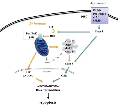
The extrinsic apoptotic pathway is triggered by the binding of first apoptosis signal receptor (Fas) or tumor necrosis factor receptor 1(TNFR 1). The Fas/Fas ligand complex then recruits death domain-containing protein (FADD), pro-caspase 8, cellular inhibitor of apoptosis protein (cIAPs), and cFLIPs to form the death-inducing signaling complex (DISC). The DISC complex directly cleaves and activates pro-caspase 8, which then triggers the activation of pro-caspase 3, the definitive enzyme for the execution of apoptosis, promoting cytosolic and nuclear alterations for cell disassembly. Caspase 3 activates the caspase-activated DNase (CAD) through cleavage of the inhibitor of CAD (ICAD), which then promotes double stranded DNA breaks and results in DNA
fragmentation7,9 (Fig 1).
Intrinsic apoptosis
Intrinsic apoptosis is controlled by mitochondrial enzymes9. After a cell is
stimulated by extracellular or intracellular stressors, pro-apoptotic members of the Bcl-2 protein family such as BAX and BAK accumulate at the mitochondria, resulting in mitochondrial outer membrane permeabilization (MOMP) and the release of cytochrome
This multiprotein complex activates caspase 9, which in turn cleaves effector caspases 3, 6, and 7. Mature caspases 3 and 7 cleave a large set of substrates resulting in the
morphological hallmarks of apoptosis, such as nuclear condensation, phosphatidylserine exposure, and the release of membrane bound vesicles without cell membrane
permeabilization12,13. Similarly, opening of the mitochondrial pore stimulates the release
1.3.2
Necrosis
Necrosis is traditionally considered to be the primary form of inflammatory cell death. Instead of the characteristic chromatin condensation, cell shrinking, and membrane blebs in apoptosis, necrosis is defined by increased cell volume, organelle swelling, lysosomal membrane permeabilization, and plasma membrane disruption.4,15 This results
in the leakage of non-membrane enclosed cytoplasmic contents including
pro-inflammatory endogenous molecules such as heat shock proteins (HSP), high mobility group box 1 (HMGB1), and uric acid, any of which can interact with the immune cells of the transplanted organ.16 Multiple forms of necrotic-like cell death exist that display these
morphological hallmarks.
Necroptosis
Necroptosis is currently the most investigated pathway of regulated necrosis. This regulated pathway features the necrotic morphology, but is mediated by receptor- interacting protein kinase (RIPK) and can be inhibited by Necrostatin-1 (Nec-1).
Necroptosis is induced by death receptors that include Fas and TNFα receptor. TNFα is a pro-inflammatory molecule with important immunoregulatory roles in mammalian immunity and cellular homeostasis.15,16
Upon stimulation of TNFR by TNFα, TNFR associated death domain (TRADD) is recruited to the plasma membrane, which in turn recruits RIPK1, cellular inhibitors of apoptosis protein-1 and 2 (cIAP1/2), linear ubiquitin chain assembly complex (LUBAC), and TNF receptor-associated factor 2 and 5 (TRAF2/5) to form a receptor-bound
complex I that initiates downstream signaling events16,17. The polyubiquitination of
complex then recruits mixed-lineage kinase domain-like protein (MLKL) that ultimately induces cell rupture and necroptosis.17,18,19
Ferroptosis and oxytosis
Ferroptosis is an iron dependent form of PCD characterized by decreased mitochondrial membrane densities, reduction of mitochondria crista, and eventual mitochondrial membrane rupture.20,21 Mechanically, the small molecule ferroptosis
inducer erastin inhibits the amino acid transporter system Xc-Cys/Glu antiporter which allows extracellular L-Cys and intracellular L-Glu exchange. This process leads to lipid peroxidation accumulation and increase in iron metabolite ROS. Glutathione (GSH) peroxidase 4 (GPX4), HSP, and nuclear factor erythroid 2 related factor 2 negatively regulate ferroptosis by limiting ROS production and cellular Fe uptake, whereas positive regulators such as NAPH oxidase and tumor protein p53 promote ROS mediated lipid peroxidation.8,22
Similarly, inhibition of Xc-Cys/Glu antiporter results in oxytosis, where
downstream activation of 12-lipoxygenase initiates mitochondrial ROS and cyclic GMP (cGMP). cGMP induces cGMP channel on the plasma membrane, stimulating Ca2+ influx
and lysosomal membrane permeabilization. Through inhibition of Xc-Cys-glu antiporter, ferroptosis and oxytosis utilize iron and calcium metabolism – respectively – to induce regulated necrosis.8,23
Parthanatos
Parthanatos, named after “Thanatos”, the personification of death in Greek mythology, involves the accumulation of poly (ADP-ribose) polymerase (PARP) and nuclear translocation of mitochondrial-associated apoptosis-inducing factor (AIF), leading to large-scale fragmentation of DNA and cell death.24,25 The process depends on
the PARP activation which depletes cellular energy through reduction of nicotinamide adenine dinucleotide (NAD+), an essential co-factor of glycolysis and TCA cycle.
astrocyte cell death. Under normal physiological conditions, PARP1 localizes in the nucleus and responds to DNA damage. If DNA damage is severe, however,
overactivation of PARP promotes parthanatic cell death.26 Studies have shown PAR
induces AIF release from the mitochondria and translation to the nucleus upon their physical interaction26,27, leading to cell death, and the implication that, along with
manifesting through the necrotic pathway, apoptotic mechanisms also contribute to parthanatic cell death.
1.4 Mitochondria regulated cell death
1.4.1
Mitochondria in endothelial cell death
As frontline defenders against vascular disease, microvascular endothelial cells not only power the cardiomyocytes with oxygen delivery and energetic substrates, but also play a role in preventing cardiovascular diseases post transplantation such as atherosclerosis and cardiac vasculopathy.28 Furthermore, endothelial cells play a crucial
role in modulating mitochondrial function in the heart where endothelial cells outnumber cardiomyocytes by nearly 3-fold.5 As proteins involved in mitochondrial homeostasis can
be involved in both apoptosis and necroptosis, it is important to place an emphasis on better understanding the mechanisms of mitochondrial regulated cell death in order to reduce injury an improve function in cardiac tissue.
1.4.2
Mitochondria plays a role in apoptosis and necroptosis
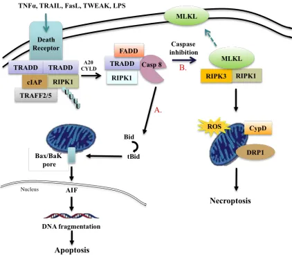
Mitochondria are known foremost as the primary cellular- energy- producing organelles, but they also play a central role in cell death. Activation of death receptors can lead to mitochondrial dysfunction events that can induce both apoptosis and necroptosis.29,30 Stimulation of either death pathway depends on the availability of
caspases – recruitment of adaptor proteins, such as TRADD, will lead to activation of caspase 8 and cleavage of B-cell lymphoma protein BID to induce mitochondrial pore permeabilization. This permeabilization results in DNA fragmentation and apoptosis; in the absence or inhibition of caspase 8, however, the TRADD adaptor protein complex recruits RIPK1, which forms the necrosome complex with RIPK3. The RIPK1/3
Fig 2. Mitochondria participates in apoptosis and necroptosis
1.4.3 BCL-2 family regulated mitochondrial dysfunction and
apoptosis
The B-cell lymphoma 2 (BCL-2) family proteins mediate mitochondrial apoptosis and cell survival. The pro-apoptotic members include BCL-2 associated X protein (BAX), BCL-2 homologous antagonist killer (BAK), BCL-2 interaction death promoter (BAD), BH3 interacting-domain death agonist (BID), and BCL-2 interaction mediator of cell death (BIM).31 Following a death signal, the pro-apoptotic members translocate to the
outer mitochondrial membrane (OMM) to be activated by BID, inducing
permeabilization of the OMM. This permeabilization stimulates the release of pro-apoptotic factors such as cytochrome c from their inner membrane space. The cytosolic cytochrome c induces the formation of the apoptosome that activates effector caspases to initiate apoptosis31,32
1.4.4 Mitochondrial permeability transition dysfunction
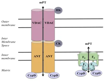
The mitochondrial permeability transition pore (mPTP) is a protein complex suggested to span both the outer and the inner mitochondrial membranes, and is presumed to perform important physiological roles.33,34 The precise molecular
composition of this pore remains highly controversial, but is thought to comprise of the voltage-dependent anion channel (VDAC) on the outer membrane and adenine nucleotide translocase (ANT) on the inner membrane (30), spastic paraplegia 7 (SPG7), phosphate carrier (PiC), ATP synthase, and cyclophilin D (CypD).35 Recently, the role of VDAC
and ANT was questioned, where mitochondrial PT deficient in VDAC and ANT still led to MPT onset.36 Furthermore, sensitivity to MPT inhibitor CsA (cyclosporine A) was still
displayed in VDAC and ANT null mitochondria.36
In normal conditions, the inner membrane channel opening is regulated to prevent membrane potential dissipation and proton gradient loss. When stimulated by
intracellular signals unrelated to cell death, brief opening of mPTP (described as
“flickering”) causes transient mitochondrial membrane potential variations necessary for cellular homeostasis such as mitochondrial Ca2+ extrusion during normal conditions.37
During Ca2+ overload and oxidative stress conditions, however, prolonged opening of the
which may result in cell death (30-34). Activation of OMM allows the release of the pro-apoptotic factors such as cytochrome c into the cytosol (31). These events are regulated by the Bcl-2 protein family; pro-apoptotic proteins Bax and Bak allow membrane permeabilization, while the anti-apoptotic members inhibit this process.32,38,39
The involvement of mitochondria, MPTP, and Bax/Bak as downstream necroptotic mediators is still a point of contention. It has been suggested that mitochondria play a role in regulated necrosis, where Bax/Bak generate a level of permeability in the outer mitochondrial membrane in non-oligomerized forms.40 In
Fig 3. Molecular components of MPTP
mPTP plays an important role in cell death by opening up the mitochondrial membrane through the formation of transporter channel proteins. Under apoptotic stimuli, prolonged pore opening allows for entry of small molecules, such as Ca2+ and protons, causing a
loss of potential, osmotic imbalance, and mitochondrial swelling.
1.5 The role of cyclophilin in mitochondrial physiology
1.5.1
The role of cyclophilin in permeability transition
Although an agreement on the precise MPT pore components is yet to be reached, the soluble protein CypD is the only unequivocally-established component of the pore complex to date.44 CypD is a peptidylprolyl isomerase that is critical in regulating MPTP
opening. CypD resides in the mitochondrial matrix, which translocates to the inner mitochondrial membrane during oxidative stress. CypD-deficient mitochondria were resistant to calcium overload and protective against cardiac ischemia reperfusion injury, whereas increased CypD level results in excessive ROS generation following ischemia injury in rats.45
CypD is then suggested to mediate a conformational change in the adenine
nucleotide translocase and triggers mPTP opening, mitochondrial swelling, and release of apoptotic effectors.46 Recently, it is proposed that CypD binds to ATP synthase, the
rotary enzyme responsible for ATP synthesis. The lateral stalk links the catalytic F1 and
the membrane bound F0 portion together. More specifically, it is proposed that CypD
may directly interact with the OSCP subunit of the lateral stalk, where, following separation of individual lateral stalk subunits, CypD was found exclusively in the immunoprecipitation with OSCP antibody.47,48
1.5.2
The role of cyclophilin in immune regulation
Aside from directly regulating the MPTP pore, Cyclophilin is also important in regulating immune function through its interaction with cyclosporine (CsA).49 CsA is a
cyclic polypeptide metabolized by a number of fungi, and its effective
immunosuppressive properties revolutionized the field of transplantation since its
discovery in 1972.49,50 Binding of CsA with Cyclophilin A inhibits calcineurin function –
a calcium/calmodulin-dependent serine threonine protein phosphatase that activates the T cell promoter IL-2 and inhibits the overall immune response.52 More specifically,
1.6 Are mitochondria directly involved in programmed
necrosis?
The involvement of mitochondria and mPTP as downstream mediators in the necroptosis pathway is still a point of contention. While it is well-established that mitochondria are the platforms for apoptosis execution, the prospect of their direct involvement in programmed necrosis remains controversial, and contradictory experiments point towards either mitochondrial-dependent or –independent forms of necrosis or necroptosis.
Evidence for the role of mitochondria in necroptosis:
Many studies have suggested that necrosome signalling might involve ROS generation from the mitochondria.53 In addition, studies demonstrated necrotic death was
inhibited in endothelial cells with the mitochondrial antioxidant MnSOD.54,55 RIPK1,
RIPK3, and/or MLKL are translocated to the mitochondria during necroptosis and thus result in ROS production.56 In particular, RIPK3 recruits MLKL to the necrosome
complex which is suggested to promote MLKL translocation to the mitochondria to induce cell death – where MLKL expression is increased in mitochondrial fraction following treatment of TNFα and pan-caspase inhibitor.
Genetics experiments confirmed the involvement of mPTP opening during PCD where CyP-D KO in MEFs (mice embryonic fibroblast cells) resulted in a partial rescue of necroptotic cell death 57,58in vitro,57,58 and that mPT pore is primarily involved in
necrotic cell death instead of apoptosis.59 In addition, studies have shown that siRNA
mediated knockdown of mitochondrial fission molecule Drp1 were able to inhibit TNFα mediated necroptosis in HeLa and HT-29 cells.60 Furthermore, Drp1 depletion decreased
death in rat renal tubule epithelial cells after TNFα treatment.61 These studies all suggest
that mitochondria play a pivotal role in the execution of the necroptotic pathway.
Evidence against the role of mitochondria in necroptosis:
necroptosis was still observed.62 Another study also questioned the downstream
RIPK3-mitochondrial association, where RIPK3 or CypD ablation were protective individually, but double knockout mice exhibited even greater protection,46 suggesting that the
necrosome and the MPT pore may be distinct pathways. However, CypD deletion did not further attenuate myocardium ischemia reperfusion injury (IRI) to that conferred by RIPK1 inhibition in the heart,63 which raises the question of whether tissue-and cell
specific differences may contribute to the coupling of MPT pore to necroptosis.
1.7 Rationale
Our previous studies have demonstrated that RIPK3-medidated necroptosis in donor heart and kidney grafts can promote inflammatory injury and transplant rejection, and that the inhibition of RIPK3 expression attenuated necrosis and reduced early graft injury and rejection64,70. The down-stream pathway of necroptosis is not clear. It is
controversial whether mitochondria are involved in necroptosis. In our current study, we aim to determine the role of CypD-regulated mPT in necroptosis in cardiac endothelial cells and in mouse heterotopic transplantation model. This will provide rationale to design an effective inhibition strategy to promote transplant tolerance.
1.8 Hypothesis
We hypothesize that Cyp-D inhibition can inhibit mPT, thus preventing cell necroptotic death, and can trigger the release of proinflammatory CDAMP, preventing graft injury in heart transplants and thus prolonging graft survival,.
1.9 Objectives
1. To determine if the inhibition of CypD function in cardiac endothelial cells can prevent necroptotic death.
Chapter 2
2
Materials and Methods
2.1 Animals
Male inbred C57BL/6 (B6), BALB/c mice and B6. Cyp-D-/- mice (Jackson Lab)
were maintained at the animal facility at Western University. All experimental procedures were approved by the Animal Use Committee of Western University.
2.2 Heterotopic cardiac transplantation and post-operative
monitoring
Donor cardiac grafts were perfused with saline to remove blood after being anesthetized with the mixture ketamine/xylazine according to the approved animal protocol. The inferior vena cava and dorsal aorta were then clamped above and below the cardiac graft. The graft was then removed, and intra-abdominal heterotopic cardiac transplants were performed in our microsurgery laboratory in accordance with an
approved protocol. The recipient received sirolimus (rapamycin, LCL laboratories, USA) from day 0 to day 9 post-transplantation (1 mg/kg mouse).Pulsation of the cardiac graft was monitored daily. Cessation of pulsation was defined as the endpoint of rejection.
2.3 Histology and Immunohistochemistry
Grafts were collected on day 28, perfused with PBS, cut transversely, and then either frozen using Tissue-Tek® O.C.T. Compound (Sakura® Finetek) or fixed with 5% formalin for paraffin embedding. Paraffin sections were used for hematoxylin and eosin (H&E) staining.
2.4 Cell death assay
B6 and Cyp-D-/- microvascular endothelial cells (MVEC) were isolated and
developed as described previously (18-20).Cells were grown in complete Endothelial Growth Media-2 (EGM-2 medium) supplemented with fetal bovine serum and growth factors (Lonza, USA).
Human TNFα (hTNFa, 100 ng/ml; PeproTech, USA) was used to induce cell death. Smac-mimetic (100 nM; Selleckchem, USA) was added to suppress the function of Inhibitor of Apoptosis Proteins (IAP). This leads to caspase activation and inhibition of RIPK1 polyubiquitination, and promotes apoptotic cell death. Caspase-mediated apoptosis was inhibited by caspase-8 inhibitor Z-Ile-Glu-Thr-Asp-Fluoromethylketoe (IETD, 10-30 μM; APExBIO, USA). Caspase-3 inhibitor
Z-Asp-Glu-Val-Asp-Fluoromethylketoe(DEVD, 10-30 μM;) and capase-9 inhibitor Z-Leu-Glu-His-Asp-Fluoromethylketoe (LEHD, 10-30μM, APExBIO) were used. RIPK1 inhibitor
necrostatin-1s (Nec-1s, 10 μM; Merck Millipore, USA) or MLKL inhibitor GW806742X (50-1000 nM, Synkinase, USA) were added to inhibit necroptosis. Mitochondrial
permeability transition events were inhibited with S-15176 (Sigma-Aldrich, USA). Dose of S-15176 (1-20 μg/ml) was optimized and 16 µg/ml was chosen for the study.
Cyclosporin A (CsA) and control FK-506 (Sigma-Aldrich) were used for Cyp-D inhibition. Dose of CsA (1-15 µg/ml) was optimized and 10 µg/ml was used for the study. ROS inhibitors are 1-oxyl-2,2,6,6-tetramethyl-4-hydroxypiperidine (Tempol, 1-10 μM, Sigma-Aldrich, USA) and N-acetyl-L-cysteine (NAC, 1-10 μM, Sigma-Aldrich). Dose was optimized and 10 μM was chosen for the study.
2.5 Immunoblot analysis
MVEC were grown to a confluent monolayer and treated as described in cell death assay. Cells were trypsinized, centrifuged at 300 g for 5 minutes and 50μL nuclear lysis buffer (20 mM HEPES, 0.4 mM NaCl, 1 mM EDTA, 1 mM EGTA, 1 mM DTT, 1 mM PMSF) was added to each sample followed by a 30 minutes incubation at 37.5°C. The nuclear fraction was collected by centrifugation at 10,000 g for 15 minutes at 4°C.
Equal amount lysates were loaded for gel electrophoresis. Protein was transferred to a nitrocellulose membrane using electrophoresis blotting system (BioRad, USA). 5% skim milk and 0.1% Tween-20 in Tris buffered saline was used for blocking.
The phosphorylated MLKL (pMLKL), Phospho S345 MLKL protein, was detected using rabbit anti-mouse pMLKL antibody (Abcam, USA). Protein was
visualized using secondary anti-rabbit IgG horseradish peroxidase (HRP)-linked antibody (Cell Signaling Tech. USA) and chemiluminescent substrate (EMD Millipore, USA). Anti-β-actin antibody (Sigma-Aldrich, USA) was used as loading control.
2.6 shRNA-mediated Cyp-D RNA silencing and Real-time
PCR
Cyp-D shRNA (Dharmacon, USA) was transfected into MVEC using Lipofectamine (Invitrogen, USA). Puromycin (Sigma-Aldrich, USA) was used for antibiotic selection to eliminate untransfected MVEC. Cyp-D gene silencing was confirmed by real-time PCR.
Total RNA from wildtype or shRNA transfected MVEC was extracted from tissue or cells by Trizol extraction (Invitrogen, USA). cDNA was generated from RNA using Superscript II (ThermoFisher, USA). Primers are: Cyp-D: CTC CAA CTC CAA GAA
CCC GC and TAA AAC AAT TCG GCC AAC TCG C; b-Actin: CCA GCC TTC CTT
2.7 Statistical Analysis
Data was analyzed using the Student’s t-test for unpaired values. The Mantel-Cox log rank test was used to determine graft survival differences. Differences were
Chapter 3
3
Results
3.1 Mitochondrial permeability participates in MVEC
necroptosis
We used wild type and RIPK3 deficient endothelial cells to determine the effects of inflammatory cytokines on cell death. TNFα, Smac-mimetic, and caspase-8 inhibitor IETD or RIPK1 inhibitor Nec-ls were added to wild type B6 and RIPK3-/- MVEC
cultures. In wild type B6 MVEC, treatment of TNFα increased cell death compared to untreated cells. The addition of caspase-8 inhibitor IETD enhanced TNFα induced cell death (Figure 4A), suggesting necroptotic death.64 Necroptosis in MVEC was inhibited
by the addition of RIPK1 inhibitor Nec-1s or RIPK3 deficiency in MVEC (Figure 4A). To determine if mitochondrial dysfunction events play a role in necroptosis, mitochondria transition permeability inhibitor S-15176 was added to MVEC. S-15176 prevents
collapse of the electrochemical gradient across the mitochondrial membrane and inhibits release of apoptotic molecules.65,66 We found that the addition of S-15176 to
TNFα+IETD treated MVEC significantly reduced necroptosis (Figure 4B).
Opening of mPTP is largely regulated by Cyp-D. Cyp-D may regulate apoptotic cell death as well necrotic cell death.67 It has been shown that Cyp-D deficiency in mouse
embryonic fibroblast cells resulted in a partial rescue of necroptotic cell death in vitro.57
However, another study showed that RIKP1/3 and mPTP/Cyp-D mediate two distinct death pathway.46 We studied the role of Cyp-D in MVEC necroptosis. Initially, we used
CsA, a calcineurin inhibitor and classical immunosuppressive drug in clinical
transplantation that can bind to Cyp-D with high affinity and inhibit mPTP opening. CsA inhibited TNFa-induced necroptotic cell death upon simultaneous caspase-8 inhibition (Figure 1C). In contrast, FK506, a non-Cyp-D binding and calcineurin-inhibiting
Figure 4. Mitochondrial permeability participates in MVEC necroptosis
(A) B6 MVEC and RIPK3-/- MVEC were plated on 96 well plate in triplicates and
treated with 100 ng/mL TNFα (T), 10 nM Smac mimetic (S) with or without 30μM IETD(I) and 10 μM Nec-1s (N) with addition of SYTOX green. Cell death was quantified by SYTOX fluorescent intensity with IncuCyte ZOOM live imaging system. Data at 24 hours were shown as mean ± SD and representative of at least 3 independent
Figure 5. Cyp-D deficiency protects MVEC from necroptosis
3.2 ROS and Caspases-3 and -9 do not contribute towards
MVEC necroptosis
Next we studied the down-stream mechanism of Cyp-D mediated necroptosis. Opening of mPTP results in ROS release and activation of caspase-9 and its downstream caspase-3. Recent studies showed that ROS play a crucial role in necrosis and that inhibition of ROS prevents TNFα-induced necroptotic cell death.68,69 In our study,
Figure 6. ROS, caspase-3 and caspase-9 do not participate in MVEC necroptosis.
(A) B6 MVEC were plated on 96 well plate in triplicates and treated with 100 ng/ml hTNFα (T), 100 nM Smac mimetic (S) with or without 30 μM IETD (I), 10 μM Nec-1s (N), caspase-3 inhibitor DEVD (30 μM) or caspase-9 inhibitor LEHD (30 μM). (B) ROS inhibitors NAC (10 μM) and Tempol (10 μM) were added to inhibit ROS activities. Cell death was quantified by SYTOX fluorescent intensity with IncuCyte ZOOM live imaging system. Data at 24 hours were shown as mean ± SD and
3.3 Cyp-D mediated necroptosis is linked to MLKL
activation
MLKL is phosphorylated by RIPK3 and then migrates to lipid rafts where it forms a pore, ultimately resulting in cell membrane rupture- a characteristic feature of necroptosis.6 To investigate if Cyp-D mediated necroptosis is linked to MLKL activation,
MLKL inhibitor GW806742Xwas added and was observed topartially inhibit
necroptosis in MVEC (Figure 7A). MLKL siRNA silencing in MVEC (Figures 7B&C) prevented necroptotic cell death (Figure 7D).
Next, we analyzed expression of phosphorylated MLKL (pMLKL), which is the active form of MLKL, by western blot. pMLKL increased significantly in B6 MVEC under necroptosis inducing conditions, which can be inhibited by Cyp-D inhibitor CsA or by Cyp-D deficiency (Figure 7E), suggesting that Cyp-D is necessary for MLKL
Figure 7. Cyp-D-regulated necroptosis is linked to downstream MLKL activation
(A) B6 MVEC necroptotic death was induced as in Figure 1. MLKL inhibitor
GW806742X(GW, 50-1000 nM) was added. Cell death was quantified by SYTOX
fluorescent intensity with IncuCyte ZOOM live imaging system. Data at 24 hours were shown as mean ± SD and representative of 3 independent experiments. (B) MLKL was silenced by pooled MLKL siRNAs (Santa Cruz). MLKL knockdown was confirmed by real-time PCR 10 hours later. B-actin amplification was used as the control. Data at 24 hours were shown as mean ± SD and representative of 3 independent PCR. (C) MLKL protein inhibition was confirmed 40 hours after by western blot using anti-total MLKL and loading control anti-b-actin. The same result was obtained in a repeated experiment. (D) MLKL knockdown protects MVEC from necroptotic death. 24 hours after siRNA silencing, necroptosis was induced in MVECs . Cell death was measured by 7-AAD positivity using flow cytometry. Data is a representative average of 3 independent cell death experiments. (E) Analysis of MLKL phosphorylation. Necroptosis was induced in B6 and Cyp-D-/- MVEC as described in Figures 1-3. Cells were collected 6 hours later
3.4 Cyp-D deficiency in donor cardiac grafts attenuates
rejection
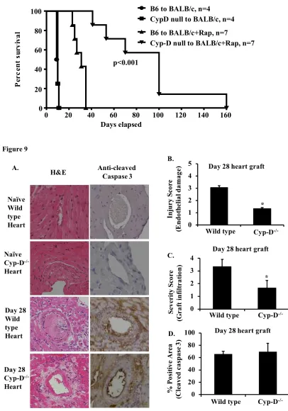
Our data indicate that Cyp-D may be an effective target to inhibit cell death. To determine if Cyp-D deficiency in donor cardiac grafts can improve transplant survival, we performed heterotopic transplantation of wildtype B6 or Cyp-D-/- hearts into BALB/c
mice followed by a brief period of immunosuppression spanning 0-9 days with sirolimus (rapamycin) (18). Cyp-D-/- grafts survived significantly longer post transplantation
compared to wild type C57BL/6 grafts (89+43 versus 29+6, n=7, p<0.0001) (Figure 8A).
To assess graft injury, we collected the cohort grafts 28 days post-transplantation. Naïve B6 hearts do not appear to be histologically different from naïve Cyp-D hearts (Figure 9A). B6 grafts showed severe lymphocyte infiltration and endothelial damage when compared to the Cyp-D-/- grafts collected 28 days post-transplantation (Figures
9B&C). However, tissue apoptosis was not significantly different between wild type and Cyp-D-/- grafts as defined by anti-cleaved caspase-3 immunohistochemistry staining
Days elapsed
P
e
r
c
e
n
t
s
u
r
v
i
v
a
l
0 20 40 60 80 100 120 140 160
0 20 40 60 80 100
p<0.001
B6 to BALB/c, n=4
CypD null to BALB/c, n=4 B6 to BALB/c+Rap, n=7
Figure 8. Cyp-D deficiency in donor cardiac grafts attenuated transplant rejection.
B6 or Cyp-D-/- cardiac grafts were heterotopically transplanted into abdomens of
BALB/c mice, followed by rapamycin (Rap) treatment (1 mg/kg, day 0-9). Grafts were palpated and scored daily and considered rejected when pulsation ceased. (n=4-7, ***p<0.001, Mantel-Cox log-rank test).
Figure 9. Cyp-D deficiency in donor grafts inhibits endothelium damage.
(A) B6 or Cyp-D-/- cardiac grafts were transplanted into BALB/c mice as in
Figure 5. Recipients (n=4 per group) were euthanized 28 days post-transplantation and grafts were collected for H&E staining and anti-cleaved caspase-3 immunohistochemistry as detailed in the Methods. Microscope magnification=200 fold. Scale bar=100 µM. (B)
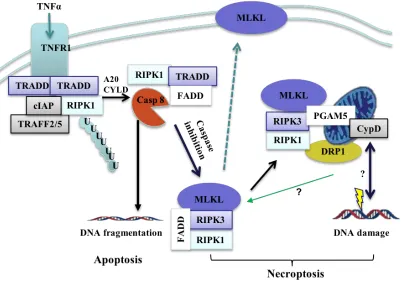
Figure 10. Proposed mechanism of cell death.
Upon stimulation of TNFR by TNFα, TNFR associated death domain (TRADD) is recruited to the plasma membrane, which in turn recruits RIPK1, cellular inhibitors of apoptosis protein-1 and 2 (cIAP1/2), and TNF receptor-associated factor 2 and 5
(TRAF2/5) to form a receptor-bound complex I that initiates downstream signaling events. The deubiquitination of RIPK1 via A20 or Cylindromatosis (CYLD) activity causes complex I to dissociate from the membrane to form a cytosolic complex II with recruitment of FADD and caspase-8 (Casp-8). Caspase-8 will initiate apoptosis via cascades of caspases including caspase-3 (Casp-3), and cleave RIPK1 and RIPK3. Inhibition of caspase-8 allows the formation of RIPK1-RIPK3 necrosome which recruits and phosphorylates MLKL that ultimately induces cell membrane rupture and
Chapter 4
4
Discussion
4.1 Summary
Solid organ transplantation remains a vital therapy for end stage heart failure. Historically, post-operative care was emphasized; however, the alleviation of cellular injury and subsequent inflammation gained more attention over the last few years. We have demonstrated previously that programmed cell death, particularly RIPK3-mediated necroptosis, can promote inflammation and tissue injury in donor heart and kidney grafts, and inhibition of RIPK3 attenuated graft rejection.64,70,71 However, necroptosis molecular
pathways downstream of RIPK3 remain unknown. In this study, we demonstrated that mitochondria are the critical component in the downstream necroptotic pathway; specifically, inhibition of the mPTP regulator molecule Cyp-D attenuated MVEC necroptosis in vitro, and Cyp-D deficient cardiac allografts showed prolonged survival compared to wild-type C57BL/6 grafts post transplantation. Our study thus demonstrates that mPTP participates in RIPK3 mediated necroptosis and contributes to inflammatory injury in cardiac allografts, and that Cyp-D can be an important therapeutic target for long-term graft survival.
4.2 MVECs necroptosis is mediated by mitochondrial
dependent mechanisms
It has been demonstrated the over the years that necroptosis is initiated by death receptors such as TNFα, which leads to the downstream RIPK1/3 activation and
formation of the necrosome. This complex has been proposed to induce necroptosis via mitochondrial independent or dependent pathways.30
The mitochondrial independent pathway involves MLKL mediated cellular membrane rupture. Upon its phosphorylation by RIPK3, MLKL undergoes
turn promote necroptosis by activating RIPK1 autophosphorylation, therefore creating a positive feedback loop for necroptosis induction.72,73 In our study, however, ROS
inhibition did not result in significant reduction in cell death (Fig 6B), suggesting that mitochondrial permeability may be a more significant contributor in death induction.
MLKL induced mitochondrial permeability increase induces cytochrome c
release, causing the formation of the supramolecular apoptosome complex with deoxyATP, APF1, and pro-caspase 9.72,73Activated caspase 9 then activates the
downstream executer, caspase 3. Caspase 3 is key in promoting cellular disassembly and nuclear fragmentation by activating CAD through inactivating the inhibitor of CAD (ICAD).74,75 Following translocation to the nucleus, CAD homodimerizes and creates
double-stranded DNA breaks. In contrast, our study showed that inhibition of caspase 3 or 9 did not prevent necroptosis (Fig 6A). This suggests that other downstream
mechanisms may be responsible for mitochondrial dependent necroptosis induction, such as mitochondrial molecules AIF and endonuclease G.76,77,78 As AIF and endonuclease G
have been characterized as apoptogenic factors, it would be interesting to investigate whether they also participate in MVEC necroptosis.
4.3 The role of Cyp-D in organ injury
The involvement of mitochondria molecules as downstream RIPK mediators is still a point of contention. While it is a well-established fact that mitochondria are platforms for apoptosis execution, the prospect of direct mitochondrial involvement in programmed necrosis remains controversial, and contradictory experiment results point towards either mitochondrial-dependent or -independent forms of necroptosis.72 Our
study demonstrates that Cyp-D, as an essential component of mitochondrial permeability transition regulator with an important role in apoptotic induction, also participates in necroptosis and thus may be an important therapeutic target.
Genetics experiments confirmed that targeting Cyp-D protected cells from
necrosis, where CyP-D knockout in MEFs (mice embryonic fibroblast cells) resulted in a partial rescue of necroptotic cell death 57in vitro, and that MPT pore is primarily involved
in necrotic cell death instead of apoptosis.80 We found that Cyp-D deficiency in donor
caspase 3 expression was not significantly different between WT and Cyp-D null grafts indicate that Cyp-D deficiency did not alter apoptotic tissue injury long-term. The data suggests that Cyp-D dependent mitochondrial permeability contributes to DAMPs release and augments inflammatory injury in the graft tissue.
4.4 Mediators of RIPK1/3/MLKL and
mitochondrial-dependent MVEC necroptosis
Even though our data suggested that mitochondrial permeability transition and its regulator Cyp-D participates in necroptosis, the precise mechanism of MLKL
participation in the mPTP pathway following TNF and caspase 8 inhibition is controversial. In Figure 7 we illustrated several possible MLKL pathways in the necroptotic pathway. After caspase 8 inhibition, it is known that MLKL translocates to the plasma membrane to induce membrane rupture and DAMP release. Alternatively, it has been proposed that RIPK3 recruits MLKL and mitochondrial protein phosphatase PGAM5 – a molecule suggested to be essential for mitochondrial fragmentation. PGAM5 recruits dynamin related protein Drp1 and dephosphorylates the serine 637 site of Drp1, activating its GTPase.60 Drp1 then translocate to mitochondrial outer membrane division
sites and causes mitochondrial fragmentation.60
Drp1 activation has been shown to be involved in apoptosis, where under apoptotic stimuli, Drp1 induced Bax oligomerization and mitochondrial cytochrome c
release81. Furthermore, Drp1 knockout Purkinje neurons displayed no signs of apoptotic
death82. Interestingly, Drp1 has also been implicated to play a role in necroptosis, where
it regulates mitochondrial division. Recent studies demonstrated that Drp1 mediated mitochondrial division is mediated by RIPK1 and RIPK360. Thus the
RIPK3/MLKL/PGAM5/DRP1 is implicated to induce mitochondria - dependent
or if it is activated through indirect pathways – for instance, others have suggested that Cyp-D mediate Drp1 function and augment Drp1 recruitment to the mitochondria.
4.5 Strategy to prevent graft injury in transplantation
As novel immunosuppression methods have arisen over the past several years, strategies that target intra-graft death and inflammation still hold great potential for investigation. Currently, siRNA therapy has been utilized to target apoptotic molecules such as caspases 3 and 8 during IRI in mice.83 However, when used on porcine kidneys,
caspase 3 siRNA delivery augmented inflammatory response and kidney tissue damage instead,84 indicating a crucial need for a thorough knowledge of the complex
interconnected pathway mechanisms of apoptosis and necroptosis.
The mPT pore and its regulators have been recognized as drug targets for IRI and neurodegenerative disorders, where Cyp-D knockout mice showed improved outcomes in disease models compared to wildtype mice. The Cyp-D inhibitor CsA is a potent
immunosuppressive which disrupts calcineurin function and downstream immune cell activation. However, the 17 different subtypes of Cyp-D in the human genome remain poorly understood, and a comprehensive understanding of the similarities and differences between Cyp-D subgroups is necessary for specific and effective drug design.85
Nevertheless, it would be interesting to determine which Cyp-D family would the most effective target in the prevention of cell death without compromising mitochondrial function, and to investigate delivery of that specific Cyp-D shRNA via viral vectors into heart grafts to promote long-term tolerance.
4.6 Limitations
models, where regions of genetic variability may exist due to the presence of “flanking” genes incorporated into the genetic background87.
In terms of immunological study, it may be beneficial to do an initial investigation into how immune response regulates cell survival (i.e. apply mixed lymphocyte reaction), in addition to mitochondrial dysfunction in vitro beforehand, so as to better anticipate in vivo transplantation results. To improve upon our in vivo model, we may consider siRNA delivery into hearts88, 89, although it is for short-term silencing and therefore may not be
able to potentiate long-term protection in heart grafts post transplantation. CRISPR/cas9-based strategy, on the other hand, may be a more promising area to explore.
Alternatively, we may consider viral particle delivery technology to achieve high efficiency tissue delivery.
4.7 Conclusion
In this study, we demonstrated that mPTP plays an important role in necroptotic death of cardiac cells and graft rejection. Additionally, inhibition of Cyp-D attenuated RIPK3- downstream MLKL phosphorylation, leading to prolonged survival when compared with wild type grafts. Our study shows that Cyp-D is an effective target in mitigating
necroptosis in MVEC and preventing cardiac graft rejection. As Cyp-D deficiency prevents mPTP function without completely impeding mitochondrial function, targeting Cyp-D may offer exciting therapeutic potential in the prevention of long-term graft injury.
4.8 Overall project significance
Since long-term survival of transplants has not been greatly improved by post-transplant core strategies that solely target the immune response, a focus on
the mitochondria is a promising tactic in an effort to prevent regulated necrosis and ultimately, to promote transplant survival.
4.9 Future Directions
While it is known that mPT downstream mechanisms is classically associated with pro-apoptotic and necrotic stimuli, it is interesting that caspase 3 and ROS do not significantly contribute to MVEC death. This suggests that other mediators may be at play for necrotic DNA damage which is independent of caspase ROS, for instance, apoptosis-inducing factor and endonuclease G (EndoG). After released into the cytosol, AIF and EndoG translocate to the nucleus an is responsible for large scale DNA
References
(1) Tonsho, M., Michel, S., Ahmed, Z et al. (2014). Heart Transplantation: Challenges Facing the Field. Cold Spring Harbor Perspectives in Medicine,4(5).
(2) Kittleson, M., Kobashigawa, J. (2017). Cardiac Transplantation: Current Outcomes and Comtemporary Controversies. Journal of the American College of Cardiology, 5(12).
(3) Pallet, N., Dieudé, M., Cailhier, J., & Hébert, M. (2012). The Molecular Legacy of Apoptosis in Transplantation. American Journal of Transplantation,12(6), 1378-1384.
(4) Hébert, M., & Jevnikar, A. M. (2015). The Impact of Regulated Cell Death Pathways on Alloimmune Responses and Graft Injury. Current Transplantation Reports,2(3), 242-258.
(5) Davidson, S. M., & Duchen, M. R. (2007). Endothelial Mitochondria:
Contributing to Vascular Function and Disease. Circulation Research,100(8), 1128-1141.
(6) Peter, M. E. (2011). Programmed cell death: Apoptosis meets necrosis. Nature,471(7338), 310-312.
(7) Taylor, R. C., Cullen, S. P., & Martin, S. J. (2008). Apoptosis: Controlled demolition at the cellular level. Nature Reviews Molecular Cell Biology,9(3), 231-241.
(8) Berghe, T. V., Linkermann, A., Jouan-Lanhouet, S., Walczak, H., & Vandenabeele, P. (2014). Regulated necrosis: The expanding network of non-apoptotic cell death pathways. Nature Reviews Molecular Cell Biology,15(2), 135-147.
(9) Fulda, S., & Debatin, K. (2006). Extrinsic versus intrinsic apoptosis pathways in anticancer chemotherapy. Oncogene,25(34), 4798-4811.
(10) Tsujimoto, Y., & Shimizu, S. (2006). Role of the mitochondrial membrane permeability transition in cell death. Apoptosis,12(5), 835-840.
(11) Zhan, M., Brooks, C., Liu, F., Sun, L., & Dong, Z. (2013). Mitochondrial dynamics: Regulatory mechanisms and emerging role in renal
pathophysiology. Kidney International,83(4), 568-581.
(12) Schwarz, M., Andrade-Navarro, M. A., & Gross, A. (2007). Mitochondrial carriers and pores: Key regulators of the mitochondrial apoptotic
program? Apoptosis,12(5), 869-876.
(13) Daisy, P., & Saipriya, K. (2012). BCL-2 Family Proteins: The Mitochondrial Apoptotic Key Regulators. Current Cancer Therapy Reviews,8(2), 133-140.
(14) Yang, S., Zhao, X., Xu, H., Chen, F., Xu, Y., Li, Z., . . . Ye, J. (2017). AKT2 Blocks Nucleus Translocation of Apoptosis-Inducing Factor (AIF) and Endonuclease G (EndoG) While Promoting Caspase Activation during Cardiac
Ischemia. International Journal of Molecular Sciences,18(3), 565.
(15) Fiers, W., Beyaert, R., Declercq, W., & Vandenabeele, P. (1999). More than one way to die: Apoptosis, necrosis and reactive oxygen damage. Oncogene,18(54), 7719-7730.
(16) Brenner, D., Blaser, H., & Mak, T. W. (2015). Regulation of tumour necrosis factor signalling: Live or let die. Nature Reviews Immunology,15(6), 362-374.
differential utilization of ASK1 kinase and p73. Cell Death & Differentiation,19(2), 274-283.
(18) Dhuriya, Y. K., & Sharma, D. (2018). Necroptosis: A regulated inflammatory mode of cell death. Journal of Neuroinflammation,15(1).
(19) Yuan, J. (2015). Faculty of 1000 evaluation for Caspase-8 regulates TNF-α-induced epithelial necroptosis and terminal ileitis. F1000 - Post-publication Peer Review of the Biomedical Literature.
(20) Xie, Y., Hou, W., Song, X., Yu, Y., Huang, J., Sun, X., . . . Tang, D. (2016). Ferroptosis: Process and function. Cell Death & Differentiation,23(3), 369-379.
(21) Fearnhead, H. O., Vandenabeele, P., & Berghe, T. V. (2017). How do we fit ferroptosis in the family of regulated cell death? Cell Death and
Differentiation,24(12), 1991-1998.
(22) Sato, M., Kusumi, R., Hamashima, S., Kobayashi, S., Sasaki, S., Komiyama, Y., . . . Sato, H. (2018). The ferroptosis inducer erastin irreversibly inhibits system xc− and synergizes with cisplatin to increase cisplatin’s cytotoxicity in cancer cells. Scientific Reports,8(1).
(23) Lewerenz, J., Ates, G., Methner, A., Conrad, M., & Maher, P. (2018).
Oxytosis/Ferroptosis—(Re-) Emerging Roles for Oxidative Stress-Dependent Non-apoptotic Cell Death in Diseases of the Central Nervous System. Frontiers in Neuroscience,12.
(24) David, K. K. (2009). Parthanatos, a messenger of death. Frontiers in Bioscience,Volume(14), 1116. doi:10.2741/3297
(25) Shin, H., Kwon, H., Lee, J., Gui, X., Achek, A., Kim, J., & Choi, S. (2015). Doxorubicin-induced necrosis is mediated by poly-(ADP-ribose) polymerase 1 (PARP1) but is independent of p53. Scientific Reports,5(1).
(26) Fatokun, A. A., Dawson, V. L., & Dawson, T. M. (2014). Parthanatos:
Mitochondrial-linked mechanisms and therapeutic opportunities. British Journal of Pharmacology,171(8), 2000-2016.
(27) Andrabi, S. A., Dawson, T. M., & Dawson, V. L. (2008). Mitochondrial and Nuclear Cross Talk in Cell Death. Annals of the New York Academy of
Sciences,1147(1), 233-241.
(28) Caja, S., & Enríquez, J. A. (2017). Mitochondria in endothelial cells: Sensors and integrators of environmental cues. Redox Biology,12, 821-827.
(29) Schwarz, M., Andrade-Navarro, M. A., & Gross, A. (2007). Mitochondrial carriers and pores: Key regulators of the mitochondrial apoptotic
program? Apoptosis,12(5), 869-876.
(30) Thornton, C., & Hagberg, H. (2015). Role of mitochondria in apoptotic and
necroptotic cell death in the developing brain. Clinica Chimica Acta; International
Journal of Clinical Chemistry, 451(Pt A), 35–38.
(31) Tsujimoto, Y. (1998). Role of Bcl-2 family proteins in apoptosis: Apoptosomes or mitochondria? Genes to Cells,3(11), 697-707.
(32) Kutuk, O., & Basaga, H. (2006). Bcl-2 protein family: Implications in vascular apoptosis and atherosclerosis. Apoptosis,11(10), 1661-1675.
(33) Nicotra, A., & Parvez, S. (2002). Apoptotic molecules and MPTP-induced cell death. Neurotoxicology and Teratology,24(5), 599-605.
(35) Karch, J., & Molkentin, J. D. (2014). Identifying the components of the elusive mitochondrial permeability transition pore. Proceedings of the National Academy of Sciences,111(29), 10396-10397.
(36) Lemasters, J. J., Theruvath, T. P., Zhong, Z., & Nieminen, A.-L. (2009).
Mitochondrial Calcium and the Permeability Transition in Cell Death. Biochimica et
Biophysica Acta, 1787(11), 1395–1401. http://doi.org/10.1016/j.bbabio.2009.06.009 (37) Pastorino, J. G., Tafani, M., Rothman, R. J., Marcineviciute, A., Hoek, J. B., &
Farber, J. L.(1999). Functional Consequences of the Sustained or Transient Activation by Bax of the Mitochondrial Permeability Transition Pore. Journal of Biological Chemistry, 274(44), 31734-31739
(38) Daisy, P., & Saipriya, K. BCL-2 Family Proteins: The Mitochondrial Apoptotic Key Regulators. Current Cancer Therapy Reviews, 2012; 8(2), 133-140.
(39) Pastorino, J. G., Tafani, M., Rothman, R. J., Marcineviciute, A., Hoek, J. B., & Farber, J. L. Functional Consequences of the Sustained or Transient Activation by Bax of the Mitochondrial Permeability Transition Pore. Journal of Biological Chemistry, 1999; 274(44), 31734-31739.
(40) Karch, J., Kwong, J. Q., Burr, A. R., Sargent, M. A., Elrod, J. W., Peixoto, P. M., . . . Molkentin, J. D. (2013). Bax and Bak function as the outer membrane component of the mitochondrial permeability pore in regulating necrotic cell death in
mice. ELife,2. doi:10.7554/elife.00772
(41) Tischner, D., Manzl, C., Soratroi, C., Villunger, A., & Krumschnabel, G. Necrosis-like death can engage multiple pro-apoptotic Bcl-2 protein family members. Apoptosis, 2012; 17(11), 1197-1209.
(42) Lomonosova, E., & Chinnadurai, G. BH3-only proteins in apoptosis and beyond: an overview. 2008; Oncogene,27.
(43) Gauba, E., Guo, L., & Du, H. (2017). Cyclophilin D Promotes Brain
Mitochondrial F1FO ATP Synthase Dysfunction in Aging Mice. Journal of
Alzheimer’s Disease : JAD, 55(4), 1351–1362.
(44) Thomas, B., Banerjee, R., Starkova, N. N., Zhang, S. F., Calingasan, N. Y., Yang,
L., … Starkov, A. (2012). Mitochondrial Permeability Transition Pore Component Cyclophilin D Distinguishes Nigrostriatal Dopaminergic Death Paradigms in the
MPTP Mouse Model of Parkinson’s Disease. Antioxidants & Redox Signaling, 16(9),
855–868.
(45) Schneider, M. D. (2005). Cyclophilin D: Knocking On Deaths Door. Science Signaling,2005(287).
(46) Linkermann A, Brasen J, Darding, H, et al. Two independent pathways of
regulated necrosis mediate ischemia-reperfusion injury. Proc. Natl. Acad. Sci. U.S.A. 2013; 110: 12024-12029
(47) Beutner, G., Alanzalon, R. E., & Porter, G. A. (2017). Cyclophilin D regulates the
dynamic assembly of mitochondrial ATP synthase into synthasomes. Scientific
Reports, 7, 14488.
(48) Beck, S. J., Guo, L., Phensy, A., Tian, J., Wang, L., Tandon, N., . . . Du, H. (2016). Deregulation of mitochondrial F1FO-ATP synthase via OSCP in Alzheimer’s disease. Nature Communications,7, 11483.
(49) Kim, S. Y., Shim, M. S., Kim, K.-Y., Weinreb, R. N., Wheeler, L. A., & Ju,
(50) Kwong, J., & Molkentin, J. (2015). Physiological and Pathological Roles of the Mitochondrial Permeability Transition Pore in the Heart. Cell Metabolism,21(2), 206-214.
(51) Nacev, B. A., Low, W.-K., Huang, Z., Su, T. T., Su, Z., Alkuraya, H., … Liu, J.
O. (2011). A Calcineurin-Independent Mechanism of Angiogenesis Inhibition by a
Nonimmunosuppressive Cyclosporin A Analog. The Journal of Pharmacology and
Experimental Therapeutics, 338(2), 466–475.
(52) Liu J, Farmer JDJ, Lane WS, Friedman J, Weissman I, Schreiber SL. Calcineurin is a common target of cyclophilin-cyclosporin A and FKBP-FK506
complexes. Cell. 1991;66:807–815.
(53) Vanlangenakker, N, Vanden Berghe, T, Bogaert P, et al. cIAP and TAK1 protect cells from TNF-induced necrosis by preventing RIP1/RIP3 dependent reactive oxygen species production. Cell Death Differ. 18, 656-665
(54) Zhan M, Brooks, C, Liu, F, et al. Mitochondrial dynamics: regulatory
mechanisms and emerging role in renal pathophysiology. Kidney Int. 2013; 83(4): 568-581
(55) Holley, A. K., Dhar, S. K., Xu, Y., & St. Clair, D. K. (2012). Manganese
superoxide dismutase: beyond life and death. Amino Acids, 42(1), 139–158.
(56) Brouns, S.J., Jore, M.M, Lundgren M. et al. Small CRISPR RNA guide antiviral defense in prokaryotes. Science 321, 960-964
(57) Karch J, Kanisicak O, Brody M, et al. Necroptosis interfaces with MOMP and the MPTP in mediating cell death PLoS ONE 2015; 10(6) e0130520
(58) Tsujimto Y, and Shimizu S. Role of mitochondrial membrane permeability transition in cell death. Apoptosis 2007; 12:835-840
(59) Bains, CP, Kaiser, RA, Purcell, NH, et al. Loss of cyclophilin D reveals a critical role for mitochondrial permeability transition in cell death. Nature. 2005; 434: 658-662
(60) Wang Z, Jiang H, Chen S, et al. The mitochondrial phosphatase PGAM5 functions at the convergence point of multiple necrotic death pathways. Cell. 2012; 148: 228-243
(61) Zhang L, Jiang F, Chen Y, et al. Necrostatin-1 attenuates ischemia injury induced cell deth in rat tubular cell line NRK-52E through decreased DRP1 expression. Int. J. Mol. Sci. 2013; 14: 24742-24754
(62) Tait SW, Oberst A, Quarato G, et al. Widespread mitochondrial depletion via mitophagy does not compromise necroptosis. Cell Rep. 2013; 5(4): 878-85
(63) Lim S. Y., Davidson S. M., Mocanu M. M., Yellon D. M., Smith C. C. (2007). The cardioprotective effect of necrostatin requires the cyclophilin-D component of the mitochondrial permeability transition pore. Cardiovasc. Drugs Ther. 21, 467–469 10.1007/s10557-007-6067-6
(64) Pavlosky A, Lau A, Su Y, Lian D, Huang X, Yin Z et al. RIPK3-Mediated Necroptosis Regulates Cardiac Allograft Rejection. Am J Transplant
2014;14(8):1778-1790.
(66) Elimadi A, Jullien V, Tillement JP, Morin D. S-15176 inhibits mitochondrial permeability transition via a mechanism independent of its antioxidant properties. Eur J Pharmacol 2003;468(2):93-101.
(67) Baines CP, Kaiser RA, Purcell NH, Blair NS, Osinska H, Hambleton MA et al. Loss of cyclophilin D reveals a critical role for mitochondrial permeability transition in cell death. Nature 2005;434(7033):658-662.
(68) Vanden Berghe T, Vanlangenakker N, Parthoens E, Deckers W, Devos M, Festjens N et al. Necroptosis, necrosis and secondary necrosis converge on similar cellular disintegration features. Cell Death Differ 2010;17(6):922-930.
(69) Shindo R, Kakehashi H, Okumura K, Kumagai Y, Nakano H. Critical
contribution of oxidative stress to TNFalpha-induced necroptosis downstream of RIPK1 activation. Biochem Biophys Res Commun 2013;436(2):212-216.
(70) Lau A, Wang S, Jiang J, Haig A, Pavlosky A, Linkermann A et al. RIPK3-mediated necroptosis promotes donor kidney inflammatory injury and reduces allograft survival. Am J Transplant 2013;13(11):2805-2818.
(71) Kwok C PA, Lian D, Jiang J, Huang X, Yin Z, Liu W, Haig A, Jevnikar A, Zhang , ZX. Necroptosis is Involved in CD4+ T-cell Mediated Microvascular Endothelial Cell Death and Chronic Cardiac Allograft Rejection. Transplantation
2017;101(9):2026-2037.
(72) Marshall KD, Baines CP. Necroptosis: is there a role for mitochondria? Front Physiol 2014;5:323.
(73) Kinnally KW, Peixoto PM, Ryu SY, Dejean LM. Is mPTP the gatekeeper for necrosis, apoptosis, or both? Biochim Biophys Acta 2011;1813(4):616-622. (74) Li LY, Luo X, Wang X. Endonuclease G is an apoptotic DNase when released
from mitochondria. Nature 2001;412(6842):95-99.
(75) Bahi N, Zhang J, Llovera M, Ballester M, Comella JX, Sanchis D. Switch from caspase-dependent to caspase-independent death during heart development: essential role of endonuclease G in ischemia-induced DNA processing of differentiated cardiomyocytes. J Biol Chem 2006;281(32):22943-22952.
(76) Baritaud M, Boujrad H, Lorenzo HK, Krantic S, Susin SA. Histone H2AX: The missing link in AIF-mediated caspase-independent programmed necrosis. Cell Cycle 2010;9(16):3166-3173.
(77) Cabon L, Galan-Malo P, Bouharrour A, Delavallee L, Brunelle-Navas MN, Lorenzo HK et al. BID regulates AIF-mediated caspase-independent necroptosis by promoting BAX
(78) Li Z, Fan EK, Liu J, Scott MJ, Li Y, Li S et al. Cold-inducible RNA-binding protein through TLR4 signaling induces mitochondrial DNA fragmentation and regulates macrophage cell death after trauma. Cell Death Dis 2017;8(5):e2775. (79) Wang H, Sun L, Su L, Rizo J, Liu L, Wang LF et al. Mixed lineage kinase
domain-like protein MLKL causes necrotic membrane disruption upon phosphorylation by RIP3. Mol Cell 2014;54(1):133-146.
(80) Bains, CP, Kaiser, RA, Purcell, NH, et al. Loss of cyclophilin D reveals a critical role for mitochondrial permeability transition in cell death. Nature. 2005; 434: 658-662
(82) Kageyama, Y., Zhang, Z., Roda, R., Fukaya, M., Wakabayashi, J., Wakabayashi, N., . . . Sesaki, H. Mitochondrial division ensures the survival of postmitotic neurons by suppressing oxidative damage. 2012; The Journal of Cell Biology,197(4), 535-551.
(83) Zhang, X. et al. (2006). Prevention Of Renal Ischemic Injury By Silencing The Expression Of Renal Caspase 3 And Caspase 8. Transplantation,82(Suppl 2), 307.
(84) Yang B, Hosgood SA, Nicholson ML. Naked small interfering RNA of caspase-3 in preservation solution and autologous blood perfusate protects isolated ischemic porcine kidneys. Transplan- tation 2011; 91: 501–507.
(85) Davis, T. L., Walker, J. R., Campagna-Slater, V., Finerty, P. J., Paramanathan, R.,
Bernstein, G., … Dhe-Paganon, S. (2010). Structural and Biochemical Characterization of the Human Cyclophilin Family of Peptidyl-Prolyl Isomerases. PLoS Biology, 8(7), e1000439.
(86) Pietra BA, Wiseman A, Bolwerk A, et al. CD4 T cell-mediated cardiac allograft rejection requires donor but not host MHC class II. J Clin Invest. 2000; 106(8): 1003-10
(87) Eisener-Dorman AF, Lawrence DA, Bolivar VJ. Cautionary insights on knockout
mouse studies: the gene or not the gene?. Brain Behav Immun. 2008;23(3):318-24.
(88) Zhang, Z., Beduhn, M. E., Zheng, X., Min, W., & Jevnikar, A. M. Preventing Tissue Injury Using siRNA. RNA Interference Methods in Molecular Biology, 2010; 341-355.
Curriculum Vitae
Name: Ingrid Gan
Post-secondary 2009-2014 The University of Western Ontario
Education and London, Ontario, Canada
Degrees: Hon. Spec. Biology
2009-2012 The University of Western Ontario London, Ontario, Canada
Piano Performance Diploma
Honours and 2017 2nd place oral presentation winner
Awards: Infection and Immunity Research Forum, UWO
2016 Poster of distinction recipient
American Transplant Congress, Chicago
2014 Dean’s Honor List, UWO
2010 Fred Pattison Piano Competition winner, UWO
Related Work Research Assistant
Experience: The University of Western Ontario 2016
Publications: Gan I, Jiang J, Lian D, et al. Mitochondrial permeability regulates cardiac endothelial cell necroptosis and cardiac allograft
rejection. Am J Transplant. 2018;00:1–13.
Zhang, Z.-X., Gan, I., Pavlosky, A., Huang, X., Fuhrmann, B., & Jevnikar, A. M. (2017). Intracellular pH Regulates TRAIL-Induced
Apoptosis and Necroptosis in Endothelial Cells. Journal of
Platform Mitochondrial membrane permeability plays a critical role for
presentations: endothelial cell necroptosis and cardiac allograft rejection. Canadian Society of Transplantation 2016 Annual Scientific
Meeting, Sep 27th, 2017, Halifax, Nova Scotia
Mitochondrial permeability regulates endothelial cell necroptotic death and cardiac allograft rejection. Infection and Immunity
Research Forum, Oct 27th, 2016, Western University
Mitochondrial permeability regulates endothelial cell necroptotic death and cardiac allograft rejection. Canadian Society of
Transplantation 2016 Annual Scientific Meeting, Oct 16th, 2016,
Quebec City, Quebec