Western University Western University
Scholarship@Western
Scholarship@Western
Electronic Thesis and Dissertation Repository
12-11-2018 6:00 PM
A Randomized Controlled Trial of a Modified Cystoscopy
A Randomized Controlled Trial of a Modified Cystoscopy
Technique
Technique
Khalil Hetou
The University of Western Ontario Supervisor
Power, Nicholas E.
The University of Western Ontario Graduate Program in Surgery
A thesis submitted in partial fulfillment of the requirements for the degree in Master of Science © Khalil Hetou 2018
Follow this and additional works at: https://ir.lib.uwo.ca/etd
Part of the Investigative Techniques Commons, Other Analytical, Diagnostic and Therapeutic Techniques and Equipment Commons, Theory, Knowledge and Science Commons, and the Urology Commons
Recommended Citation Recommended Citation
Hetou, Khalil, "A Randomized Controlled Trial of a Modified Cystoscopy Technique" (2018). Electronic Thesis and Dissertation Repository. 5968.
https://ir.lib.uwo.ca/etd/5968
This Dissertation/Thesis is brought to you for free and open access by Scholarship@Western. It has been accepted for inclusion in Electronic Thesis and Dissertation Repository by an authorized administrator of
Abstract
Introduction
Pain, anxiety and embarrassment are well documented feelings in patients undergoing ambulatory diagnostic cystoscopy. In this study, we explored utilizing Peak-end theory in improving pain perception in patients undergoing diagnostic cystoscopy.
Materials and Methods
We conducted a randomized clinical trial at the London Health Sciences Center for patients undergoing an ambulatory diagnostic cystoscopy for the first time. Males and females as well as allocation ratios were 1:1. The control arm received a standard cystoscopy. In the intervention arm the cystoscope was left for additional 2 minutes without further manipulation in the bladder before scope removal. The primary outcome was VAS pain scores after cystoscopy in both arms.
Results
We present the preliminary results of 54 patients out of 61 patients recruited thus far in this ongoing study after exclusion of 7 patients. Baseline characteristics were balanced between the two arms. Mean VAS scores were lower in the intervention arm but not statistically significant (17.2 mm vs. 12.0 P=0.30). Post-cystoscopy anxiety scores were lower in the intervention group but were only statistically significant in the males’ subgroup (3.4 vs. 0.96, P= 0.013).
Conclusions:
Co-Authorship Statement:
Khalil Hetou is the first author for all chapters of this thesis. Dr. Nicholas Power is the senior author for all chapters of this thesis.
Chapter 2 was co-authored by Ailsa Gan (co-first author) who helped in designing the methodology of the study as well as collected data. In addition, Dr. Joseph Chin and Dr. Jonathan Izawa co-authored chapter 2 in acknowledging their significant contributions in this study as co-investigators who recruited patients.
Dedication:
To my parents: Hala and Chaker, as well as to my sisters: Ghaidaa, May, Serin and Salam who have been always supportive and inspirational.
Acknowledgments
Dr. Nicholas Power, who shared with me this research idea and generously supported my application to the Masters of Science in Surgery program and agreed to supervise this work,
Faculty of the Masters of Science in Surgery Program at Western University, who I learned a lot from in the last year and they showed continuous support,
Ailsa Gan, a medical student at the University of Western Ontario, who not only helped in designing the study but also in extracting and reviewing data,
The wonderful nursing staff at the urology clinic at Victoria hospital: Zelda, Karin, Joyce, Elizabeth, Heather, Sarah, Jasmine and Horace. This work would not be possible without their help and support,
The great urology research coordinators at Victoria Hospital: Catherine, Wendy, Amanda, Stephanie, Kaydee, Corrine and Helen for their support and the urology administrative staff at Victoria Hospital: Carol, Tracey and Karen,
Dr. Joseph Chin and Dr. Jonathan Izawa: great consultants and mentors, who
Table of Contents
Abstract ………..i
Key words ………...ii
Co-Authorship Statement ……….iii
Dedication ………....iv
Acknowledgments ………v
List of tables ……….vi
List of figures ………...vii
List of appendices ………..viii
1. Chapter 1: Introduction ………1
1.1Cystoscopy from the urologist’s perspective ……….1
1.1.1 History of Cystoscopy ………1
1.1.2 The current cystoscope and its properties………...7
1.1.3 The current technique of the outpatient cystoscopy………....8
1.1.4 Clinical applications of the cystoscope………..11
1.1.5 Summary of Cystoscopy from urologist’s perspective………..15
1.2Cystoscopy form patient’s perspective………...16
1.2.1 Before cystoscopy………...16
1.2.2 During cystoscopy………..17
1.2.3 After cystoscopy……….18
1.2.4 Summary of cystoscopy form patient’s perspective………...19
1.3Current Approaches in the Literature to Alleviate Pain Associated with Cystoscopy……….19
1.3.1 Assessment of Pain associated with Cystoscopy in the Literature….19 1.3.2 Pharmacological interventions………...21
1.3.3 Non-pharmacological interventions………...23
Table of Contents (Continued)
1.4Peak-end Theory……….28
1.4.1 Types of Experience Evaluation……….28
1.4.2 Retrospective Global Evaluation and its features………...28
1.4.2.1Peak-end Rule………...29
1.4.2.2Duration neglect………31
1.4.2.3Violations of Monotonicity………...33
1.4.3 The Peak-end Theory: Characteristics and potential applications…..34
1.5The purpose of this study and specific aims………35
1.5.1 Purpose Statement………....35
1.5.2 Specific aims………....36
1.6 Hypothesis………...36
1.7 Significance………37
2. Chapter 2: Materials and Methods………...39
2.1 Study Design……….39
2.2 Inclusion and Exclusion Criteria………...40
2.3 Outcomes………...41
2.4 Sample Size Calculation………41
2.4.1 Effect size and what VAS difference is clinically significant……….41
2.5 Methodology……….43
2.6 Statistical Analysis………47
2.6.1 Visual Analogue Scale in statistical analysis………..47
Table of Contents (Continued)
3. Results………..50
4. Discussion……….56
4.1Overview……….56
4.2Limitations………...57
4.2.1 Variables that are difficult to assess………....57
4.2.2 Risk of bias………..58
4.2.3 Potential indirect costs……….58
5. Conclusions………..59
6. Appendix………..60
7. References………67
List of Tables:
1. Table 1: Summary of current pharmacological interventions in alleviating pain associated with cystoscopy.
2. Table 2: Summary of current non-pharmacological interventions in alleviating pain associated with cystoscopy.
3. Table 3: Baseline patients’ characteristics in both males and females. 4. Table 4: Educational level in both males and females.
5. Table 5: Baseline patients’ characteristics in the female group in both arms. 6. Table 6: Educational levels females in both arms.
7. Table 7: Baseline patients’ characteristics in the male group in both arms 8. Table 8: Educational levels males in both arms.
9. Table 9: Frequency of reported pain category in both arms including males and females in each arm.
10.Table 10: Frequency of reported pain category in both arms in the females’ subcategory.
11.Table 11: Frequency of reported pain category in both arms in the females’ subcategory.
12.Table 12: Mean VAS scores in combined group as well as median VAS scores in gender stratified sub-groups
List of Figures:
1. Figure 1: Bozzini’s original light conductor with specula. 2. Figure 2: Optical tube after Max Nitze.
3. Figure 3: Woodcut of the first urethroscope and cystoscope built on the principle of the platinum wire filament based on Nitze’s design.
Permission for figures 1, 2 and 3 were obtained after direct communication with the European Association of Urology office and citations are provided.
4. Figure 4: The flexible cystoscope. (Permission is granted by citation only)
5. Figure 5: Schematic diagram showing a flexible cystoscope inserted through the urethral meatus in male and female. (Permission is granted by citation only)
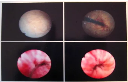
6. Figure 6: Cystoscopy images of the bladder and urethra. (Permission is granted by citation only)
7. Figure 7: Urine test strip used as part of initial assessment of microhematuria.
(Permission is granted by citation only)
8. Figure 8: Microscopic hematuria: Red blood cells in a urine sample seen under the microscope. (Permission is granted by citation only)
List of Appendices:
1. Approval letter from the Research Ethics Committee.
2. Letter of information and Consent Form
3. Pre-cystoscopy survey.
4. Post-cystoscopy survey.
List of Abbreviations:
1. VAS: Visual Analogue Scale. 2. BMI: Body Mass Index.
3. TURBT: Transurethral Resection of the Bladder Tumor. 4. LUTS: Lower Urinary Tract Symptoms
Chapter 1
Introduction
1.1 Cystoscopy from the urologist’s perspective
1.1.1 History of cystoscopy
Visualizing the internal organs through natural orifices like mouth and urethral meatus
using instruments from distance (endoscopy) has been attracting physicians since ancient
times in an attempt to understand patient’s symptoms and signs. Currently, endoscopy is
much more differentiated and there is just about a special scope for each hollow organ:
colonoscopy for colon, otoscopy for the ear and cystoscopy for the urinary bladder (1, 2).
Cystoscopy is a commonly performed procedure in urology clinics worldwide. It enables
urologists to visualize the urethra and the bladder by introducing a scope through the
urethral meatus. Before the invention of cystoscopy, diagnosing bladder tumors or stones
was done using invasive methods. Open surgical exploration of the bladder through lower
abdominal incisions was one of the common diagnostic tools to explore the bladder.
Blind insertion of sounds and clamps into the urethra was a common method of trying to
caused patients increased morbidity and were very low yield in clarifying patients’ lower
urinary tract symptoms (3).
It is well accepted that the German-Italian Phillip Bozzini was the first to use the
“Lichtleiter”, which is a German word that means the ‘light conductor’, to visualize
multiple body cavities including the urethra (4). In 1806, Bozzini used an in-built
beeswax candle with adjustable viewing ports (figure1). The device that Bozzini invented
was not specific for one organ; he used it to visualize urethra, ear, gunshots tracts, etc.
Since 1806, endoscopy has gone through many technical modifications accompanied
with advances in medical and mechanical knowledge. Endoscopy was also challenged at
the beginning by some scientists from the medical community who believed that it was
inferior to open surgical exploration of internal organs including the urinary bladder (5).
Developers from the urologic community focused on 2 critical components: better light
Figure 1: Bozzini’s original light conductor with specula. Courtesy of the History office at the European Association of Urology.(Int.Nitze-Leiter Research Society for Endoscopy/Nitze-Leiter
Collection). http://history.uroweb.org/history-of
But it was not before 1879, when Maximilian Nitze developed the first usable endoscope
used specifically to visualize the urethra and the bladder, the cystoscope. “A lucky break
led me to the right path.” Those were Nitze’s own words when he described the
coincidence as he was cleaning an eye-piece lens and was able to see an inverted image
of Matthäus Church in Berlin across the street from his lab through that lens. That
coincidence led him to the idea of creating a form of telescope to enable him to visualize
internal organs (4). See Figure 2. The light source in the telescope he developed was
attached on the distal part of the scope, offering maintained illumination throughout the
procedure regardless of the angle the operator is working on. Moreover, he developed a
water channel that ran through the scope to cool it down. Concepts of current rigid
cystoscopy are based on Nitze’s model (figure 3).
Figure 2: Optical tube after Max Nitz. Courtesy of the History office at the European Association of Urology. From the Int. Nitze-Leiter Research Society for Endoscopy, Vienna/Reuter
Collection.
Figure 3: Woodcut of the first urethroscope and cystoscope built on the principle of the platinum wire filament based on Nitze’s design. Courtesy of the History office at the European Association
of Urology. From the collection of the Int. Nitze-Leiter Research Society for Endoscopy.
A revolutionary step in the development of cystoscopes was the use of fiber optics (7). In
1951, a physicist named Harold Hopkins applied fiber optic technology to cystoscopies
following its success in gastrointestinal tract visualization. A German company named
Storz bought this patent. Its first flexible fiber optic cystoscope was introduced at the
International Society of Urology in Munich in 1967. With this new system urologists
worldwide have been using flexible cystoscopy with much better illumination and image
definition than older cystoscopies. (Figure 4)
Figure 4: Flexible cystoscope with fiber-optic technology.Photograph taken by Michael Reeve, 25 April 2005. {{GFDL}}.
Although still in use, fiber optics are vulnerable to break. In 1970, Boyle and Smith developed the
charge-couple device (8). A sensor at the tip of the scope converts the optical image into a digital
signal that can be transferred via the shaft of the endoscope to an image processor where a video
signal is generated and displayed on a monitor. This latest advancement improved image
resolution and durability of the cystoscope.
1.1.2 The current cystoscope and its properties
There are two types of cystoscopes currently used worldwide in the urological
community: the rigid and the flexible cystoscopes (9).
The current rigid cystoscope as described earlier is based on Nitze’s model. It is
composed of an optical lens, bridge sheath and obturator. Optical lenses are offered in
different angles to serve different purposes. Bridges can accommodate more than one
working channel. Because of that, the rigid cystoscope is more commonly used in the
operating room where diagnostic, therapeutic and interventional procedures are usually
needed and done through those working channels. In addition, because of their rigidity,
they are easier to handle with one hand which gives the surgeon the ability to use the
The flexible cystoscope however is more commonly used in the outpatient setting
because it causes less pain than rigid cystoscopy due to its thinner diameter and
non-metallic material (10-12). Moreover, use of rigid cystoscopies was found to be associated
with higher levels of anxiety prior to cystoscopy (13).
Although a single working channel can be incorporated into the flexible cystoscope, it is
more commonly used for diagnostic purposes in the outpatient clinic (14). Regardless of
its type, the cystoscope must be attached to a light source and irrigation source. Irrigation
helps with opening the collapsed urethra and helps visualize the bladder.
Visualization during cystoscopy historically has been done by the surgeon directly
looking in to the lens of the cystoscopy. With recent technological advances, now a days
a camera is attached to the lens and its cable is attached to a video-endoscopic unit
consisting of a fixed tower, monitor, light source and printer (14)
1.1.3 The current technique of the outpatient cystoscopy:
For a rigid cystoscope, the patient is usually positioned in lithotomy position both in
males and females. With the flexible cystoscope the lithotomy position is not necessary.
The male patient is usually positioned in a supine position where as the female patient is
the urethral opening (meatus). After disinfection of external genitalia, a lubricating gel is
applied into the urethra. The tip of the cystoscope is then administered to the meatus.
Irrigation helps open the collapsed urethra. It also helps the surgeon to inspect the urethra
for possible diseases as well as navigating the way to the bladder. Once in the bladder
irrigation, helps inflate the bladder walls, stretching all folds that could harbor possible
disease (15).
Figure 5: Diagram showing a cystoscopy for a man and a woman. Author: Cancer Research UK. 30 July 2014 (released by CRUK).
https://commons.wikimedia.org/wiki/File:Diagram_showing_a_cystoscopy_for_a_man_and_a_w
Cystoscopy is performed in a systematic fashion. After insertion of the scope through the
urethra, inspection of the ureteral orifices is done. Shape and color of urine efflux out of
the orifices should be noted. Then inspection of all bladder walls is done. Advantage of
the flexible cystoscope is its ability to deflect in different angles giving the surgeon the
ability to visualize difficult areas of the bladder like the anterior bladder neck and anterior
bladder wall (figure5). After the end of the procedure, the bladder is emptied, and the
cystoscope is removed (15).
1.1.4 Clinical applications of the cystoscope
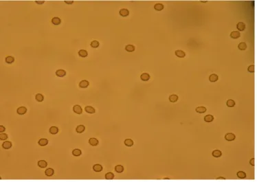
Blood in the urine is called hematuria. It could be gross or microscopic. Microscopic
hematuria can be tested using urine dipstick and microscopic urinalysis. Dipstick color
change indicating blood in urine is based on the fact that hemoglobin has a peroxidase
like activity which catalyzes an oxidative reaction with the dipstick substance which
changes color correlated with the amount of oxidation (16). See figure 7.
Figure 7: A urine test strip photo. Author: Author: J3D3. 24 February 2012.
Figure 8: Microscopic hematuria: Red blood cells in a urine sample seen under the microscope.
Author: Bobjgalindo. 19 February 2005.
https://commons.wikimedia.org/wiki/File:MicroHematuria.JPG
Microscopic hematuria is defined as having more than 3 red blood cells (RBCs) per high
power field (HPF) (16). See figure 8. Whether it is gross or microscopic, persistent
hematuria after ruling out infection and other benign causes is an indication for referral to
the urology clinic for further assessment (17). If confirmed, a cystoscopy is usually
performed in the outpatient setting. The main aim of cystoscopy in the hematuria setting
To date, cystoscopy has remained the gold standard of diagnosing bladder cancer. No
other investigation available is able to replace cystoscopy. The Canadian, American and
European urological guidelines recommend cystoscopy as the gold standard for
diagnosing bladder cancer. (18-19).
Bladder cancer is the fifth most common cancer in Canada. About 9,000 Canadians are
diagnosed with bladder cancer each year (20). The most common sign of bladder cancer
is asymptomatic presence of blood in the urine. The confirmation of bladder cancer
diagnosis is done by a diagnostic cystoscopy. If confirmed, the patient will undergo a
procedure called trans-urethral resection of the bladder tumor (TURBT) (21). According
to the pathological assessment after resection, a bladder tumor is classified as either
muscle invasive or muscle invasive. 75% of patients with bladder cancer have
non-muscle invasive bladder cancer (20). Depending on the microscopic appearance of the
tumor cells and recurrence history, patient with non-muscle invasive bladder cancer
might need adjuvant intravesical treatments to reduce recurrence and progression of the
disease.
Because of its high recurrence rates (60-70%), surveillance strategies with regular
cystoscopies have been developed by urologists worldwide and are recommended by all
urological guidelines to assess for possible recurrence after TURBT and allow early
intervention. Meaning that, every patient with non-muscle invasive bladder cancer
success of the intravesical treatments. The surveillance duration in cases of no recurrence
might reach up to 5 years according to the European guidelines.
The standard of care for patients with muscle invasive bladder cancer is neo-adjuvant
chemotherapy followed by radical surgical removal of the bladder, pelvic lymph nodes,
and urinary diversion. However, in a sub-set of patients who are high risk for surgery due
to age and/or comorbidities, a bladder preserving approach is undertaken which includes
TURBT and a combined chemo-radiation regimen. Following this combined treatment;
cystoscopy is of cardinal importance to assess treatment success as well as surveillance to
assess for recurrence (22).
The lining urothelium of the bladder is in continuity with the lining urothelium of the
ureter and the renal pelvis. Patients with upper tract urothelial cancer are at increased risk
of developing bladder cancer. Therefore, surveillance cystoscopies are indicated in
patients with previously treated upper tract urothelial cancer (23-24).
In addition to cystoscopy’s role in malignant diseases, it plays a critical role in the
evaluation of benign conditions that might affect the urinary tract. Persistent lower
urinary symptoms (LUTS) including urgency, frequency, nocturia and dysuria in the
absence of urinary tract infection is an indication for diagnostic cystoscopy not only to
rule out bladder cancer but also to help in the assessment of potential diagnoses of benign
bladder conditions like interstitial cystitis, chronic pelvic pain syndrome, and urethral
1.1.5 Summary of cystoscopy from urologist’s perspective
Visualization of the lower urinary tract has undergone a significant evolution since the
last century. Flexible cystoscopy is the most common type of cystoscope used in the
ambulatory setting. It is usually performed while the patient is awake and with the use
transurethral lidocaine-based lubricating gel. From a urologist’s perspective, cystoscopy
is an excellent tool to visualize the lower urinary tract. Over the years, cystoscopy has
remained in all urological guidelines as the irreplaceable gold standard diagnostic and
surveillance tool for bladder cancer as well as other benign and malignant urological
diseases.
In the following section, we will explore cystoscopy from the patient’s perspective. Since
flexible cystoscopy is the type of cystoscopy investigated in this study, our critical
1.2 Cystoscopy from patient’s perspective.
1.2.1 Before cystoscopy
Like any other medical procedure, urologists approach patients requiring cystoscopy by
explaining the indication based on their symptoms and signs. This is followed by
explaining the steps of the procedure to the patient, potential side effects and what
measures the urologist will perform to alleviate the pain which most commonly include
lidocaine gel during the informed consent process.
At this point it might be clear and expected that patients feel uncomfortable, embarrassed
and anxious as they anticipate an unpleasant painful procedure in a sensitive body area.
This assumption has been investigated and confirmed. Yerlikaya et al performed a
prospective clinical trial that compared pain perception in females after a diagnostic
cystoscopy to that after urodynamic study (28). Urodynamic studies (UDS) is a dynamic
investigation of the transport, storage and evacuation of urine. UDS in this study included
introducing a Foley’s catheter into the bladder and filling the bladder to certain points to
assess for filling pressure and leaking pressure points as well as a rectal catheter that
helped in assessing abdominal pressure. The study did not only show pain perception
from cystoscopy is higher than that associated with urodynamic studies but also pain
anticipation was even higher than pain scores obtained after cystoscopy. Patient’s anxiety
before cystoscopy although logically expected has been examined and conformed too.
pain in patients undergoing cystoscopy and urodynamic studies (29). In the cystoscopy
group, 93.7 % (Age 18-50 years) reported a degree of anxiety before cystoscopy. Older
patients reported less anxiety levels. In addition, anxiety and embarrassment were
statistically correlated.
1.2.2 During cystoscopy:
Pain associated with cystoscopy has been the focus of many urologists for a long time.
The first reported article in PubMed goes back to 1946 when Felber published an abstract
titled “Does cystoscopy need to be painful?” (29). Since then, many articles have been
published about patient’s pain and discomfort associated with cystoscopy and potential
interventions to manage that pain.
Studies investigating pain associated with cystoscopy usually use the Visual Analogue
Scale to give pain a value. VAS values in studies assessing pain associated with
cystoscopy are variable. Highest mean VAS scores associated with cystoscopy reported
by Walker et al was 6.6 in the control group (31). However, other studies like Cornel et al
reported mean VAS score as low as 1.55 in the control group (32). Biardeu et al in their
study including 101 patients who received flexible cystoscopy for the first time, 59,4 %
reported some level of pain. Moreover, pain during different steps of the procedure was
3.1 compared to pain during gel installation 0.7 +/- 2.1 and cystoscope removal 0.7 +/-
2.1. The pain peak-phase was confirmed by another study by Poletajew et al. In their
comparative analysis, peak-pain levels were recorded at the time of insertion of the scope
in both study arms (33)
Greenstein et al study in pain associated with diagnostic cystoscopy included the largest
population sample in the literature. He examined pain associated with cystoscopy in
1,320 patients. In subgroup analysis that included only patients who had flexible
cystoscopy, they found that pain scores were significantly lower in first timers compared
to repeaters. In addition, his results confirmed the fact discussed earlier that flexible
cystoscopy compared to rigid cystoscopy caused less pain (34).
1.2.3 After cystoscopy
No long-term study examined correlation between higher pain perception after
cystoscopy and less compliance for future repeat cystoscopies. However, one study
explored patient’s willingness to repeat cystoscopy at the time of first cystoscopy. Kwon
et al asked participants about their willingness to repeat cystoscopy in their comparative
interventional trial (35). They found that patients who had higher VAS scores were less
willing to repeat cystoscopy. Although no long-term data exists, this finding suggests
1.2.4 Summary of patient’s perspective
Patients planned to undergo ambulatory flexible cystoscopy for the first time anticipate
pain and have anxiety about the procedure even before the initiation of the procedure.
Cystoscopy even in its flexible form is associated with pain, anxiety and embarrassment
during the procedure. Pain levels associated with cystoscopy were reported using VAS
scores in the literature and were variable among studies. Males reported average higher
VAS scores than females. However, most patients report a certain level of pain and
anxiety associated with the procedure. Higher VAS scores were associated with less
willingness to repeat the procedure in one study that might impact patient’s compliance if
surveillance cystoscopy is indicated.
1.3 Current Approaches in the Literature to Alleviate Pain Perception
Associated with Cystoscopy
1.3.1 Assessment of Pain associated with Cystoscopy in the Literature
Pain and discomfort associated with ambulatory cystoscopy has been acknowledged by
the urologic community. This is demonstrated by the literature published from different
associated with cystoscopy that highlight the ongoing efforts to improve patients’
experience during this unpleasant procedure.
Studies that examined pain associated with flexible cystoscopy used the visual analogue
scale (VAS) to assess patients’ pain associated with the procedure. This test has
established performance across clinical specialties in assessing pain (36). The scale is
graded from 1 to 10. 1, representing the least amount of pain and 10 representing severe
amount of pain. The patient will draw a line between these 2 values to represent the
amount of pain he/she felt during the procedure. The test has many forms that follow the
same concept. Although there are other tests that assess pain like Likert and Borg scales,
the VAS showed superiority over those tests (37).
Current approaches in management of pain and discomfort associated with cystoscopy
are divided into pharmacological and non-pharmacological approaches. Pharmacological
approaches include interventions that include a pharmacological product given to the
patient that aims to relieve the pain associated with cystoscopy. A non-pharmacological
approach includes a form of distraction that could be utilized during cystoscopy with the
1.3.2 Pharmacological interventions
The most commonly used pharmacological intervention before the initiation of
cystoscopy is delivering a lidocaine-based gel through the urethra. Many preparations
have been made available in the market for this purpose with different amounts of gel and
lidocaine concentrations. Furthermore, the use of the transurethral lidocaine-based gel is
variable among urologists regarding amounts used, indwelling time and penile clamping
after introduction of the gel.
Our literature search demonstrated conflicting results about the efficacy of transurethral
lidocaine gel in alleviating pain associated with cystoscopy (38, 39). David et al reported
the most recent systemic review and meta-analysis (40). The review included 4
randomized clinical trials with a total patient population of 411 male patients. Patients
were randomized to either receiving transurethral lidocaine gel (intervention) or
transurethral plain gel (placebo). Although 3 of the 4 studies included showed no
evidence of VAS score differences between the 2 arms, meta-analysis showed that
patients in the lidocaine gel arm were 1.7 times less likely to develop moderate to severe
pain (score of 3 or higher). In addition, the study did highlight its limitations which
include mainly variability of the amount of gel introduced and dwelling times of gel in
the urethra before cystoscopy start.
instillation of lidocaine-based gel was more painful for patients than plain gel suggesting
an irritating effect of lidocaine itself to the urothelium that adds negatively to the overall
patient experience (41).
Other studies examined whether a longer indwelling time of the lidocaine gel increases
this intervention’s efficiency. Losco et al performed a prospective comparative trial
where he randomized men to either undergo flexible cystoscopy with insertion of
lidocaine-based gel either immediately prior to cystoscope insertion or after a 3-min
interval. This modified gel insertion technique however showed no significant differences
in VAS scores (42).
Although transurethral lidocaine-based gel is the most commonly used pharmacological
intervention, researchers investigated other pharmacological interventions. Nadeem et al
investigated the role of a diclofenac suppository taken by the patient 1 hour before
cystoscopy in addition to lubricating plain gel compared to use of only plain lubricating
gel (43). 60 patients were randomized to either group. The intervention arm had
statistically significant lower VAS scores, which might question the value of lidocaine in
the lubricating gel. The indications for cystoscopy in both groups in this study were
variable ranging from first timers, to repeaters and cystoscopy with stent removal which
The previously cited study by Poletajew et al was a randomized controlled trial. They
proposed that the inefficiency of transurethral gel is that it does not reach the posterior
urethra. For that, a catheter was inserted after transurethral gel administration to aid in
distributing the gel material to the posterior urethra before the initiation of cystoscopy.
There were no statistically significant differences between the 2 arms in VAS scores at
the time of cystoscopy, but patient’s perception of the procedure was better in the
intervention arm. Other outcomes confirmed the fact that was reported by other studies
that the number of previous cystoscopies is inversely related with VAS scores.
1.3.3 Non-pharmacological interventions
The most investigated non-pharmacological interventions in alleviating patients’ pain and
discomfort during cystoscopy was the use of music as a method of distraction. A pair of
headsets is offered to the patient and he/she listens to music of his/her choosing. In
another setting, music is played in the cystoscopy suite. Kyriakides et al reported the
most recent systemic review and meta-analysis examining the role of music in different
urological procedures including diagnostic flexible cystoscopy (44).
In this meta-analysis, three randomized clinical trials were included in the cystoscopy
setting. Anxiety and VAS-pain scores were significantly lower with the use of music
the only weakness in those trials due to lack of blinding which is unavoidable in such
studies.
Allowing the patient to visualize the cystoscopy examination on the monitor while lying
during the cystoscopy procedure was investigated as non-pharmacological intervention to
alleviate pain associated with cystoscopy. Two randomized control trials from Soomro et
al and Zhang et al showed statistically significant lower VAS scores in the intervention
arms (45, 46). However, another previously cited randomized clinical trial by Cornel et al
showed no significant difference between the 2 arms. No systematic review was found in
the literature that examined this intervention.
Less commonly investigated interventions include hand-holding during cystoscopy. The
previously cited study by Kwon et al in their randomized clinical trial assigned a nurse
that held the patient’s hand during cystoscopy in the intervention arm. 81 patients were
randomized to either a standard cystoscopy or with hand-holding. VAS scores were
significantly lower in the intervention group.
With the rapid development of virtual reality and its applications, investigators at the
Triple Army Medical Center in the United States examined the role of VR in lowering
patients’ pain perception with cystoscopy. In their randomized clinical trial Walker et al
(cited earlier) randomized a total of 45 patients to either standard cystoscopy with the use
of VR helmet as a distraction during cystoscopy or standard cystoscopy alone. This study
Hruby et al investigated the effect of transcutaneous electrical stimulation in the lower
abdominal area in improving patients’ pain associated with cystoscopy (47). In their
randomized control trial that included 148 patients, no significant difference in VAS
scores in the electrical stimulation group compared to placebo.
Another randomized clinical trial investigated increasing the pressure of the irrigation
during flexible cystoscopy on pain perception. Pressure was manipulated by increasing
the height of the irrigation fluid bag from the bed. Higher irrigation pressure was
associated with lower discomfort and pain scores than patients who received lower
irrigation pressure during flexible cystoscopy (48).
1.3.4 Summary of current interventions
Interventions in the literature that were investigated to alleviate pain associated with
cystoscopy were either pharmacological or non-pharmacological. Our literature search
showed variable outcomes in the most commonly used pharmacological intervention
which is transurethral lidocaine-based gel. Only the use of music during cystoscopy
were less rigorously studied and most of them showed no impact on patients’ pain
perception.
The past and recent publications reviewed, show the huge efforts from the international
urological community in exploring ways to improve patients experience during
cystoscopy. Even the use of more personal, expensive and complicated technology and
medications were considered by those investigators to alleviate pain associated with
cystoscopy. This reflects the keen interest in improving overall patients’ experience with
diagnostic cystoscopy.
An optimal intervention would be that of low costs, minimal side effects and be simple to
perform. Before we introduce the intervention investigated in this study, we will in the
following section describe how we assess experiences in different ways and how
understanding that assessment might open the door for a modified technique in
cystoscopy that might improve patient’s pain perception.
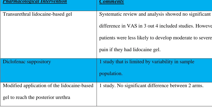
Tables 1 and 2 summarize current interventions published in the literature with the intent
Pharmacological Intervention Comments
Transurethral lidocaine-based gel Systematic review and analysis showed no significant
difference in VAS in 3 out 4 included studies. However,
patients were less likely to develop moderate to severe
pain if they had lidocaine gel.
Diclofenac suppository 1 study that is limited by variability in sample
population.
Modified application of the lidocaine-based
gel to reach the posterior urethra
1 study. No significant difference between 2 arms.
Table 1: Summary of current pharmacological interventions in alleviating pain associated
with flexible cystoscopy.
Non-pharmacological Intervention Comments
Music Systematic review and meta-analysis showed
evidence of VAS score decrease with music.
Visualizing the procedure 3 RCTs, 2 showed significant decrease and the 3rd
did not show any difference. No systematic review
and meta-analysis available.
Patient’s hand-holding during the procedure Only 1 study that showed lower VAS scores in the
intervention arm.
Virtual reality Only 1 study that showed no significant difference.
Increasing irrigating pressure during the
procedure.
Only 1 study that showed lower VAS scores in
the intervention arm.
Table 2: Summary of current non-pharmacological interventions in alleviating pain
1.4 The Peak-end Theory
1.4.1 Types of Experience Evaluation
There are 2 types of experience evaluation (49). Instant evaluation, which is providing a
specific intensity of an effect at a specific moment: like asking a person how are you right
now? And the other type of evaluation is remembered, which is a retrospective evaluation
of a certain experience, for example: How was the movie?
The 2 types have distinct features that have been investigated. In instant evaluation, there
is a moment to moment feedback that is recorded which gives an accumulative overall
effect of that experience. Whereas in remembered feedback, there is a global evaluation
of the entire experience. It could be a short visit to the urologist or an evaluation of a
1-week long vacation.
The significance of differentiating between these 2 types of evaluation comes from the
fact that when patients are asked about their pain after an unpleasant medical procedure
they provide a form of retrospective remembered global evaluation. Although there is
some controversy of memory’s weight in impacting future decisions (50) retrospective
evaluation is the type of evaluation that people depend at least partly on for future
decisions. The impact of retrospective global evaluation today goes beyond
social media platforms. Sharing personal global evaluations might have impact on others
planning to be part of the same or similar experience (51, 52).
1.4.2 Retrospective Global Evaluation
The remembered retrospective global evaluation has been examined in multiple animal
and human experiments in the literature. 3 distinct features of this type of evaluation were
identified: Peak-end rule, duration neglect and violation of monotonicity.
1.4.2.1 Peak-end rule
Humans evaluate life experiences whether it was pleasant or not on daily basis whether it
was their vacation or a movie that they saw. One would expect that their retrospective
evaluations are based on an average of all the individual moments of that vacation or the
individual clips of that movie. But, when we recall a movie or any specific experience,
we do not recall each clip or moment. We would rather recall certain clips and moments.
Kahneman et al examined further remembered evaluation and its characteristics and
the peak event and the event at the end of that experience. This is based on 2 known
psychological biases in memory: peak and recency biases. High intensity incidents within
an experience outweigh less intense moments. We tend also to remember the most recent
moments rather than the more remote ones. The accuracy of the Peak-end rule was
challenged by Kahneman et al in multiple experiments (53, 54).
In his experiment with Redelmeier, Kahneman explored the Peak-end rule in the medical
setting. They hypothesized that this theory might have an application on pain perception
associated with an unpleasant medical procedure. They performed a prospective clinical
trial where they randomized over 600 participants into either standard colonoscopy
(control) or to standard colonoscopy extended by leaving the scope for 3 minutes in the
rectum at the end of the colonoscopy. Thus, creating a less aversive end to the procedure.
Pain assessments during the procedure (instant evaluations) as well as 1 hour after the
procedure (global, remembered evaluation) were measured with ten-point intensity scale.
Not only did patients in the intervention arm have statistically significant lower VAS
scores but also compliance rates with repeat colonoscopies if indicated were higher in the
intervention arm. Participants’ recollections were strongly associated with the momentary
instant peak and end moments (55).
The peak-end rule extend applies even for longer- term memories. In another experiment,
Kahneman recruited patients with clinically confirmed rheumatoid arthritis to evaluate
their pain status 7 times a day. These assessments were compared with their overall pain
instant evaluation reported during an episode and of the evaluation reported at its end
predicted subsequent global evaluations at least as accurately as the average of instant
assessment.
The peak-end rule opened the doors for Kahneman and his colleagues to explore the next
2 features of retrospective global evaluation.
1.4.2.2 Duration neglect
In his experiment with Redelmeier, Kahneman noted that although the procedure was
longer in duration in the intervention arm and the scope was left in the rectum, it was
ended with less aversive event compared to the actual colonoscopy which includes scope
manipulation and irrigation. This according to authors contributed to the study resulting
in lower VAS scores in the intervention are. Longer duration did not apparently influence
participants’ retrospective evaluations.
In another experiment, Kahneman and colleagues recruited 32 college students to 2
unpleasant experiences. The one shorter in duration included students emerging their
hands into bowl of cold water (14 C0) for 60 seconds. Then in the longer experiment they
kept their hands into bowl of cold water for 60 seconds and then the water was warmed to
15 C0 (still experientially cold) and they kept their hands for extra 30 seconds. Students
experiment would they prefer to repeat, the majority chose the longer experiment as they
felt it was more comfortable.
Although 15 C0 was still unpleasant for the human body, it is less unpleasant than 14 C0.
Having a less aversive ending affected how those students evaluated retrospectively their
experiences. In addition, this experiment confirmed the suggested feature of remembered
evaluation which duration neglect is and further enforced the concept of the peak-end
rule. Participants evaluated their experiences based on the average between peak and end
phases regardless of the duration (56).
Kahneman examined this phenomenon in a type of evaluation that involved non-tactile
stimulus. With his previously cited study, with Fredrickson (53), Kahneman made
participants view aversive film clips and pleasant film clips that varied in duration and
intensity. They provided real-time ratings of affect during each clip and global
evaluations of each clip when it was over. Another group of students viewed these same
clips and later and ranked them retrospectively by either pleasant or unpleasant.
Retrospective evaluations appear to be determined by a weighted average of "snapshots"
1.4.2.3 Violations of monotonicity
Monotonicity from a psychological perspective is one of the principles in decision
making. If we were to decide between two alternatives but one has a higher outcome than
the other, than that option is deemed better. As logical as it might sound, Birnbaum in his
study explored monotonicity and when people violate it in decision making (57). In his
experiment he asked undergraduate students to choose between guaranteed amounts of
money and gambles. Each of the 30 gambles was presented for comparison with 2 groups
of money amounts. Means of those amounts were intentionally put higher in group 2 than
in 1 to investigate the impact of contextual effects. Instructions clearly stated that they
should prefer amounts of money if they exceeded the most a gamble could offer and
prefer a gamble to any amount less than the least amount a gamble would offer. Despite
instructions promoting monotonicity satisfaction, 70% of participants showed at least one
violation of monotonicity. 50% violated it more often than they satisfied it and 25 %
satisfied it more than they violated it. Subgroup analysis for the 2 groups were made and
showed difference percentages of choosing gamble over a sure amount of money. He
concluded that comparison judgments are not always simple and straight forward
comparison of values but are instead influenced by the distributions that form the context
In the experiment described earlier by Kahneman (56), students were asked which
experiment they would repeat. They were asked to make a choice between two similar
alternatives. Both experiments were considered aversive experiences (cold water).
Following monotonicity, one would expect more students will choose the shorter in
duration experiment with less total pain. However, the clear majority chose the longer in
duration experiment because of its less aversive ending.
1.4.3 The Peak-end theory: potential applications
As noted earlier patient’s pain perception after an unpleasant medical procedure is based
on retrospective global evaluation rather than instant evaluations. When a person
retrospectively evaluates an experience, whether a pleasant or an unpleasant one, he/she
will not replay the whole experience in his/her mind to evaluate. Because of peak and
recency biases, only specific snapshots will be remembered, and the evaluation will be
based on the average of those specific snapshots. Moreover, as described earlier
retrospective evaluation appears to be independent of duration.
Retrospective evaluation and Peak-end theory interpretation of global evaluation added a
new way of looking into how seniors evaluate their life in general and quality of life (58).
Peak-end theory motivated research in business development to explore ways to improve
overall customer satisfaction (59, 60).
Kahneman and Redelmeier were the first scientists to explore this theory application in
the medical setting to improve overall patient experience in colonoscopy. However, the
use of intravenous opioids is part of the standard of care in patients undergoing this type
of procedure. This might make it challenging to delineate the real effect of their
intervention on post procedure VAS scores. To the best of our knowledge, Peak-end
theory concepts have not yet been examined in unpleasant medical procedure where
intravenous opiates are not traditionally used.
1.4 The purpose of this study and specific aims
1.5.1 Purpose Statement
The purpose of this research is to evaluate the feasibility and efficiency of modifying the
cystoscopy procedure by prolonging the end-phase with less aversive maneuvers based
on the peak-end theory to improve patient’s pain perception after ambulatory diagnostic
1.5.2 Specific aims
1. Examine the feasibility and effectiveness of the peak-end theory in the flexible
cystoscopy setting on lowering pain perception scores.
2. Assessment of pain associated with flexible cystoscopy at our institution.
1.5 Hypothesis
We hypothesized that prolonging the end phase of the cystoscopy with a less aversive
phase by leaving the cystoscope for an additional 2 minutes in the bladder without further
manipulation will lead to lower post interventional VAS scores than in patients receiving
1.7 Significance
Cystoscopy has been and is still currently the gold standard diagnostic tool in many
urologic diseases, mainly bladder cancer. In addition, it is irreplaceable by any other
investigational diagnostic modality. Recurrence is a hallmark feature of bladder cancer
making cystoscopy a frequent surveillance procedure that millions of patients around the
world must undergo for years.
Pain perception associated with flexible cystoscopy is an ongoing clinical challenge for
urologists worldwide. This is evident by the amount of literature published on pain
associated with the procedure as well as investigational trials for interventions that could
lead to lower pain perception associated with the procedure. Scientists considered simple
and inexpensive as well as complex and very expensive interventions with minimal yield
most of the times.
Improving patients’ pain perception will improve their overall experience. In addition,
future decisions depend on their evaluation of the experience that they had during
cystoscopy. Therefore, by improving patients’ experience with flexible cystoscopy, we
decrease the chances for noncompliance where a repeat cystoscopy is warranted.
Currently, people share their experiences not only with their families and friends but also
on multiple social medical platforms. In addition, more people are researching procedures
might be shared with people that might need cystoscopy in the future. Improving
patients’ experiences will act as an indirect positive impact on people in whom
cystoscopy is warranted but are concerned with the pain and discomfort associated with
it.
The peak-end theory concept offers a very low-risk profile intervention to improve a
patient’s perception. In addition, there are no direct costs that the healthcare provider, the
Chapter 2
Materials and Methods
2.1 Study Design
This is a randomized prospective single blinded study collecting quantitative data (pain
perception as measured using a visual analogue scale) after a diagnostic only flexible
cystoscopy for patients who have not had cystoscopy before. The study compared a
modified technique based on the Peak-End theory’s concepts by prolonging the end phase
with a less aversive maneuver.
The study has been approved by the University of Western Ontario Research Ethics
Board and approved by Lawson Research Institute Board. Recruitment started in
September 2017. Co-investigators at the Urology Clinic at the Victoria Hospital of
London Health Sciences Center recruited patients on the day of their scheduled
cystoscopy.
The only surgeons who performed cystoscopies for the purposes of this study are Drs.
Power, Izawa, and Chin. The 3 surgeons were neither involved in the randomization
process nor in data extraction and analysis. Data collection was performed by the nursing
2.2 Inclusion and Exclusion Criteria
Inclusion criteria were: 1. all patients with ages from 18 to 60 years old planned to have
an ambulatory diagnostic flexible cystoscopy for the first time. 2. Males and females
were included. To assess for potential confounders previously described in the literature
we have designed recruitment to include equal numbers of males and females in each arm
and only first timers.
Exclusion criteria included:
1. Patients who underwent cystoscopy before
2. Patients with congenital or acquired urinary tract anomalies.
3. Patients with chronic pain or on chronic pain medications.
4. Cystoscopies that involved additional interventions along with
cystoscopy were excluded.
5. Patients with previous pelvic or urethral surgeries or radiation were
excluded.
6. Patients planned to undergo rigid cystoscopy were excluded.
7. Patients currently taking medication for chronic pain (e.g. opioids,
TCAs) will be excluded from the study.
2.3 Outcomes
The primary outcome of this study is to compare the mean VAS scores after flexible
cystoscopy for the first time in the standard cystoscopy group with the modified group in
the ambulatory setting at our institution.
Secondary outcomes include:
1. Anxiety levels before flexible cystoscopy
2. Anxiety levels after flexible cystoscopy
2.4 Sample Size Calculation
2.4.1 Effect size and VAS score difference.
An important component of sample size calculation is effect size. As clinicians we are
interested in not only a statistically significant difference in VAS scores between the 2
arms, but also a difference that is clinically relevant.
In our literature search, most studies looking into clinically significant VAS score
evaluation in the emergency department for acute pain rather than a retrospective
evaluation after the procedure. Based on those studies, a clinically significant difference
varied from 12 mm to 30 mm on the 100 mm VAS (61, 62). Sadovsky et al suggested
that a clinically significant VAS difference depends on the severity of the basal VAS
score. Kelly et al, however found that basal VAS scores do not really affect clinically
significant VAS scores (63).
In this study, VAS scores were evaluated in a retrospective manner. As we have
discussed earlier, there are some key differences between retrospective and instant
assessments of experiences. In the Redelmeier et al study, a 5 mm difference was noted
between the 2 arms of the study in the retrospectively collected data.
We assumed an expected mean VAS score of 3.48 cm with standard deviation of 1.53,
based on the Greenstein et al study. Simulating the retrospective effect of this technique
on patients described in Redelmeier study, we assumed an effect change of 0.5 cm on
VAS score scale. Considering a power of 0.8 and 0.05 level of significance, we proposed
a sample size of 296 patients with 1:1 allocation ratio.
If an effect size of 10 mm was considered, a total of 79 patients will suffice. Based on our
difference noted in the Redelmeier et al trial, we planned an interim analysis after
recruiting 79 patients. If results showed significant difference of 10 mm or more then we
will terminate the study.
Because of slow recruitment we submitted the analysis of only 61 patients. However,
future analysis will remain as planned in the statistical design.
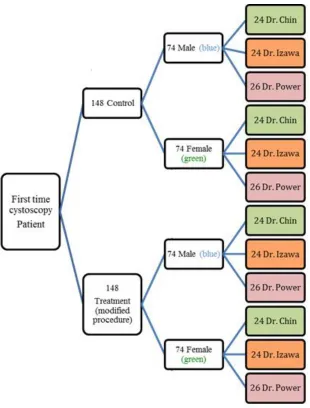
2.4 Methodology
Sealed study packages have pre-randomized study numbers for both interventions. Study
numbers were randomized to both arms using the Medsharing® mobile application.
Because we had a preliminary sample size of more than 100, simple randomization
sequence of study numbers was performed. Randomization, allocation process and
creation of the master list were done by Khalil Hetou. Each study number was
pre-randomized and its allocated arm was noted in the master list. Study number allocations
were not known to the investigators. Equal number of packages was allocated in each
arm. Gender stratification was performed by labeling equal numbers of males and
Patient assignment to in investigator was predetermined so that each investigator
performs similar number of intervention and controls as well as of males and females. 2
investigators had exactly the same number of patients assigned. Only one investigator
had 8 more patients than the other 2 investigators. Please see figure 9.
The study packages were sealed and put in a safe place at the urology clinic where
access is only granted to official personal. Each investigator was assigned 2 boxes. One
box labeled for males that has equal numbers of intervention and control
(pre-randomized) study packages. The other box labeled for females that have equal numbers
of intervention and control (pre-randomized) study packages. All study packages were
identical from the outside except for the study number. Investigators were blinded to the
master list.
The study package included a sealed envelope with instruction to the investigator about
which arm this patient was randomized to. Instruction envelope shall be opened only at
the time of cystoscopy.
In addition, the study package contained a letter of information, consent form, a
pre-intervention survey, post-pre-intervention survey and a debriefing form.
296 closed envelopes were created. Envelopes were gender stratified with 1:1 male to
female ratio. 148 blue envelopes for men and 148 green envelopes for women were
created. Once the patient was deemed a candidate for the trial and consented, the gender
matched closed envelope will be attached to his/her file. Dr. Chin will receive 96 (48 blue
and 48 green) closed envelopes. Dr. Izawa will get the same amount and type of
Once patients have been informed and have completed the consent form, they were given
a short pre-procedure survey to fill assessing demographic, history of cystoscopy (to
confirm first-time cystoscopy), and pre-cystoscopy anxiety levels. This survey confirmed
that the participant is first timer in cystoscopy. In addition, age, level of education,
pre-intervention level of anxiety and BMI were collected and analyzed to assess for potential
confounders.
The standard group had a cystoscopy performed by the current standard technique
defined in chapter 1. Modified intervention included the routine procedure with the
addition of a two-minute period prior to removal of the scope from the urethra during
which no activity was to occur. During this period, the physician-initiated discussions of
findings and directions of care while leaving the scope in-situ without further
manipulation or irrigation. In routine care, this discussion began after removal of the
scope. In modified care, the discussions began during the two-minute period of inactivity
and continue after the scope has been removed. For accurate measurement of the
2-minute period in the modified group, a digital clock was installed in each cystoscopy
room to help the surgeon and nursing staff to determine time to remove the scope
accurately.
As previously discussed, duration neglect is one of the hallmark features of the Peak-end
theory. Although variability in the length of the procedure between patients was
expected, it was not analyzed. Moreover, no maximum or minimum limit was given for
After the procedure, patients were asked to rate the pain and anxiety they experienced
during the procedure on two-100 mm Visual Analogue Scales. After they completed the
post-interventional survey, patients received a debriefing form that includes specifics of
the procedure, information and links about the peak-end theory that they can follow and
Figure 9: Schematic diagram showing assignment of patients in each arm and to the 3
2.5 Statistical analysis
2.6.1 Visual Analogue Scale in statistical analysis
There are some disagreements in the literature on how to handle VAS scores in statistical
analysis and how VAS measurements behave in different statistical tests (60, 64).
However, according to our literature search, only one study by D. Franklin et al (65)
investigated VAS score in an objective manner using simulating software that challenged
VAS measurements in different parametric and non-parametric tests. The goal of that
study was to evaluate the effectiveness of several statistical tests to compare VAS
measurements among groups using a computer simulation.
Data was collected from two studies published by the University of Iowa hospitals and
clinics’ obstetric anesthesia department. They examined whether women in the early
epidural analgesia group had better obstetric outcome than women who had intravenous
morphine or oxytocin. VAS measurements were taken in a standard fashion from women
in all these 3 groups.
VAS measurements were randomly selected with replacement from the measured VAS
distributions. Then various statistical tests were run. These two steps were repeated 3999
Null-hypothesis in this study was true, meaning that there was no difference among the
groups. None of the statistically tests suggested significant difference exists. T-test and
ANOVA showed greater power than other parametric tests. A five category VAS has as
high power as the corresponding continuous VAS.
A possible drawback of this study included being a single center study and the data
distribution was close to normal distribution, which is not necessarily true for data from
other centers or in a multi-center setting. Therefore, conclusions could not be applied if
>16% of patients in any group ranked their pain as 0 or 10 cm.
Authors concluded that t and ANOVA are good choices to compare VAS measurements
among groups. However, these results may not be reliable if >16% of patients rank their
pain at one of the two extremes.
2.6.2 Statistical Analysis overview
The principal analysis will be comparing VAS scores between standard cystoscopy and
the modified cystoscopy. All statistical evaluations were performed using STATA IC for
Mac version 15.0 software (StataCorp. 2017. Stata Statistical Software: Release 15.
College Station, TX: StataCorp LLC). A test of normality using Shapiro-Wilk test was
applied to the data in both arms and subcategories to determine the use of either
The outcome variable examined (continuous vs. categorical) further dictated the type of
Chapter 3
Results
Because of unexpected slow recruitment, results submitted with this thesis represent
preliminary data on 61 patients recruited from September 2017 till July 2018.
Recruitment is still on-going, and an interim analysis will be performed after including 79
patients.
Out of 61 patients, 7 patients were excluded due to one of the following reasons: 1.
Repeat cystoscopy (n=4). 2. Withdrew consent (n=1). 3. Post-procedure survey was
unfilled (n=2). None of the patients were excluded because they were not able to
complete the cystoscopy in both arms. Only per-protocol analysis was performed.
Of included patients in the analysis, 33 cystoscopies were performed by Dr. Chin (13
modified and 20 standard), 19 cystoscopies were performed by Dr. Power (13 modified
and 6 standard) and 2 cystoscopies were performed by Dr. Izawa (both were standard)
54 patients were included in the current data analysis. 27 patients in the standard arm and
27 in the modified arm. Age in both arms as well as mean BMI and mean pre-cystoscopy
anxiety in the standard arms passed the test of normality. However, BMI and pre-anxiety
enough participants (>15). Test of proportion was used to calculate the difference of
education in both groups.
Baseline characteristics for both the intervention and standard arms were well balanced
and are summarized in Tables 1 and 2.
Table 3: Baseline characteristics between the 2 arms including males and females
Educational Level Standard Arm Modified Arm P-Value
Below High School 0 1 0.30
High School 9 8 0.70
College/University 15 15 0.89
Post-graduate 3 2 0.67
Table 4: Educational level comparison between the 2 arms including males and females.
One missing value in the standard arm
Variable Standard Arm Modified Arm P-Value
Gender Male (%) 15 (55.5) 13(48.1) 0.90
Gender Female (%) 12 (44.4) 14 (52.0) 0.90
Mean age (SD) 58.3 (14.6) 56.7 (16.2) 0.71
Mean BMI (SD) 26.7 (4.7) 29.5 (7.7) 0.12
Gender stratified analysis was performed for baseline characteristics. Age as well mean
pre-cystoscopy anxiety scores passed the normality tests. Means and t-tests were used to
compare these two variables. However, data in the BMI intervention group did not pass
the normality test. Therefore, we compared medians of BMI in both arms using.
Baseline characteristics comparison showed no significant differences between the 2
arms in females and is summarized in tables 3 and 4.
Variable Standard Modified P-Value
Mean age (SD) 52.8 (13.6) 46.5 (15.7) 0.30
Median BMI 22.85 25.8 0.31
Mean Pre-cystoscopy anxiety (SD) 4.33 (2.7) 4.83 (3.09) 0.65
Table 5: Baseline patients’ characteristics female group in both arms
Educational Level Standard Arm Modified Arm P-Value
Below High School 0 1 0.95
High School 3 4 0.84
College/University 7 8 0.45
Post-graduate 2 1 0.45