Scholarship@Western
Scholarship@Western
Electronic Thesis and Dissertation Repository
10-10-2018 1:00 PM
The Impact of Free Radical Stabilization Techniques on In Vivo
The Impact of Free Radical Stabilization Techniques on In Vivo
Property Changes in Highly Cross-Linked Polyethylene Acetabular
Property Changes in Highly Cross-Linked Polyethylene Acetabular
Liners
Liners
Michael M. Decker .
The University of Western Ontario
Supervisor Brent Lanting
The University of Western Ontario Robert Klassen
The University of Western Ontario Graduate Program in Surgery
A thesis submitted in partial fulfillment of the requirements for the degree in Master of Science © Michael M. Decker . 2018
Follow this and additional works at: https://ir.lib.uwo.ca/etd
Part of the Orthopedics Commons
Recommended Citation Recommended Citation
Decker, Michael M. ., "The Impact of Free Radical Stabilization Techniques on In Vivo Property Changes in Highly Cross-Linked Polyethylene Acetabular Liners" (2018). Electronic Thesis and Dissertation
Repository. 5769.
https://ir.lib.uwo.ca/etd/5769
This Dissertation/Thesis is brought to you for free and open access by Scholarship@Western. It has been accepted for inclusion in Electronic Thesis and Dissertation Repository by an authorized administrator of
i
Highly cross-linked polyethylene (HXLPE) was introduced into total hip arthroplasty (THA) to reduce long-term wear-related complications. However, HXLPE production and in vivo oxidation can alter mechanical behavior. Mechanical failure of HXLPE liners at the implant rim have been reported. The purpose of this thesis is to determine if
thermal free radical stabilization techniques used in HXLPE production alter the mechanical properties, physical properties and oxidative stability of liner rims after extended in vivo time.
Retrieved remelted, single annealed and sequentially annealed HXLPE liner rims were mechanically tested using a validated microindentation technique. Oxidation and crystalline phase composition were measured. Results demonstrated remelted liner rims had a decrease in mechanical properties but were oxidatively stable, whereas single and sequentially annealed liners demonstrated oxidation and increased crystallinity despite stable mechanical properties. This suggests mechanical properties change in vivo for certain implants, but this is not due to in vivo oxidation or altered crystallinity.
Keywords
ii
Co-Authorship Statement
Chapter 1
▪ Michael Decker – Author
▪ Brent Lanting – Assistance with manuscript preparation
Chapter 2
▪ Michael Decker – Author
▪ Brent Lanting – Assistance with manuscript preparation
Chapter 3
▪ Michael Decker – Author
Chapter 4
▪ Michael Decker – Author
▪ Brent Lanting – Assistance with manuscript preparation
Chapter 5
▪ Michael Decker – Author, study design, data analysis and interpretation
▪ Brent Lanting – Assistance with manuscript preparation, study design and data interpretation
▪ Matthew Teeter – Assistance with manuscript preparation and data interpretation
▪ Robert Klassen – Assistance with sample testing and data interpretation
▪ Aria Khalili – Sample testing, assistance with study design
Chapter 6
▪ Michael Decker – Author, study design, data analysis and interpretation
iii
▪ Mary Jane Walzak – Sample preparation, sample testing, assistance with data interpretation and study design
▪ Matthew Teeter – Assistance with manuscript preparation and data interpretation
▪ Robert Klassen – Assistance with manuscript preparation, sample testing and data interpretation
▪ Aria Khalili – Sample testing, assistance with study design
Chapter 7
▪ Michael Decker – Author
iv
Acknowledgments
There are a number of people who should be acknowledged for the role they played in the completion of this thesis. I have been extremely fortunate to be surrounded by a
supportive and knowledgeable team. To Dr. Lanting, I cannot thank you enough for your support and encouragement not only in this work but throughout fellowship. You have always made yourself available and willing to provide guidance. You’ve provided me
support in my professional and personal life and I am fortunate to call you a mentor. To Dr. Teeter, thank you for your support in navigating and accessing the retrieval
laboratory as well as your assistance in interpreting and presenting the data for this project. To Dr. Klassen, thank you for providing me access to your laboratory and staff for our mechanical testing and your thoughtful input in our data interpretation. To Aria Khalili, thank you for all of your hard work throughout the year in getting mechanical testing completed. To Mary Jane Walzak, thank you for all of your work through Surface Science Western. Your help in sample preparation, testing, study design and
interpretation of our results cannot be understated. You have helped me obtain an understanding of material testing that I hope to continue to expand. To Bryn Zomar, thank you for your help in getting appropriate ethics approvals and your support. To Lyndsay Somerville, thank you for your support as well as your assistance in obtaining the textbook I needed to build knowledge on this subject. You have all been essential to this thesis and I cannot thank you enough.
I would like to thank all of the consultants in the Arthroplasty division for their time, patience, and dedication to my training. My year here was unforgettable and I thank you all for allowing me to be a part of this fellowship. Your contributions to the implant retrieval laboratory allowed me to complete this work, and I thank you for that as well.
v
Table of Contents
Abstract ... i
Co-Authorship Statement... ii
Acknowledgments... iv
Table of Contents ... v
List of Abbreviations ... ix
List of Tables ... xi
List of Figures ... xii
List of Appendices ... xvii
Chapter 1 ... 1
1 Total Hip Arthroplasty: An Introduction ... 1
Anatomy and Biomechanics of the Hip Joint ... 1
Hip Arthritis and Treatment Options ... 3
Total Hip Arthroplasty ... 4
1.3.1 Implant Design ... 4
1.3.2 Implant Fixation ... 6
1.3.3 Implant Positioning ... 7
1.3.4 Implant Longevity ... 10
Research Objectives ... 12
References ... 13
Chapter 2 ... 19
2 Highly Cross-Linked Polyethylene: The Impact of Free Radical Stabilization on Implant Properties ... 19
Introduction ... 19
vi
Properties ... 23
The Impact of Oxidation on First Generation HXLPE Acetabular Liners ... 24
2.4.1 Oxidation and Mechanical Properties of HXLPE Acetabular Liners in Experimental Models ... 24
2.4.2 Oxidation and Mechanical Properties of Retrieved Annealed HXLPE Acetabular Liners ... 26
2.4.3 Oxidation and Mechanical Properties of Retrieved Remelted HXLPE Acetabular Liners ... 27
2.4.4 Mechanical Failure of First Generation HXLPE Acetabular Liners ... 29
Second Generation HXLPE: Sequential Annealing and Antioxidant Stabilization ... 33
2.5.1 Sequentially Annealed HXLPE ... 33
2.5.2 Antioxidant Stabilized HXLPE... 35
Conclusions ... 36
References ... 36
Chapter 3 ... 43
3 Thesis Outline and Summary ... 43
Chapter 4 ... 45
4 Methodology for the Assessment of Physical and Mechanical Properties of Highly Cross-Linked Polyethylene ... 45
Indentation Testing ... 46
Assessment of Oxidation ... 49
Assessment of Crystallinity ... 52
Summary ... 54
References ... 55
vii
5 A Comparison of Hardness Changes Between Retrieved Highly Cross-Linked
Polyethylene Bearings with Different Free Radical Stabilization Techniques ... 62
Introduction ... 62
Materials and Methods ... 64
Results ... 67
Discussion ... 71
References ... 74
Chapter 6 ... 79
6 Effects of Free Radical Stabilization on Changes in Mechanical and Structural Properties of Retrieved Highly Cross-Linked Polyethylene Acetabular Liners ... 79
Introduction ... 79
Materials and Methods ... 81
6.2.1 Implant Selection ... 81
6.2.2 Microindentation Testing ... 82
6.2.3 Sample Preparation and Fourier Transform Infrared Spectroscopy Analysis... 83
6.2.4 Raman Spectroscopy Analysis ... 84
6.2.5 Scanning Electron Microscopy ... 85
6.2.6 Statistical Analysis ... 86
Results ... 86
Discussion ... 93
References ... 97
Chapter 7 ... 103
7 Discussion ... 103
Discussion and Conclusions ... 103
viii
References ... 106
Appendix A - Patient Data for Retrieved HXLPE Liners (Chapter 5) ... 111
Appendix B - Patient Data for Retrieved HXLPE Liners (Chapter 6) ... 114
Appendix C – Ethics Approvals ... 115
Appendix D – Permissions for Tables and Figures ... 118
ix
List of Abbreviations
ASTM – American Society for Testing and Materials
C – Celsius
cm – Centimeter
CoCr – Cobalt Chromium
DOI – Dartmouth oxidation index
DSI – Depth-sensing indentation
F – Force
FTIR – Fourier transform infrared spectroscopy
g – Gram
gf – Gram-force
HV – Vickers hardness
HXLPE – Highly cross-linked polyethylene
IR – Infrared
IRL – Implant retrieval laboratory
ISO – International Organization for Standardization
keV – thousand electronvolt
kgf – kilogram-force
kGy – Kilogray
x
nm – Nanometer
OI – Oxidation Index
SEM – Scanning electron microscopy
THA – Total hip arthroplasty
TVI – Transvinylene index
UHMWPE – Ultra-high molecular weight polyethylene
xi
List of Tables
Table 5-1 - Summary of retrieval and control implant data ... 66
Table 5-2 - In vivo and Ex vivo Times (years) by thermal stabilization ... 67
Table 5-3 - Correlation between Vickers Hardness (HV), in vivo and ex vivo time ... 68
Table 6-1 - Summary of retrieval and control implant data ... 82
Table 6-2 - In vivo and ex vivo times (years) for each thermal stabilization group ... 86
Table 6-3 - Vickers Hardness, oxidation, and crystalline phase percentage data from retrieved and control liner rims for each thermal stabilization group ... 91
Table 6-4 - Vickers Hardness, oxidation, and crystalline phase percentage data from remelted retrieved and control liner rims based for samples with and without detectible levels of oxidation ... 92
xii
List of Figures
Figure 1-1 - Extrapelvic (A) and intrapelvic (B) schematic views of the anterior and posterior columns of the pelvis. (Permissions from Callaghan JJ, Rosenberg AG, Rubash HE, Clohisy J, Beaule P, DellaValle C. The Adult Hip: Hip Arthroplasty Surgery.
Wolters Kluwer Health) ... 1
Figure 1-2 - Anterior (A) and posterior (B) views of the ligaments comprising the hip joint capsule (permissions from Callaghan JJ, Rosenberg AG, Rubash HE, Clohisy J, Beaule P, DellaValle C. The Adult Hip: Hip Arthroplasty Surgery. Wolters Kluwer Health; 2015) ... 2
Figure 1-3 - Free body diagram of the joint reactive force (JRF) in the hip created by the combined forces of body weight (W) and abductor force (Ab). (from Mirza SB, Dunlop DG, Panesar SS, Naqvi SG, Gangoo S, Salih S. Basic science considerations in primary total hip replacement arthroplasty. Open Orthop J 2010, doi:
10.2174/1874325001004010169) ... 3
Figure 1-4 - The components of a common THA design, in this case a metal femoral head articulating on a modular polyethylene acetabular liner (Permissions from Total Hip Arthroplasty (THA). OrthopaedicsOne Clerkship. In: OrthopaedicsOne - The
Orthopaedic Knowledge Network. Created Dec 13, 2010 21:12. Last modified Dec 14, 2010 09:10 ver.3. Retrieved 2018-06-17, from https://www.orthopaedicsone.com/x/-oDYAg)... 5
Figure 1-5 - Examples of different methods of fixation in THA, including fully
cementless (A), fully cemented (B), hybrid fixation (C) and reverse hybrid fixation (D). Note that the blue regions represent bone cement. (permissions from Pivec R, Johnson AJ, Mears SC, Mont MA. Hip arthroplasty. Lancet Lond Engl. 2012;380(9855):1768-1777. doi:10.1016/S0140-6736(12)60607-2) ... 7
xiii
Impingement with total hip replacement. J Bone Joint Surg Am. 2007;89(8):1832-1842. doi:10.2106/JBJS.F.01313) ... 9
Figure 1-7 - As femoral head diameter increased, the distance the femoral head must travel to dislocate, or the “jump distance” increases. (Permission from Cho YJ, Nam DC,
Jung K. Arthroplasty in Femoral Head Osteonecrosis. Hip Pelvis. 2014;26(2): 65-73. doi:10.5371/hp.2014.26.2.65) ... 9
Figure 1-8 - Example of a failed HXLPE acetabular liner at the rim of the implant
(permissions from Tower SS. Rim Cracking of the Cross-Linked Longevity Polyethylene Acetabular Liner After Total Hip Arthroplasty. J Bone Joint Surg Am. 2007;89(10):2212. doi:10.2106/JBJS.F.00758) ... 11
Figure 2-1 - Representation of the three major phases of UHMWPE: Crystalline phase, amorphous phase, and the third interphase. (Permission from Pezzotti G. Raman spectroscopy of biomedical polyethylenes. Acta Biomater. 2017 Jun 1;55:28–99. DOI: 10.1016/j.actbio.2017.03.015) ... 21
Figure 2-2 - Bolland’s cycle demonstrating the oxidation of hydrocarbons such as polyethylene (Permission from Costa L, Bracco P. Mechanisms of Cross-Linking,
Oxidative Degradation, and Stabilization of UHMWPE. In: Kurtz SM, editor. UHMWPE Biomaterials Handbook (Third Edition), Oxford: William Andrew Publishing; 2016 [p. 467–87.) ... 22
Figure 2-3 - Accelerated aging of retrieved HXLPE liners with various free radical stabilization techniques demonstrates loss of oxidative stability in both remelted and annealed liners. (Permissions from Rowell SL, Reyes CR, Malchau H, Muratoglu OK. In vivo Oxidative Stability Changes of Highly Cross-Linked Polyethylene Bearings: An Ex vivo Investigation. J Arthroplasty. 2015;30(10):1828-1834.
doi:10.1016/j.arth.2015.05.006)... 29
xiv
Report. J Bone Joint Surg Am. 2008;90(11):2499-2504. doi:10.2106/JBJS.G.01304) ... 31
Figure 2-5 - Example of a retrieved fractured annealed HXLPE liner at the implant rim. (Permissions from Hara D, Nakashima Y, Yamamoto T, et al. Late failure of annealed highly cross-linked polyethylene acetabular liner. J Mech Behav Biomed Mater.
2013;28:206-212. doi:10.1016/j.jmbbm.2013.08.003) ... 32
Figure 4-1 – Vickers Microindentation with a square-based diamond tip indenter. The Indenter is loaded into the sample surface with a predefined loading force and dwell time, leaving a residual surface indentation. The resultant diagonal lengths (d1 and d2) can be
measured under an optical microscope. (Courtesy TWI Ltd. Hardness Testing Part 1 - Job Knowledge 74,
https://www.twi-global.com/technical-knowledge/job-knowledge/hardness-testing-part-1-074/ (accessed 24 June, 2018)) ... 48
Figure 4-2 – Schematic diagram of an interferometer configured for Fourier transform infrared spectroscopy (Courtesy Wikimedia Commons Public Domain,
https://commons.wikimedia.org/w/index.php?curid=25333405, accessed 02 July, 2018) ... 50
Figure 4-3 – Chemical structure of polyethylene. (Courtesy Wikimedia Commons Public Domain, https://commons.wikimedia.org/w/index.php?curid=1018160 (accessed 02 July 2018)) ... 51
Figure 5-1 - Vickers Microindentation with a square-based diamond tip indenter. The Indenter is loaded into the implant rim surface with a predefined loading force and dwell time, leaving a residual surface indentation. The resultant diagonal lengths (d1 and d2) can
be measured under an optical microscope. (Courtesy TWI Ltd. Hardness Testing Part 1 - Job Knowledge 74,
https://www.twi-global.com/technical-knowledge/job-knowledge/hardness-testing-part-1-074, (accessed 24 June, 2018)) ... 66
Figure 5-2 - No statistically significant correlation was noted between Vickers hardness and in vivo time for remelted HXLPE liner rims. No individual HXLPE group
xv
Figure 5-3 – A statistically significant correlation was noted between Vickers hardness and ex vivo time for remelted HXLPE liner rims. No individual HXLPE group
disproportionately impacted the correlation results. ... 69
Figure 5-4 - Comparison of hardness values for control and retrieved liner rims grouped according to their thermal stabilization. Note that statistically significant differences were found in Vickers hardness between retrieved remelted liner rims compared to both single and sequentially annealed liner rims, as well as between remelted control and retrieved liner rims. ... 70
Figure 6-1 - Vickers Microindentation with a square-based diamond tip indenter. The Indenter is loaded into the implant rim surface with a predefined loading force and dwell time, leaving a residual surface indentation. The resultant diagonal lengths (d1 and d2) can
be measured under an optical microscope. (Courtesy TWI Ltd. Hardness Testing Part 1 - Job Knowledge 74,
https://www.twi-global.com/technical-knowledge/job-knowledge/hardness-testing-part-1-074 (accessed 24 June, 2018)) ... 83
Figure 6-2 – Representative implant with removed cross section and indication of locations assessed. Note that the blue arrows indicate the location and direction of the FTIR scans performed both at the articular surface and the rim of the removed cross sections. ... 84
Figure 6-3 - Oxidation Indices of remelted liner rims with detectable oxidation. Note that all remelted control liners and remaining remelted retrieved liners demonstrated OI < 0.1 throughout the rim... 87
Figure 6-4 - Oxidation Indices of single annealed retrieved and control liner rims. All samples, including the control liner, had a rim OI > 0.1 ... 88
Figure 6-5 - Oxidation Indices of sequentially annealed liner rims with detectable
oxidation. The control liner demonstrated low but detectable oxidation ... 89
xvi
max
rim with OImax=3.96 and in vivo time of 6.79 years ... 90
Figure 6-7 - SEM image of a sample demonstrating evidence of surface microcracking along the rim in a remelted liner with no detectable oxidation in the middle of the rim .. 93
xvii
List of Appendices
Appendix A - Patient Data for Retrieved HXLPE Liners (Chapter 5) ... 111
Appendix B - Patient Data for Retrieved HXLPE Liners (Chapter 6) ... 114
Appendix C - Ethics Approvals ... 115
1
Chapter 1
1
Total Hip Arthroplasty: An Introduction
Anatomy and Biomechanics of the Hip Joint
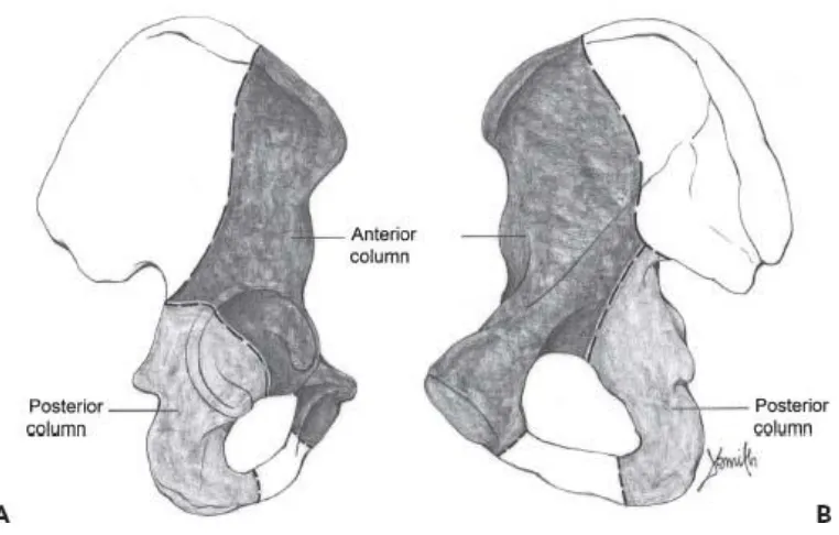
The bony hip joint is a ball and socket type joint comprised of two parts: the head of the femur articulating inside the acetabulum of the bony pelvis. The acetabulum is created by the fusion of the ischium, ilium, and the pubis. The acetabulum is supported by the thick anterior and posterior columns of the pelvis (figure 1-1). These structures are responsible for transmission of forces from the trunk to the lower extremities through the hip joint (1).The acetabulum is hemispherical with an equatorial axis angled approximately 45 degrees abducted in the coronal plane and 15 degrees anteverted in the sagittal plane (2), and provides nearly circumferential coverage of the femoral head. This amount of coverage supports a substantial range of motion, while maintaining joint stability.
Figure 1-1 - Extrapelvic (A) and intrapelvic (B) schematic views of the anterior and
Rubash HE, Clohisy J, Beaule P, DellaValle C. The Adult Hip: Hip Arthroplasty
Surgery. Wolters Kluwer Health)
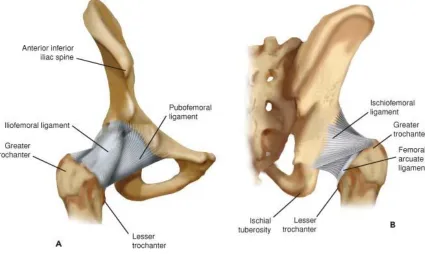
Though the hip joint is highly congruent, it relies on other anatomic structures to increase joint stability. The acetabular labrum and joint capsule act as static stabilizers of the joint. The labrum lies on the outer acetabular rim circumferentially, deepening the
femoroacetabular articulation and increasing joint congruence. The hip joint capsule attaches to the acetabulum on the outside of the labrum and to the femur along the intertrochanteric ridge, and is comprised of the iliofemoral, pubofemoral, and ischiofemoral ligaments (figure 1-2).
Figure 1-2 - Anterior (A) and posterior (B) views of the ligaments comprising the
hip joint capsule (permissions from Callaghan JJ, Rosenberg AG, Rubash HE,
Clohisy J, Beaule P, DellaValle C. The Adult Hip: Hip Arthroplasty Surgery.
Wolters Kluwer Health; 2015)
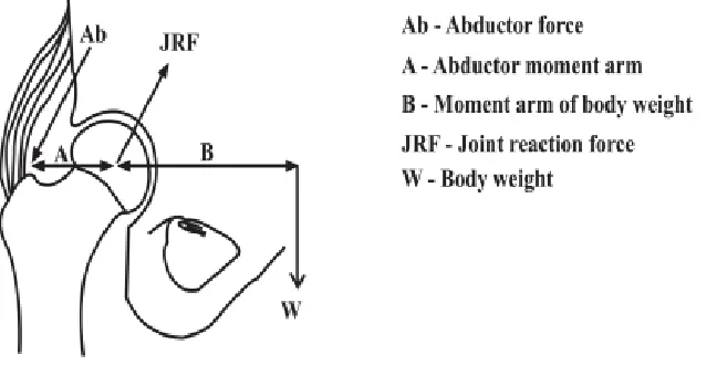
stance, the hip abductors must generate a force 2.5 times the body weight to counter the force of gravity and maintain a level pelvis. The summed forces of body weight and the hip abductors creates a resultant joint reactive force in the hip joint, which is
approximately 3 times body weight during single leg stance and may be as high as 10 times body weight when lifting, running, or jumping (3) (figure 1-3). As the body’s center of gravity is posterior to the axis of the hip joint in the sagittal plane, forces placed on the joint from a position of hip flexion can lead to posteriorly directed force creating torsion on the proximal femur that can be as high as 0.9 times body weight (3).
Figure 1-3 - Free body diagram of the joint reactive force (JRF) in the hip created
by the combined forces of body weight (W) and abductor force (Ab). (from Mirza
SB, Dunlop DG, Panesar SS, Naqvi SG, Gangoo S, Salih S. Basic science
considerations in primary total hip replacement arthroplasty. Open Orthop J 2010,
doi: 10.2174/1874325001004010169)
Hip Arthritis and Treatment Options
Arthritis is a term used to describe inflammation in a joint. Arthritis of the hip can be the product of a number of pathologic processes including autoimmune diseases,
gender, obesity, low bone density, muscle weakness, and joint laxity (6).With no current treatments available to slow, stop or reverse the process of osteoarthritis, medical
management focuses on treatment of symptomatic patients with both nonsurgical and surgical modalities. Recent recommendations by the American College of Rheumatology for nonsurgical treatment of symptomatic hip osteoarthritis include participation in cardiovascular and/or resistance based exercises, aquatic exercises, weight loss, participation in self-management programs, manual therapy with supervised exercises, psychosocial interventions, thermal agents, use of walking aids, oral acetaminophen, oral non-steroidal anti-inflammatories, tramadol, and intraarticular corticosteroid injections (7). However, should nonoperative treatment fail to adequately control symptoms and a patient’s function is significantly impaired, surgical intervention with total hip
arthroplasty (THA) would be indicated (8).
Total Hip Arthroplasty
The mainstay of surgical treatment for osteoarthritis of the hip is total hip arthroplasty (THA) (9).Although there were prior attempts at design and implantation of THA, it was the low friction THA introduced and refined by Sir John Charnley in the 1960’s that revolutionized surgical treatment of symptomatic hip arthritis(10,11). In this technique, a metal femoral stem was fixed into the femoral medullary canal and a high-density
polyethylene acetabular cup was fixed into the acetabulum with an acrylic bonding agent. Survivorship analyses have demonstrated excellent long-term results at up to 35 years (11–13). Though the initial indications for THA proposed by Charney were more limited, they have expanded to younger, higher demand patients with improvements in implant design, fixation techniques and surgical techniques. In fact, a 1995 consensus statement published by the National Institute of Health (NIH) in the United States noted that THA was an option for “nearly all patients with diseases of the hip that cause chronic
discomfort and significant functional impairment (15).”
1.3.1
Implant Design
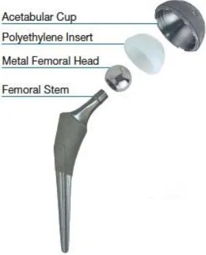
a portion of the femoral neck are removed and, after appropriate preparation of the bone, a metallic stem is inserted into the femoral medullary canal. This stem has a neck, on which a spherical femoral head is attached via a conical trunnion and bore press fit taper connection, commonly referred to as a Morse taper. On the acetabular side, the native acetabular cartilage is removed through a reaming process and replaced with either a monobloc polyethylene hemispherical cup or a metallic hemispherical shell, in which a modular bearing surface is attached. Due to the modular capability of modern implants, multiple bearing surfaces are available for THA articulation. These include cobalt
chromium (CoCr) or ceramic femoral heads and CoCr, ceramic, and ultra-high molecular weight polyethylene (UHMWPE) acetabular liners. The most commonly utilized THA bearing design is a CoCr femoral head that articulates with an UHMWPE acetabular liner (figure 1-4). This modularity of implants allows for adjustment of various parameters that can restore normal hip biomechanics.
Figure 1-4 - The components of a common THA design, in this case a metal femoral
head articulating on a modular polyethylene acetabular liner (Permissions from
Total Hip Arthroplasty (THA). OrthopaedicsOne Clerkship. In: OrthopaedicsOne -
The Orthopaedic Knowledge Network. Created Dec 13, 2010 21:12. Last modified
Dec 14, 2010 09:10 ver.3. Retrieved 2018-06-17, from
1.3.2
Implant Fixation
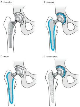
Fixation of THA components can be achieved with either a cemented (Polymethyl methacrylate) or cementless technique (figure 1-5). Modern cement fixation as introduced and studied by Charnley (10,16) relied not on adherence of the acrylic polymer to bone, but rather on the formation of a cast of the inner surface of the bone which would allow distribution of forces from the hip joint evenly over a large surface area. Cementing technique has evolved over time (17) to enhance penetration into
Figure 1-5 - Examples of different methods of fixation in THA, including fully
cementless (A), fully cemented (B), hybrid fixation (C) and reverse hybrid fixation
(D). Note that the blue regions represent bone cement. (permissions from Pivec R,
Johnson AJ, Mears SC, Mont MA. Hip arthroplasty. Lancet Lond Engl.
2012;380(9855):1768-1777. doi:10.1016/S0140-6736(12)60607-2)
1.3.3
Implant Positioning
components of a THA, specifically the length and size of the modular femoral head bearing or the size, offset, or version of the modular acetabular bearing surface.
On the femoral side, the major parameters to consider in implant placement and design are length, offset, version and head-to-neck ratio. Restoration of leg length is achieved through the depth of insertion of the femoral component, the length of the femoral neck, and the length of the femoral head. Leg length is important for normal gait mechanics. Offset refers to the distance between the center of the femoral head and the long axis of the femoral stem, and is predominantly a product of the stem design. However, if a longer or shorter femoral head is utilized, horizontal offset is increased or decreased,
Figure 1-6 - Increasing the head-to-neck ratio increases the impingement free range
of motion in THA constructs (permissions from Malik A, Maheshwari A, Dorr LD.
Impingement with total hip replacement. J Bone Joint Surg Am.
2007;89(8):1832-1842. doi:10.2106/JBJS.F.01313)
Figure 1-7 - As femoral head diameter increased, the distance the femoral head
must travel to dislocate, or the “jump distance” increases. (Permission from Cho
YJ, Nam DC, Jung K. Arthroplasty in Femoral Head Osteonecrosis. Hip Pelvis.
2014;26(2): 65-73. doi:10.5371/hp.2014.26.2.65)
on stability and impingement. Excessive anteversion or retroversion of the acetabulum, much like the femoral component, can lead to neck/acetabulum impingement and prosthetic dislocation. Excessive vertical placement of the acetabular component may lead to increased risk of dislocation, excessive edge loading of the implant and wear (21), whereas excessive horizontal placement can lead to decreased hip flexion and risk of anterior impingement and dislocation (22). However, optimal acetabular implant version and inclination remains controversial, with recommendations ranging from 0o-30o of anteversion and 30o-50o of inclination (22).
Implant position has a major impact on hip joint biomechanics and stability, as noted above, however it also plays a major role in the wear on the polyethylene bearing surface and should be considered when deciding on a THA construct. Increasing femoral head size leads to increased total surface area contact between the femoral head and
polyethylene liner, leading to increased volumetric wear (23). Relatively vertical
acetabular component positioning can cause the femoral head to articulate with the edge of the polyethylene liner superiorly, increasing contact stress over decreased contact area and increasing wear (24).
1.3.4
Implant Longevity
liners, HXLPE has proven to be successful in terms of wear and osteolysis (30–37) . However, mechanical properties of the implant are compromised at the expense of improved wear properties, specifically fatigue crack propagation resistance (29,38–40). Irradiation used to create polyethylene cross-linking leads to the formation of free radicals (44). These free radicals can react with oxygen species in vivo, leading to
polymer chain scission, recrystallization and ultimately increased brittleness (42–46). To stabilize these free radicals, implant manufacturers utilize thermal treatments such as remelting above or annealing below the material melting point. Remelting more effectively removes free radicals at the expense of mechanical properties, where
annealing less effectively removes free radicals but maintains mechanical properties (50). Furthermore, certain manufacturers utilize irradiation for implant sterilization, which can reintroduce or increase free radicals in the finished implant. Recent reports of mechanical failure of certain HXLPE acetabular liners at the rim of the implant (51–58) have raised concerns about the possibility of decreased mechanical properties on the longevity of these bearing surfaces (figure 1-8).
Figure 1-8 - Example of a failed HXLPE acetabular liner at the rim of the implant
(permissions from Tower SS. Rim Cracking of the Cross-Linked Longevity
Polyethylene Acetabular Liner After Total Hip Arthroplasty. J Bone Joint Surg
Research Objectives
Projections indicate a likely significant increase in the volume of THA procedures being performed in the coming years (27,59,60), driven by both an aging population as well as a shifting threshold for surgery towards younger patients (60). Given the anticipated increase in surgical volume as well as the significant costs and morbidity (26,28,61) associated with revision surgery, improving implant longevity remains imperative. Although HXLPE has improved polyethylene wear and wear-related revision surgery in THA (30–37) , concern exists that in vivo oxidation and reduced mechanical properties may create a new clinical problem, supported by reports of catastrophic failure of
HXLPE acetabular liners, particularly at the implant rim (51–58). Prior implant retrieval studies have found oxidation of HXLPE acetabular liners at the rim and articular surface as well as degraded mechanical properties of the articular surface (42–45). However, little is known about the mechanical properties of retrieved HXLPE liner rims after in vivo exposure. Pilot studies have been performed to validate a non-destructive method of assessing the mechanical properties of retrieved HXLPE acetabular liner rims (62), though the studies were initially limited to a single type of HXLPE liner.
References
1. Wasielewski RC, Kusuma S, Rosenberg AG. Chapter 2: Gross Anatomy of the Hip. In: The Adult Hip: Hip Arthroplasty Surgery. Vol 1. 3rd Edition. Two Commerce Square, 2001 Market Street, Philadelphia, PA 19103: Lippincott Williams & Wilkins; 2016:37-54.
2. Mirza SB, Dunlop DG, Panesar SS, et al. Basic science considerations in primary total hip replacement arthroplasty. Open Orthop J. 2010;4:169-180.
doi:10.2174/1874325001004010169
3. Canale ST, Beaty J. Campbell’s Operative Orthopaedics. Vol 1. 12th Edition. Philadelphia, PA: Elsevier Mosby
4. Felson DT. Chapter 43. Osteoarthritis. In: Imboden JB, Hellmann DB, Stone JH, eds. CURRENT Diagnosis & Treatment: Rheumatology. 3rd ed. New York, NY: The McGraw-Hill Companies; 2013.
5. Johnston Hough Jr. A. Chapter 108: Pathology of Osteoarthritis. In: Arthritis & Allied Conditions. 15th Edition. 530 Walnut Street, Philadelphia, PA 19106 USA LWW.com: Lippincott Williams & Wilkins; 2005:2169-2197.
6. Zhang Y, Jordan JM. Epidemiology of osteoarthritis. Clin Geriatr Med. 2010;26(3):355-369. doi:10.1016/j.cger.2010.03.001
7. Hochberg MC, Altman RD, April KT, et al. American College of Rheumatology 2012 recommendations for the use of nonpharmacologic and pharmacologic therapies in osteoarthritis of the hand, hip, and knee. Arthritis Care Res. 2012;64(4):465-474. doi:10.1002/acr.21596
8. Harris WH, Sledge CB. Total hip and total knee replacement (1). N Engl J Med. 1990;323(11):725-731. doi:10.1056/NEJM199009133231106
9. Learmonth ID, Young C, Rorabeck C. The operation of the century: total hip replacement. The Lancet. 2007;370(9597):1508-1519. doi:10.1016/S0140-6736(07)60457-7
10. Charnley J. Arthroplasty of the hip. A new operation. Lancet Lond Engl. 1961;1(7187):1129-1132.
11. Charnley J. The Long-Term Results of Low-Friction Arthroplasty of the Hip Performed as a Primary Intervention. Bone Jt J. 1972;54-B(1):61-76.
13. Callaghan JJ, Bracha P, Liu SS, et al. Survivorship of a Charnley total hip arthroplasty. A concise follow-up, at a minimum of thirty-five years, of previous reports. J Bone Joint Surg Am. 2009;91(11):2617-2621. doi:10.2106/JBJS.H.01201
14. Berry DJ, Harmsen WS, Cabanela ME, et al. Twenty-five-year survivorship of two thousand consecutive primary charnley total hip replacements: Factors affecting survivorship of acetabular and femoral components. J Bone Joint Surg Am. 2002 Feb;84-A(2):171-7.
15. NIH consensus conference: Total hip replacement. NIH Consensus Development Panel on Total Hip Replacement. JAMA. 1995;273(24):1950-1956.
16. Charnley J. The Bonding of Prostheses to Bone by Cement. J Bone Joint Surg Br. 1964;46:518-529.
17. August 1, 2005. The Evolution of Contemporary Cementation Techniques. https://www.healio.com/orthopedics/hip/news/online/%7B03cc1461-32e7-46b6-a282-cadd462d0264%7D/the-evolution-of-contemporary-cementation-techniques. Accessed November 5, 2017.
18. Junnila M, Laaksonen I, Eskelinen A, et al. Implant survival of the most common cemented total hip devices from the Nordic Arthroplasty Register Association database. Acta Orthop. 2016;87(6):546-553. doi:10.1080/17453674.2016.1222804
19. Troelsen A, Malchau E, Sillesen N, et al. A review of current fixation use and registry outcomes in total hip arthroplasty: the uncemented paradox. Clin Orthop. 2013;471(7):2052-2059. doi:10.1007/s11999-013-2941-7
20. Kienapfel H, Sprey C, Wilke A, et al. Implant fixation by bone ingrowth. J Arthroplasty. 1999;14(3):355-368.
21. Schmalzried TP, Guttmann D, Grecula M, et al. The relationship between the design, position, and articular wear of acetabular components inserted without cement and the development of pelvic osteolysis. J Bone Joint Surg Am. 1994;76(5):677-688.
22. Widmer K-H, Zurfluh B. Compliant positioning of total hip components for optimal range of motion. J Orthop Res Off Publ Orthop Res Soc. 2004;22(4):815-821. doi:10.1016/j.orthres.2003.11.001
23. Elfick AP, Hall RM, Pinder IM, Unsworth A. Wear in retrieved acetabular
components: effect of femoral head radius and patient parameters. J Arthroplasty. 1998;13(3):291-295.
25. Clarke A, Pulikottil-Jacob R, Grove A, et al. Joint Registries. NIHR Journals Library; 2015. https://www.ncbi.nlm.nih.gov/books/NBK273990/. Accessed November 5, 2017.
26. Bozic KJ, Kurtz SM, Lau E, et al. The epidemiology of revision total hip arthroplasty in the United States. J Bone Joint Surg Am. 2009;91(1):128-133. doi:10.2106/JBJS.H.00155
27. Kurtz S, Ong K, Lau E, et al. Projections of primary and revision hip and knee arthroplasty in the United States from 2005 to 2030. J Bone Joint Surg Am. 89(4):780-785. doi:10.2106/JBJS.F.00222
28. Gwam CU, Mistry JB, Mohamed NS, et al. Current Epidemiology of Revision Total Hip Arthroplasty in the United States: National Inpatient Sample 2009 to 2013. J Arthroplasty. 2017;32(7):2088-2092. doi:10.1016/j.arth.2017.02.046
29. Willert HG, Bertram H, Buchhorn GH. Osteolysis in alloarthroplasty of the hip. The role of ultra-high molecular weight polyethylene wear particles. Clin Orthop.
1990;(258):95-107.
30. Cooper RA, McAllister CM, Borden LS, et al. Polyethylene debris-induced osteolysis and loosening in uncemented total hip arthroplasty. A cause of late failure. J Arthroplasty. 1992;7(3):285-290.
31. Ingham E, Fisher J. Biological reactions to wear debris in total joint replacement. Proc Inst Mech Eng [H]. 2000;214(1):21-37. doi:10.1243/0954411001535219
32. McKellop H, Shen F, Lu B, et al. Development of an extremely wear-resistant ultra high molecular weight polythylene for total hip replacements. J Orthop Res.
1999;17(2):157-167. doi:10.1002/jor.1100170203
33. Paxton EW, Inacio MCS, Namba RS, et al. Metal-on-conventional polyethylene total hip arthroplasty bearing surfaces have a higher risk of revision than metal-on-highly crosslinked polyethylene: results from a US registry. Clin Orthop.
2015;473(3):1011-1021. doi:10.1007/s11999-014-4105-9
34. Jacobs CA, Christensen CP, Greenwald AS, et al. Clinical performance of highly cross-linked polyethylenes in total hip arthroplasty. J Bone Joint Surg Am. 2007;89(12):2779-2786. doi:10.2106/JBJS.G.00043
35. Lachiewicz PF, Soileau ES, Martell JM. Wear and Osteolysis of Highly Crosslinked Polyethylene at 10 to 14 Years: The Effect of Femoral Head Size. Clin Orthop. 2016;474(2):365-371. doi:10.1007/s11999-015-4319-5
37. Glyn-Jones S, Thomas GER, Garfjeld-Roberts P, et al. The John Charnley Award: Highly crosslinked polyethylene in total hip arthroplasty decreases long-term wear: a double-blind randomized trial. Clin Orthop. 2015;473(2):432-438.
doi:10.1007/s11999-014-3735-2
38. Hanna SA, Somerville L, McCalden RW, et al. Highly cross-linked polyethylene decreases the rate of revision of total hip arthroplasty compared with conventional polyethylene at 13 years’ follow-up. Bone Jt J. 2016;98-B(1):28-32.
doi:10.1302/0301-620X.98B1.36527
39. Garvin KL, White TC, Dusad A, et al. Low wear rates seen in THAs with highly crosslinked polyethylene at 9 to 14 years in patients younger than age 50 years. Clin Orthop. 2015;473(12):3829-3835. doi:10.1007/s11999-015-4422-7
40. Mall NA, Nunley RM, Zhu JJ, et al. The incidence of acetabular osteolysis in young patients with conventional versus highly crosslinked polyethylene. Clin Orthop. 2011;469(2):372-381. doi:10.1007/s11999-010-1518-y
41. Gencur SJ, Rimnac CM, Kurtz SM. Fatigue crack propagation resistance of virgin and highly crosslinked, thermally treated ultra-high molecular weight polyethylene. Biomaterials. 2006;27(8):1550-1557. doi:10.1016/j.biomaterials.2005.09.010
42. Ansari F, Ries MD, Pruitt L. Effect of processing, sterilization and crosslinking on UHMWPE fatigue fracture and fatigue wear mechanisms in joint arthroplasty. J Mech Behav Biomed Mater. 2016;53:329-340. doi:10.1016/j.jmbbm.2015.08.026
43. Baker DA, Bellare A, Pruitt L. The effects of degree of crosslinking on the fatigue crack initiation and propagation resistance of orthopedic-grade polyethylene. J Biomed Mater Res A. 2003;66(1):146-154. doi:10.1002/jbm.a.10606
44. Costa L, Bracco P. 26 - Mechanisms of Cross-Linking, Oxidative Degradation, and Stabilization of UHMWPE. In: Kurtz SM, ed. UHMWPE Biomaterials Handbook (Third Edition). Oxford: William Andrew Publishing; 2016:467-487.
doi:10.1016/B978-0-323-35401-1.00026-0
45. Currier BH. Evaluation of Oxidation and Fatigue Damage of Retrieved Crossfire Polyethylene Acetabular Cups. J Bone Joint Surg Am. 2007;89(9):2023.
doi:10.2106/JBJS.F.00336
46. Wannomae KK, Bhattacharyya S, Freiberg A, et al.In vivo oxidation of retrieved cross-linked ultra-high-molecular-weight polyethylene acetabular components with residual free radicals. J Arthroplasty. 2006;21(7):1005-1011.
doi:10.1016/j.arth.2005.07.019
47. Kurtz SM, Hozack WJ, Purtill JJ, et al. 2006 Otto Aufranc Award Paper:
48. Kurtz SM, Hozack W, Marcolongo M, et al. Degradation of mechanical properties of UHMWPE acetabular liners following long-term implantation. J Arthroplasty. 2003;18(Supplement):68-78. doi:10.1016/S0883-5403(03)00292-4
49. Muratoglu OK, Bragdon CR. 15 - Highly Cross-Linked and Melted UHMWPE. In: Kurtz SM, ed. UHMWPE Biomaterials Handbook (Third Edition). Oxford: William Andrew Publishing; 2016:264-273. doi:10.1016/B978-0-323-35401-1.00015-6
50. Manley MT. 16 - Highly Cross-Linked and Annealed UHMWPE. In: Kurtz SM, ed. UHMWPE Biomaterials Handbook (Third Edition). Oxford: William Andrew Publishing; 2016:274-292. doi:10.1016/B978-0-323-35401-1.00016-8
51. Blumenfeld TJ, McKellop HA, Schmalzried TP, et al. Fracture of a Cross-Linked Polyethylene Liner. J Arthroplasty. 2011;26(4):666.e5-666.e8.
doi:10.1016/j.arth.2010.07.009
52. Duffy GP, Wannomae KK, Rowell SL, et al. Fracture of a Cross-Linked Polyethylene Liner Due to Impingement. J Arthroplasty. 2009;24(1):158.e15-158.e19. doi:10.1016/j.arth.2007.12.020
53. Furmanski J, Anderson M, Bal S, et al. Clinical fracture of cross-linked UHMWPE acetabular liners. Biomaterials. 2009;30(29):5572-5582.
doi:10.1016/j.biomaterials.2009.07.013
54. Hara D, Nakashima Y, Yamamoto T, et al. Late failure of annealed highly cross-linked polyethylene acetabular liner. J Mech Behav Biomed Mater. 2013;28:206-212. doi:10.1016/j.jmbbm.2013.08.003
55. Moore KD, Beck PR, Petersen DW, et al. Early Failure of a Cross-Linked Polyethylene Acetabular Liner: A Case Report. J Bone Joint Surg Am. 2008;90(11):2499-2504. doi:10.2106/JBJS.G.01304
56. Tower SS. Rim Cracking of the Cross-Linked Longevity Polyethylene Acetabular Liner After Total Hip Arthroplasty. J Bone Joint Surg Am. 2007;89(10):2212. doi:10.2106/JBJS.F.00758
57. Waewsawangwong W, Goodman SB. Unexpected Failure of Highly Cross-Linked Polyethylene Acetabular Liner. J Arthroplasty. 2012;27(2):323.e1-323.e4.
doi:10.1016/j.arth.2011.04.010
58. Ast MP, John TK, Labbisiere A, Robador N, et al. Fractures of a single design of highly cross-linked polyethylene acetabular liners: an analysis of voluntary reports to the United States Food and Drug Administration. J Arthroplasty.
2014;29(6):1231-1235. doi:10.1016/j.arth.2013.12.022
60. Ravi B, Croxford R, Reichmann WM, et al. The changing demographics of total joint arthroplasty recipients in the United States and Ontario from 2001 to 2007. Best Pract Res Clin Rheumatol. 2012;26(5):637-647.doi:10.1016/j.berh.2012.07.014
61. Bozic KJ, Kamath AF, Ong K, et al. Comparative Epidemiology of Revision Arthroplasty: Failed THA Poses Greater Clinical and Economic Burdens Than Failed TKA. Clin Orthop. 2015;473(6):2131-2138. doi:10.1007/s11999-014-4078-8
Chapter 2
2
Highly Cross-Linked Polyethylene: The Impact of Free
Radical Stabilization on Implant Properties
Introduction
Total hip arthroplasty (THA) has proven to be an extremely successful surgical procedure in producing significant pain reduction and improved physical function in patients with end stage arthritis of the hip(1). Sir John Charnley was the first to introduced UHMWPE into his low friction THA design in 1962 (2), and it remains the most common bearing surface used in THA today. However, the success of conventional UHMWPE has been limited by the long term material wear, which can lead to instability, osteolysis, aseptic implant loosening and need for revision surgery (3–5). Furthermore, demand for THA in young and active patients has also increased the demand for a more durable bearing surface (6). These factors facilitated the development of highly cross-linked polyethylene (HXLPE) bearing surfaces. Clinical and in vitro studies have confirmed a significant reduction in wear rates compared to conventional UHMWPE, along with a significant reduction in osteolysis and wear-related revision surgery rates (7–13).
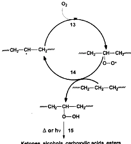
Cross-linking in UHMWPE is produced when the material is irradiated. Radiation energy leads to bond cleavage in polyethylene chains and development of highly reactive free radicals. In an inert environment, these free radicals combine to create the desired effect of cross-linking. However, if free radicals are exposed to oxygen they can react and create a cyclic, self-perpetuating oxidation cycle that leads to significant material degradation and changes in the mechanical properties of the implant (14).
polyethylene chains, and though free radicals are significantly reduced they are not completely eliminated. These residual free radicals can be oxidized when exposed to air or oxygen rich joint fluid, leading to mechanical property degradation. Moreover, in vivo oxidation of remelted polyethylene has been detected (15–19), leading to the discovery of alternative mechanisms for in vivo oxidation in the absence of residual free radicals (15,18,20–24). Second generation HXLPE liners have been introduced with new stabilization processes, including thermal sequential annealing and antioxidant doping, with the aim of improving the balance of wear, mechanical properties, and oxidative stability.
Reports of mechanical failure of first generation HXLPE liners (25–32) along with concerns regarding the oxidative stability and mechanical properties of first and second generation HXLPE implants has led to both experimental testing and retrieval analysis to better understand the behavior of these implants in vivo. Here, a review the literature regarding the oxidative stability and mechanical properties of first and second generation HXLPE implants is presented.
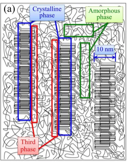
UHMWPE and the Development of HXLPE
Figure 2-1 - Representation of the three major phases of UHMWPE: Crystalline
phase, amorphous phase, and the third interphase. (Permission from Pezzotti G.
Raman spectroscopy of biomedical polyethylenes. Acta Biomater. 2017 Jun 1;55:28–
99. DOI: 10.1016/j.actbio.2017.03.015)
In order to produce medical grade UHMWPE implants, a number of precise
manufacturing processes must take place. Ethylene gas is converted to polymer form as a powder or resin using a converter and following ASTM standard F648 and ISO standard 5834-1 (35). The resin is then consolidated by either ram bar extrusion, compression sheet molding, or direct compression molding techniques under elevated temperatures and pressures. The final implant is then fabricated by machining the consolidated material into its final component shape and size. Each of these phases in manufacturing, many of which are proprietary, has the potential to change the chemical and
microstructural properties of UHMWPE. Once the final implant has been machined, it requires packaging and sterilization. Up until the 1990’s, UHMWPE implants were
Radiation sterilization imparts significant energy into polyethylene chains which can lead to the cleavage of C-C and C-H covalent bonds, forming highly reactive free radicals. In an inert environment without other reactive species, free radicals in the amorphous regions can recombine to form cross-links within the polyethylene chains, whereas those formed in the crystalline region remain trapped (20). These remnant free radicals are long living and if are exposed to oxygen can form alkyl free radicals, leading to a
self-perpetuating oxidation process known as Bolland’s cycle (figure 2-2) (37). This oxidation
cycle leads to chain scission and a reduction in the molecular mass of UHMWPE, with a subsequent increased proportion of the crystalline phase (36,38) through the development of thin crystalline lamellae in the amorphous region (14). The end result is an increase in material brittleness, most notably in the subsurface region of the implant 1-2mm below the outer surface. This increased brittleness could potentially increase the risk of fatigue damage (36).
Figure 2-2 - Bolland’s cycle demonstrating the oxidation of hydrocarbons such as
polyethylene (Permission from Costa L, Bracco P. Mechanisms of Cross-Linking,
UHMWPE Biomaterials Handbook (Third Edition), Oxford: William Andrew
Publishing; 2016 [p. 467–87.)
Prior to this discovery, the oxidative process occurred during implant storage on the shelf prior to implantation due to sterilization of the implant in air and storage in oxygen permeable packaging. Manufacturers transitioned to oxygen barrier packing and sterilization with new methods including gamma irradiation in an inert environment or sterilization in gas plasma or ethylene oxide. It was soon discovered that wear rates in gas plasma and ethylene oxide were nearly twice that of gamma air sterilized implants (39– 41) which led to the discovery that radiation induced cross-linking played a significant factor in wear properties. Furthermore, gamma inert sterilized implants retrieved after in vivo time continued to demonstrate evidence of oxidative degradation due to residual free radicals and in vivo exposure to oxygen (42). These discoveries led to the development of the first generation of highly cross-linked polyethylene implants.
The Impact of Radiation Cross-Linking and
Thermal Stabilization on HXLPE Properties
First generation HXLPE was developed to take advantage of the wear-reducing properties imparted by crosslinking while reducing or eliminating the potential for oxidative degradation and minimize the impact on mechanical properties. Gamma or electron beam radiation is utilized to create cross-linking in polyethylene at variable doses depending on implant manufacturer. McKellop et al (43) discovered that crosslink density and crystallinity increases with increased doses of radiation, and wear rates are reduced up to radiation doses of 200 kGy. Saturation of cross-linking occurs at
approximately 100 kGy (44). However, radiation cross-linking decreases ductility, which manifests as a reduction in elongation to failure, toughness, and fatigue crack propagation resistance (45), with tensile and fracture toughness continuing to decline at radiation doses beyond 100 kGy (43,46,47). For these reasons, a dose of approximately 100 kGy represents an acceptable balance between crosslink density, wear reduction, and
In order to maintain the wear-reducing benefits of cross-linking while minimizing the potential for oxidative degradation, two major thermal free radical stabilization processes were developed as part of the first generation of HXLPE bearings. The first, known as remelting, involves heating irradiated polyethylene to some temperature above its melting point (~ 1370 C) whereas the second, known as annealing, involves heating irradiated polyethylene to a temperature below its melting point. Both then undergo a cooling and recrystallization process. The desired outcome in both of these processes is to mobilize free radicals within the crystalline region to allow cross-linking and termination of free radicals and thus avoid the potential for oxidation (14,20). When polyethylene is remelted, the rigid crystalline regions are able to mobilize and free radicals present in these regions can reconnect or cross-link, leaving undetectable levels of free radicals (48). Chain mobility and reformation of crystallites during the recrystallization process is limited by the presence of crosslinks, therefor the total crystallinity after remelting is reduced (45). As the strength of UHMWPE is dependent on its relative crystallinity, remelted HXLPE demonstrates decreased ultimate strength, yield strength, and fatigue resistance (14,45). When polyethylene is annealed, not all crystalline lamellae are melted and as such, not all free radicals are stabilized. The relative crystallinity of annealed HXLPE remains nearly unchanged, as do the mechanical properties (49,50). Though differences exist in the physical and mechanical properties of first generation HXLPE liners based on thermal free radical stabilization process, both have demonstrated superior surface wear characteristics, in vitro and in vivo, compared to conventional UHMWPE THA bearings (9).
The Impact of Oxidation on First Generation
HXLPE Acetabular Liners
2.4.1
Oxidation and Mechanical Properties of HXLPE Acetabular
Liners in Experimental Models
oxidation. The OI is calculated from FTIR by normalizing the absorption peak of carbonyl groups, formed from the oxidation of polyethylene chains, against an internal reference absorption peak for the polyethylene. McKellop et al (43) demonstrated, in an accelerated aging study, that remelted HXLPE THA bearings demonstrated no evidence of oxidation whereas untreated HXLPE liners demonstrated significant subsurface oxidation. A number of other studies directly compared annealed and remelted first generation HXLPE liners utilizing different aging protocols (38,51,52). These studies found evidence of significant oxidation in annealed liners with no detectable evidence of oxidation in remelted liners. Annealed liners also demonstrated increased wear rates compared to remelted liners (51,52). Although remelted HXLPE liners have shown undetectable levels of free radicals and no evidence of oxidation in these accelerated aging studies, recent studies have demonstrated that remelted HXLPE liners do indeed have the potential to oxidize. Medel et al (21) performed a cyclic loading and accelerated aging experimental study on remelted and annealed HXLPE. Annealed HXLPE
2.4.2
Oxidation and Mechanical Properties of Retrieved Annealed
HXLPE Acetabular Liners
Retrieval studies have provided great insight into the behavior of first generation HXLPE acetabular liners after time in vivo and have confirmed a number of findings from
experimental studies. Wannomae et al (53) assessed 14 retrieved annealed and 12 retrieved remelted liners with in vivo times up to three years. Samples were taken from non-articulating regions near the rim. The remelted liners exhibited no detectable oxidation, whereas the annealed liners demonstrated evidence of oxidation in the subsurface region, with OI ranging from range 0.22 to 5.81. Remelted liners had no significant change in crystallinity compared to controls, while annealed liners showed a significant increase in crystallinity, especially when the OI was greater than 1.0. A strong correlation was found between oxidation and crystallinity but only a weak correlation for oxidation and in vivo time. This study suggested that remelting was a superior free radical stabilization technique compared to annealing as it led to a reduced the risk of in vivo oxidation and structural property changes.
Currier et al (54) evaluated 12 annealed HXLPE liners with in vivo time up to 5.3 years for oxidation and evidence of degradation at the rim and articular surface. There was evidence of significant oxidation, with the most marked oxidation at the implant rim, and a strong relationship between rim oxidation and in vivo time. Rim cracking was also noted to correlate to in vivo time, with the crack rating correlating to oxidation. Rim delamination was found to correlate only with in vivo time when taking into account signs of impingement. The authors concluded that annealed HXLPE THA liners oxidize in vivo to a significant enough degree to compromise mechanical properties and lead to fatigue damage, especially in the setting if impingement of the femoral neck on the acetabular rim.
articular surface. Furthermore, in vivo time was found to correlate to oxidation of the unworn articular surface and the rim, but did not correlate with oxidation values at the worn regions. These studies assessed the mechanical properties of the articular surface and found no appreciable correlation between in vivo time and mechanical behavior. It is evident from these studies that oxidation of annealed HXLPE liners occurs preferentially in regions exposed to larger amounts of synovial fluid turnover. Additionally, the
mechanical properties of the implants, though only tested along the articular surface, did not appear to be impacted by in vivo time. However, rim mechanical properties were not tested, despite being the region of greatest oxidation.
MacDonald et al (15), in a large retrieval study assessing oxidation and articular surface mechanical properties of 80 annealed HXLPE acetabular liners with in vivo time ranging from 0 to 10.3 years, found moderate oxidation at the rim in annealed liners with over half of these liners demonstrating severe rim oxidation (OI>3). Rim oxidation did correlate with in vivo time. Oxidation was also found at the articular surface and was found to correlate with a reduction in ultimate load in this region of the implant. These findings imply that in vivo oxidation of annealed HXLPE THA liners leads to a
compromise in the mechanical properties, as found in the testing of the articular surface of these implants. However, rim mechanical properties were not tested and it is unclear if a similar change in mechanical properties occurs in this region given the sharp elevations in oxidation with in vivo time.
2.4.3
Oxidation and Mechanical Properties of Retrieved Remelted
HXLPE Acetabular Liners
correlation between oxidation and in vivo time. Another retrieval analysis (17) of 11 remelted HXLPE liners with in vivo time of 2 weeks to 7.2 years, also found evidence of subsurface articular surface oxidation after in vivo exposure, though ex vivo time after surgical removal was not controlled. MacDonald et al (15), in the above noted study, also assessed oxidation and articular surface mechanical properties of 160 retrieved remelted HXLPE liners with in vivo time ranging from 0 to 11.4 years. Results demonstrated detectable levels of oxidation at the articular surface with a positive correlation to in vivo time, however no such relationship was found at the rim or backside of the liner and no significant changes in articular surface mechanical properties were noted. Muratoglu et al (18) assessed oxidation, crystallinity and crosslink density of 34 remelted HXLPE liners after removal and shelf aging (ex vivo) time. Shelf aged control remelted liners
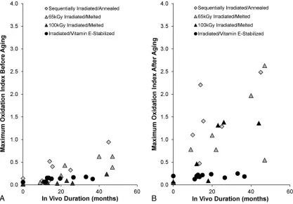
demonstrated no evidence of oxidation at 7 years. Low but detectable oxidation was found at the rim of retrieved implants immediately after surgical removal, and after shelf aging both rim and articular surface oxidation increased and were correlated with ex vivo time, but not in vivo time. In spite of this finding of low oxidation, cross link density significantly decreased and crystallinity significantly increased with ex vivo time for both the articular surface and rim. Rowell et al (19) evaluated a number of different retrieved implant types, including remelted HXLPE acetabular liners, for oxidation and cross link density after surgical removal and accelerated aging. Half of the retrieved remelted HXLPE acetabular liners demonstrated evidence of in vivo oxidation, which increased in the subsurface region with accelerated aging. Retrievals without measurable in vivo oxidation showed oxidation after ex vivo accelerated aging (figure 2-3), with subsurface peaks in the articular surface region and at the surface of unloaded regions. Acetabular rim oxidation was noted to increase with accelerated aging as well.
Figure 2-3 - Accelerated aging of retrieved HXLPE liners with various free radical
stabilization techniques demonstrates loss of oxidative stability in both remelted and
annealed liners. (Permissions from Rowell SL, Reyes CR, Malchau H, Muratoglu
OK. In vivo Oxidative Stability Changes of Highly Cross-Linked Polyethylene
Bearings: An Ex vivo Investigation. J Arthroplasty. 2015;30(10):1828-1834.
doi:10.1016/j.arth.2015.05.006)
2.4.4
Mechanical Failure of First Generation HXLPE Acetabular
Liners
Tower et al (26) reported on the mechanical failure of four remelted liners from a single manufacturer after in vivo times ranging from 7 to 27 months. In all cases, acetabular components were malpositioned vertically. Analysis of the failed implants exposed severe cracking or failure was at the rim with damage evident at the superior aspect where the polyethylene engages the locking mechanism. Cracking was found to begin at the outer edge of the implant and propagate towards the articular surface. Polyethylene thickness at the site of crack propagation was less than 4mm for each implant.
Mechanical properties were nearly identical to control samples, though lower than non-crosslinked reference polyethylene. Moore et al (27) and Waewsawangwong and
Figure 2-4 - Example of a retrieved fractured remelted HXLPE liner at the implant
rim. (Permissions from Moore KD, Beck PR, Petersen DW, Cuckler JM, Lemons
JE, Eberhardt AW. Early Failure of a Cross-Linked Polyethylene Acetabular Liner:
A Case Report. J Bone Joint Surg Am. 2008;90(11):2499-2504.
doi:10.2106/JBJS.G.01304)
Duffy et al (30) analyzed the physical properties and oxidation in a case of a retrieved, fractured remelted HXLPE liner. The implant fractured along the rim in the region of an elevated 10-degree lip, which was found to be impinging during range of motion of the hip. The implant demonstrated no evidence of oxidation, and the transvinylene index and crystallinity were normal for the radiation dose applied for cross-linking. Furmanski et al (31) performed an extensive analysis of 4 retrieved, fractured remelted HXLPE liners from four different manufacturers. Crystallinity of the liners was found to be within expected baseline range, and there was no evidence of oxidation (OI<0.1). In these samples, all fractures initiated on the outer surface of the liners at a region of stress concentration or material discontinuity. Finite element analysis further confirmed that the peak magnitude of the maximum principle tensile stress occurred near the sites of
Not all reported cases of mechanical failure of HXLPE liners have occurred in remelted liners. Hara et al (25) reported a case of liner rim fracture in an annealed HXLPE liner design with a 15 degree elevated lip (figure 2-5). The fracture initiated at the junction of the liner rim and acetabular component dome and propagated towards the articular surface near, but not at, the elevated lip. Polyethylene thickness was no less than 7.4mm throughout the liner. Oxidation was significant along the liner rim (OI = 2.34) and present but low at the articular surface (OI = 0.465).
Figure 2-5 - Example of a retrieved fractured annealed HXLPE liner at the implant
rim. (Permissions from Hara D, Nakashima Y, Yamamoto T, et al. Late failure of
annealed highly cross-linked polyethylene acetabular liner. J Mech Behav Biomed
Mater. 2013;28:206-212. doi:10.1016/j.jmbbm.2013.08.003)
Second Generation HXLPE: Sequential Annealing
and Antioxidant Stabilization
A second generation of HXLPE liners has been developed in an attempt to maintain the superior wear resistance provided by cross-linking while improving oxidative stability mechanical properties (14), and includes sequentially annealed and vitamin-E infused HXLPE.
2.5.1
Sequentially Annealed HXLPE
Sequentially annealed polyethylene was proposed as a way to produce a highly cross-linked polyethylene with the material properties of annealed liners but with little to no residual free radicals (58). In this process, sequential low dose radiation crosslinking (30 kGy) is followed by thermal annealing, and the process is repeated three total times for a total dose of 90 kGy, a dose thought to maximize cross-linking while maintaining mechanical properties. The low dose of radiation is thought to leave cross-links far enough apart to allow sufficient chain mobility for free radicals to mobilize and be extinguished during the annealing phase (59). The implants are then gas plasma sterilized.
Experimental investigation confirmed the potential benefits of sequentially annealing over single annealing used in first generation HXLPE. Dumbleton et al (58) and Wang et al (60) published on accelerated aging of sequentially annealed compared to single annealed HXLPE and other UHMWPE formulations. Before aging, free radical concentration in the single annealed HXLPE was shown to be over seven times higher and crosslink density was less than half compared to the sequentially annealed HXLPE. Furthermore, oxidation after accelerated aging was found in the subsurface of single annealed HXLPE (OI=1.1), with minimal oxidation (OI=0.05) and maintained
crystallinity found in the sequentially annealed HXLPE. Mechanical testing demonstrated the ultimate tensile strength and the amount of material elongation of sequentially
mechanical failure rates in hip simulator analysis was found to be lower in the sequentially annealed compared to single annealed HXLPE liners (58). A number of clinical studies (61–66) found excellent wear rates for sequentially annealed HXLPE.
Retrieval analysis of sequentially annealed THA liners has been limited thus far but has provided important insight into the oxidative stability and mechanical properties of these implants after in vivo exposure. In the same study by Rowell (19) noted previously, sequentially annealed HXLPE liners were assessed for oxidation and crosslink density after surgical removal. Oxidation was found at both the implant rim and articular surface, and accelerated aging induced significant increases in oxidation and pre-oxidation
products (hydroperoxides) in both regions, along with a significant reduction in crosslink density. Though the levels of oxidation at retrieval were low, this study suggests a
potentially significant loss of oxidative stability in vivo, likely do to the presence of residual free radicals. Reinitz et al (67) assessed 65 sequentially annealed acetabular liners with in vivo time ranging from 1 month to 6.4 years. Oxidation trends in these liners were similar to gamma sterilized UHMWPE, with two of the 65 liners
demonstrating subsurface white bands along the rim and articular surface with
significantly elevated oxidation indices and decreased crosslink density. Kurtz et al (68) directly compared oxidation and mechanical properties of 185 retrieved single and sequentially annealed HXLPE liners with in vivo time under five years. Oxidation was found along the rim and articular surface in both groups, however the oxidation was significantly lower for the sequentially annealed liners with the most pronounced
difference between groups found at the liner rim. Articular surface wear and mechanical properties were similar, and both groups demonstrated a 10% rate of liner rim damage. However, the damage mode of the liner rims in the sequentially annealed group was predominantly burnishing and scratching with no evidence of delamination or subsurface cracking, whereas in the single annealed group there were several samples with rim delamination and subsurface cracking.