Original Article
Differentiation and growth of kype skeletal tissues in
anadromous male Atlantic Salmon (Salmo salar)
P.-ECKHARD WITTEN*
,1and BRIAN K. HALL
21Institute of Marine Research at the University of Kiel, Germany and 2Department of Biology, Dalhousie University, Halifax NS, Canada
ABSTRACT The re-initiation of bone development in adult starving Atlantic salmon (Salmo salar) during their energetically expensive upstream migration is remarkable and deserves closer examina-tion. Dramatic alterations of the skull bones and teeth, most prominently, the development of a kype in males, are widely known but little studied or understood. We describe the microstructure and the cellular processes involved in the formation of the skeletal tissues of the kype. Fresh bone material, obtained from animals migrating upstream was subjected to radiological, histological or histochemi-cal analysis. We show that the kype is, in part, composed of rapidly growing skeletal needles arising at the tip of the dentary. Proximally, the needles anastomose into a spongiosa-like meshwork which retains connective tissue inside bone marrow spaces. Ventrally, the needles blend into Sharpey fiber bone. Skeletal needles and Sharpey fiber bone can be distinguished from the compact bone of the dentary by radiography. Rapid formation of the skeleton of the kype is demonstrated by the presence of numerous osteoblasts, a broad distal osteoid zone, and the appearance of proteoglycans at the growth zone. The mode of bone formation in anadromous males can be described as ‘making bone as fast as possible and with as little material as possible’. Unlike the normal compact bone of the dentary, the new skeletal tissue contains chondrocytes and cartilaginous extracellular matrix. Formation of the skeleton of the kype resembles antler development in deer (a form of regeneration), or hyperostotic bone formation in other teleost fishes, rather than periosteal bone growth. The type of bone formation may be understandable in the light of the animals’ starvation and the energetic costs of upstream migration. However, the structured and regulated mode of bone formation suggests that the skeleton of the kype has functional relevance and is not a by-product of hormonal alterations or change of habitat.
KEY WORDS:
Grilse, bone growth, spawning migration, kype, Sharpey fiber bone, chondroid bone
0214-6282/2002/$25.00
© UBC Press Printed in Spain www.ijdb.ehu.es
*Address correspondence to: Dr. P. Eckhard Witten. Institute of Marine Research at the University of Kiel, Düsterbrooker Weg 20, 24105 Kiel, Germany. Fax: +49 (0)404-2838-3937. e-mail: pewitten@aol.com
Abbreviations used in this paper: 1SW salmon, grilse or salmon that have spent one winter at sea before returning to spawn for the first time; BICP/NBT, 5-Bromo-4-chloro-3-indolyl phosphate/Nitro blue tetrazolium; HBQ-staining, Hall-Brunt Quadruple Stain; IgG, Immunoglobulin G; K, condition factor; PBS, Phosphate buffered saline solution; TRAP, Tartrate resistant acid phosphatase; TRIS, 2-Amino-2-hydroxymethyl-1,3-propanediol.
Introduction
Alterations of the bones of the skull and the teeth occur in both sexes of Atlantic salmon (Salmo salar) during migration upstream to spawn; morphological changes were described in detail by Tchernavin (1937, 1938a, 1944) almost two-thirds of a century ago, and more recently by Kacem et al. (1998). Although all bones of the salmon skull change during migration, and although feeding teeth are replaced by a new set of teeth known as breeding teeth, the formation of a kype (hook) on the tip of the lower jaw in male salmon is the most prominent alteration (Fig. 1)1.
The kype was the topic of scientific interest long before Tchernavin drew attention to the changes in the salmon skull. Darwin (1877) was aware that male salmon developed a kype during the breeding season. In his classic treatment, British and Irish Salmonidae, Day (1887) noted that “the cause of the existence of a knob on the lower jaw of male salmon and trout has been a fruitful cause of discussion from the early ages down to the present day” (p. 57).
Despite the fact that the Atlantic salmon is one of the best-studied teleost species, little information about the function of the kype is available. Hutchings and Myers (1987) suggested that male salmon use their kype to compete with other anadromous males
1 The origins of the term “kype” are obscure. It could derive from “kip” for a hook (1615)
(i.e., males swimming up river from the sea to spawn) and to fight “sneaker” males; see Table 1 for definitions of the terms for different life stages of Atlantic salmon. Järvi (1990) concluded from his experiments that the kype functions as an attractant to females at the spawning grounds. Other consequences of formation of a kype for the animals’ biology — impacts on feeding behavior, repetitive spawning success, survival in fresh water, and the influence of the kype on the survival of marine stages — are even less understood (Fleming, 1996). From cellular, developmental and physiological perspectives, two interrelated facts associated with formation of the kype are remarkable: initiation of de novo osteogenesis in adults and formation of new bone in a life phase when the animals starve, suffer from a lack of bone minerals, mobilize their energy reserves for gonad development, undertake a metabolically expensive upstream migration, and compete with other males for females at the spawning grounds (Hutchings and Myers, 1987, Fleming, 1996, Persson et al., 1998, Witten and Hall, 2001).
The present study profits from, and builds upon, the understand-ing of the fish skeleton contained in the fundamental work by Moss (1961a,b). Since then, considerable progress has been made, resulting in a view of fish bone which is no longer determined from the bias of studies on the mammalian skeleton. Today, similarities and differences between fish bone and mammalian bone are clearly defined (Huysseune, 2000). Furthermore, in Osteichthyeans, cartilage, bone, and teeth are not regarded as separated entities, while the occurrence of intermediate tissues, such as chondroid bone, results in an enormous variety of skeletal tissues (Hall, 1975, 1978; Benjamin, 1986, 1988a,b, 1989, 1990; Huysseune and Verraes, 1990; Smith and Hall, 1990; Benjamin and Ralphs, 1991; Hall and Miyake, 2000; Huysseune, 2000). Now, fish bone is recognized as a dynamic tissue, displaying resorption and remod-eling for growth and for metabolic purposes (Vielma and Lall, 1998; Persson et al., 1998; Witten et al., 2001). The cellular processes involved in fish bone turnover are manifold when compared to similar processes in mammalian bone (Teitelbaum, 2000). They include bone resorption by mononucleated cells, multinucleated osteoclasts, and by osteocytes, and demineralization of bone matrix (Lopez et al., 1976; Hughes et al., 1994a,b; Witten et al., 1999a; Huysseune, 2000; Kacem et al., 2000). Recent studies also demonstrate the metabolic activity of the salmon skeleton, but whether this activation mainly reflects activity in the endoskeleton or in the dermoskeleton (the scales) remains unclear (Persson et al., 1998; Kacem et al., 2000). Despite indications that bone metabolism in salmon migrating upstream is triggered by the needs for calcium (Persson et al., 1999), there is evidence that salmon bone metabolism is basically linked to the animals phosphorous demands (Vielma and Lall, 1998; Vielma et al., 1999).
Apart from a single reference to histology by Day (1887; see discussion), repeated by Tchernavin, (1944, p. 228), no informa-tion about microstructure or histology of the kype is available. Since analysis of the structure and microstructure of bone and bone cells provides information about functional and metabolic properties of the skeleton (Smith-Vaniz et al., 1995), we studied the microstruc-tural and underlying cellular processes involved in formation of the skeletal tissues of the kype. By providing basic data about the structure, growth, and dynamics of the kype, we seek both a better understanding of this unique skeletal structure and a basis for understanding its function. The present study profited, both from
access to animals from one of the world’s largest and best-studied populations of Atlantic salmon — that in the Miramichi River system in New Brunswick, Canada (Chaput, 1997; Chaput et al., 1998, 2000) — and from facilities which, for the first time, allowed processing of fresh tissue samples for various types of analysis.
Results
Gross Morphology
All 1SW male salmon (grilse or salmon that have spend one winter at sea before returning to spawn for the first time; see Table 1 for terminology) had a well-developed kype at the distal extremity of the lower jaw as an extension of the dentary bone (Fig. 1). Proximally, the kype is equipped with teeth but the teeth were partly covered by the oral epithelium when the fish were caught. Gross anatomical examination revealed that development of the kype involved morphological changes at the distal extremity of the lower jaw. The dentary is prolonged and curved dorsally. At the base of the kype, the height of the jaw increases (Fig. 2). No hard tissue is present at the tip of the kype, which is entirely composed of soft connective tissue (Fig. 2). When the mouth is closed, the kype fits into a deep cavity, located between the premaxillae. In grilse, the insertion of the kype into the upper jaw enables males to close their mouth, despite the presence of the kype (Fig. 2) Multi-sea winter salmon cannot close their mouths properly due to additional jaw alterations. Multi-sea winter salmon have been collected for a subsequent study.
Radiology
X-ray analysis of the dentary revealed that the hard tissue of the kype is composed distally of long skeletal needles that protrude distally and apically into the connective tissue of the kype (Fig. 3).
TABLE 1
DEFINITIONS OF TERMS USED TO DESCRIBE THE SALMON LIFE CYCLE
Terms Definitions
Alevin Yolk sac larvae
Fry First feeding larvae after the yolk sac has been resorbed Parr Juvenile salmon that stay for one to four years in freshwater Mature Parr =
Sneaker males Males that mature as juveniles and participate in the spawning of adult anadro-mous animals. Sneakers can stay lifelong in freshwater and become “landlocked salmon” or they can re-enter the “regular life cycle” and become anadromous salmon. Mature parr are also called precocious paar or jacks (Pacific salmon). Smolts Juvenile salmon, transferring from freshwater to saltwater
Landlocks Males and females that do not migrate to the sea and become mature in freshwater
Grilse Animals that return to their home rivers for spawning for the first time, after spending one winter in the sea (1SW = one sea winter salmon). Grilse usually have a fork length below 63 cm.
Salmon Animals that return to their home rivers for spawning for the first time after spending more than one winter at sea (MSW = multi sea winter salmon). Also animals that have survived the first spawning and that return to their home rivers for repeated spawning, after spending one or more “winters” (years) in the sea. Salmon entering fresh water also called “bright salmon”. The fork length of salmon is usually above 63 cm.
Kelts Animals that have survived spawning. Kelts stay over winter in the river and return to the sea in the spring. Kelts are also called “black salmon”.
Shorter skeletal needles that extend in a ventral caudal direction broaden both the base of the kype and extend proximally into the distal part of the dentary. Skeletal needles are distinguishable from the com-pact bone of the dentary (Fig. 3). X-rays also show the presence of unerupted teeth associated with the new hard tissue.
Structures described so far occur in all males. Individuals differ only slightly con-cerning the detailed shape of the skel-eton of the kype. Also, the arrangement of the breeding teeth on the hard tissue of the kype differs between individuals; the nature of the hard tissue is described below. Analysis of such variation will form part of a future study. The extent of skeletal alterations that develop in the jaws of males can be seen by comparing the male (Fig. 3) with the jaw of a female (Fig. 4) which only displays signs of rudi-mentary skeletal needles distally and ventrally but not apically
Fig. 1. Head of an anadromous male grilse from the Miramichi River. The animal displays a prominent kype (white arrowhead). Breeding teeth (black arrow-head) partly protrude and are partly unerupted and covered by oral mucosa. Scale bar, 3 cm.
Fig. 2. A medial section through the forehead of a male grilse. The distal end of the dentary broadens through the development of the kype (between the two black arrowheads). The kype itself fits into a deep cavity that develops between the two premaxillae and protrudes almost through the dorsal side (white arrowhead). Scale bar, 0.75 cm.
Fig. 3. X-ray of the distal one-third of the lower jaw of a typical male grilse, showing the basic shape of the hard tissue of the kype. Variations among individuals are minor. The compact bone of the dentary (black arrowhead) is clearly distinguishable from the skeletal tissue of the kype that develops ventrally and at the tip of the lower jaw (white arrowheads). The ventral hard tissue extends into and broadens the distal part of the dentary (also see Fig. 2). The new hard tissue has a needle-like structure. Breeding teeth partly insert into the skeletal tissues of the kype and are partly covered by the oral epithelium. Scale bar, 0.4 cm.
Fig. 4. X-ray of the distal one-third of the lower jaw of a female grilse to show the differences in the basic shape of the hard tissue between the sexes and to demonstrate the extend of skeletal alterations that develop in males. Females display only a rudimentary development of skeletal needles distally and ventrally on the jaws but not apically (white arrowheads). Scale bar, 0.4 cm.
Fig. 5. Diagram of the kype of a male grilse indicating the location of the different types of hard tissues that comprise the skeleton of the kype, and providing orientation for the terms apical, distal, proximal, ventral and dorsal used in the text when referring to development of the kype and its tissues. The skeleton of the kype (dark gray) encases the tip of the dentary bone (gray) like a cap.
Microstructure
A schematic overview of the tissues of the kype and their distribution is shown in Fig. 5. Sagittal sections throughout the tip of the lower jaw confirm the presence of the skeletal needles, seen on X-rays. The needles protrude distally into the connective tissue of the kype and anastomose proxi-mally to form a spongiosa-like meshwork (Figs. 5,6,7). Thick skeletal needles, developing on the ventral side of the jaw, merge into a solid mass of hard tissue (Figs. 6,8). As seen in X-rays, a border between the skeleton of the kype and the compact bone of the dentary is readily identifiable on the basis of organization of the bone structure (Figs. 5,6).
Skeletal Tissue and Cells
of proteoglycans in the zone of osteoid formation (revealed by HBQ, Toluidine Blue and AH staining; Fig. 11).
Rapid bone formation, however, does not lead to the develop-ment of typical periosteal bone. Distally, a modified form of in-tramembranous bone formation results in the development of a chondroid bone-like tissue as bone forming cells develop into osteocytes (surrounded by bone matrix and equipped with charac-teristic cell processes) and chondrocytes (Figs. 12,13, and see
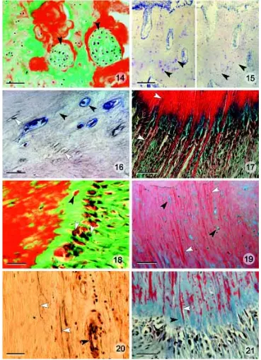
Table 2 for the features used to identify cells as chondrocytes). Basically the skeletal needles contain a mixture of bone and cartilage cells but locally, bone cells or cartilage cells can predomi-nate. Hyaline cartilage is present at the distal tip of the lower jaw, joining the left and right dentary bones (Fig. 14).
Toluidine blue-, Alcian blue/Haematoxylin-, and HBQ-staining methods provide evidence for the dual presence of osteocytes and chondrocytes within the bone matrix (Figs. 8,11,12,13). In addition,
Fig. 6. A sagittal section through the tip of the dentary. As already revealed by gross morphological examination, the tip of the kype is entirely made from connective tissue (green). Needle like bone structures (black arrowheads), seen on X-rays (Figs. 3,4) arise from the perios-teum of the lower jaw. The white arrowheads point to the border between skeleton of the kype and cortical bone of the lower jaw. Masson’s trichrome staining. Scale bar, 0.2 cm.
Fig. 7. The boundary between the bone and connective tissue of the kype (distal above).
Needle-like bone (white arrowheads) protrude distally into the connective tissue (black arrow-head) and anastomose proximally into a spon-giosa-like meshwork. Toluidine Blue staining. Scale bar, 250 µm.
Fig. 8. A view of the ventral portion of the skeletal tissue of the kype to show develop-ing solid bone (black arrowhead). The bone formation front is rich in proteoglycans (blue staining, white arrowheads). Blue spots inside the bone matrix represent chondrocytes sur-rounded by cartilage matrix (see also Fig. 13). HBQ staining. Scale bar, 250 µm.
Fig. 9. A view of the distal growth front of the kype bone (growth is from right to left) to show prominent osteoblasts (white arrow-head) at the tip of rapidly growing skeletal needles, made of cancellous bone (see also Figs. 3,4,6,7). The staining indicates a broad band of non-mineralized bone matrix (osteoid; black arrowhead) underneath the osteoblast layer. Masson’s trichrome staining. Scale bar, 150 µm.
Fig. 10. Osteoid staining reveals the pres-ence of a broad osteoid zone (blue staining, black arrowhead) at the tip of growing skeletal needles, indicating rapid bone growth. Similar orientation to Fig. 9. Osteoid Staining. Scale bar, 150 µm.
Fig. 11. Prominent expression of proteoglycans at the tip of a growing skel-etal needle (blue staining, black arrowheads) indicates rapid growth of cancellous bone. Alcian Blue/Haematoxylin staining. Scale bar, 150 µm.
Fig. 12. Part of the matrix of the skeleton of the kype contains osteocytes with characteristic osteocytic cytoplasmic processes (canaliculi; white arrowheads). Toluidine Blue staining. Scale bar, 15 µm.
the presence of cartilage-specific extracellular matrix in the bone was verified by chondroitinase and hyaluronidase digestion, which spe-cifically remove cartilaginous ground substances, in this case, in the vicinity of cells identified on histological and histochemical criteria as chondrocytes. Enzymatic digestion results in negative proteoglycan staining with Toluidine Blue and Alcian Blue/Haematoxylin (Fig. 15).
Chondrocytes inside the bone matrix could also be labeled with a monoclonal antibody directed against collagen type II, which is the typical collagen type of cartilage (Beresford, 1993). Bone cells did not react with the antibody (Fig. 17).
The periosteal hard tissue that develops ventrally in the kype is typical Sharpey fiber bone, i.e., a bony tissue with thick
collag-Fig. 14. Solid cartilage, both unmineralized (black arrowheads) and mineralized (red)
appears distally and medially at the joint be-tween the dentary bones in the left and right lower jaws. Masson’s trichrome staining. Scale bar, 60 µm.
Fig. 15. Treatment of sections with chondroitinase and hyaluronidase selec-tively removes chondroitin-6-sulphate and hy-aluronic acid respectively. (Left) Non-enzyme-treated section stained with Alcian blue/He-matoxylin staining shows proteoglycans sur-rounding cartilage cells inside the bone ma-trix. (black arrowheads). (Right) Treatment with chondroitinase before staining removes chondroitin-6-sulphate from the bone (black arrowheads). Results after Hyaluronidase treatment (not shown) are similar. Scale bar, 150 µm.
Fig. 16. Immunohistochemical demonstra-tion of collagen type II in cartilage cells,
surrounded by bone matrix. Theapplication of a monoclonal antibody directed against col-lagen type II indicates that in addition to the cartilage matrix components hyaluronic acid and chondroitin-6-sulphate, cartilage cells and the surrounding matrix (black arrowheads) contain collagen type II (black arrowheads). Small osteocyte-like cells do not display col-lagen type II (white arrowheads). Application of a monoclonal anti-collagen type II antibody. Scale bar, 25 µm.
Fig. 17. Collagen fibers (white arrowheads) continue inside the matrix of the skeleton
of the kype (Sharpey fiber bone) at the ventral side of the dentary. Osteoblasts arranged in multiple layers (black arrowheads), deposit new bone matrix in the gaps between the fibers. HBQ staining. Scale bar, 40 µm.
Fig. 18. Growth of Sharpey fiber bone. As in distal bone formation in the kype (Fig. 9), the growth of Sharpey fiber bone involves the activity of prominent osteoblasts (white ar-rowheads). Osteoblasts secrete a broad layer of non-mineralized bone matrix that contains entrapped osteoblasts (black arrowhead). Masson’s Trichrome staining. Scale bar, 25
µm.
Fig. 19. The hard tissue of the kype on the ventral side of the dentary is Sharpey fiber bone with enclosed collagen fibers (white arrowheads). This bone also displays
chon-droid bone-like features, as evidenced by the enclosed cartilage cells (black arrowheads). HBQ staining. Scale bar, 100 µm.
Fig. 20. Elastin staining indicates the presence of elastin fibers (white arrowheads) in the connective tissue of the kype. Walls of blood vessels stain also positive (black arrowhead). No elastin is detectable in the bone matrix. Verhoef’s Elastin staining. Scale bar, 200 µm.
enous fiber bundles that extend from the connective tissue, in this case of the kype, into the new bone matrix. Large osteoblasts secrete osteoid into the gaps between the fibers (Figs. 17,18). As in the deposition of skeletal needles distally, osteoblasts develop into typical osteocytes and chondrocytes (Fig. 19). Consequently, the ventral segment of the kype is Sharpey fiber bone that also displays features of chondroid bone similar to that described by Beresford (1993) as chondroid bone type one. HBQ- and Elastin-staining provide evidence that the fibers that continue from the
connective tissue into the bone matrix are made of collagen. Elastin, recently reported to occur in fish bone (Miyake et al., 2001) is restricted to the connective tissue part of the kype and to the walls of blood vessels (Fig. 20).
Mineralization
As revealed by osteoid- and Masson’s trichrome-staining, the pattern of mineralization of the skeletal tissue of the kype varies according to type and location of the developing hard tissue.
Fig. 22. Chondrocytes inside the skeleton of the kype mediate a cartilage-like miner-alization (red staining) that starts in the vicin-ity of single cells and cell groups (black arrow-head). Pairs of chondrocytes indicate that cells continue to divide prior to matrix mineraliza-tion. Osteoid staining. Scale bar, 40 µm.
Fig. 23. The compact bone of the dentary (top) is clearly separated from the skeleton of the kype (below) which is composed of both Sharpey fiber and chondroid bone. The border between compact and Sharpey fiber bone is labeled by white arrowheads. An os-teon (black arrowhead) is forming in the vicin-ity of the cortical bone. Masson’s trichrome staining. Scale bar, 150 µm.
Fig. 24. A primary osteon in the cortex of the dentary. Notice the absence of chondrocytes and Sharpey fibers in the bone matrix. The endosteal surface is covered with osteoid (blue staining, black arrowheads). Osteoid staining. Scale bar, 60 µm.
Fig. 25. A primary osteon in the cortex of the dentary. The circular arrangement of the parallel fiber bone (white arrowheads) has been visualized using polarized light micros-copy. HBQ staining. Scale bar, 60 µm.
Fig. 26. Cross linkage of skeletal needles creates bone marrow space that primarily contains connective tissue (white arrow-heads). Masson’s trichrome staining. Scale bar, 150 µm.
Fig. 27. Bone resorbing cells in salmon display typical features, also known from mammalian Osteoclasts. The cells express TRAP (red), are multinucleated and display a ruffled border (black arrowheads). Secreted TRAP appears as a red stripe at the bone surface (white arrowhead). The extracellular dephosphorylation of bone sialoprotein and osteopontin by TRAP is thought to be re-sponsible for the observed detachment of the cells from the bone surface (Ek-Rylander et al., 1994) as is the case for the right osteoclast and for cells in Fig. 28. TRAP staining. Scale bar, 40 µm.
Fig. 28. An alternative way of creating bone marrow space is by bone resorption by multi-nucleated osteoclasts (black arrowheads). Along with the osteoclasts, blood vessels (white arrowheads) are present in the bone marrow. Osteoid staining. Scale bar, 60 µm.
Skeletal needles display a mineralization front that starts beneath the broad osteoid zone (Fig. 11). In the ventral Sharpey fiber bone, collagen fibers are the sites of first mineral deposition (Fig. 21). Cartilage and cartilaginous parts of the skeleton of the kype undergo a cartilage-like pattern of mineralization, starting in the vicinity of chondrocytes (Fig. 22).
Bone Matrix
The kype skeletal tissue is made from primary cancellous, chondroid-like bone distally (Figs. 11-15) and from Sharpey fiber bone with chondroid bone-like features ventrally (Figs. 17-22). These tissues clearly differs from the compact bone of the dentary, which consists of parallel-fibered bone, structurally organized in osteons (Figs. 23-25). Compact bone contains neither chondrocytes nor Sharpey fibers (Fig. 24). The border between the compact bone of the dentary and the skeletal tissue of the kype is charac-terized by an abrupt change of bone type. The chondroid-like bone and the chondroid-like Sharpey fiber bone in direct contact with the compact bone (Figs. 6,23). The abrupt structural changes allows cortical bone and skeletal needles of the kype to be discriminated in (Figs. 3,4).
Remodeling
The skeletal tissue of the kype displayed evidence of continuous growth at the time the salmon were caught. Growth includes
development of cross connecting distal skeletal needles into a spongiosa-like formation (Figs. 6,7) which creates small bone marrow spaces containing connective tissue (Fig. 26). Concur-rently, proximal parts of the skeleton of the kype are converted into compact bone (Fig. 21), which involves the creation of bone marrow space by osteoclastic bone resorption (Figs. 27-29), but not by cross connection of bone needles. From the analysis of serial sections we interpret this as a typical bone remodeling sequence involving the resorption of primary bone by numerous multinucleated and TRAP active osteoclasts (Figs. 27,28) and it’s replacement by secondary lamellar bone (Fig. 29). Lamellar bone then forms the inner wall of forming osteons. Through bone remodeling, both the skeleton of the kype and the bone of the dentary increase and grow.
Discussion
The morphological alterations observed in the lower jaws of Atlantic salmon migrating upstream from the Miramichi river match the morphological descriptions of the phenomenon provided by Day (1987), Tchernavin (1938a) and Kacem et al. (1999) for Atlantic salmon migrating in Britain, Scotland and France. Hence, there is reason to assume that the alterations in the jaws of males found in this study are typical for Atlantic salmon in other locations, irrespective of population or genetic background.
Despite precise descriptions of the reinitiation of bone growth and morphological changes by Tchernavin (1938a) and Kacem et al. (1999), the hard tissue(s) of the kype have not been analyzed in detail. Darwin (1909, p. 510) discusses it as a cartilaginous projection, citing Yarrell’s History of British Fishes as the source. Day (1887) cites Gadow as having sectioned a kype, stained it with carmine, and described it as consisting “entirely of fibrous connec-tive tissue, without any traces of cartilaginous cells in it.” Day goes on: “Therefore the hook cannot be looked upon either as an outgrowth of the bones of the lower jaw, or as a sort of horny excrescence like horns, nails...” (p. 57). In the few subsequent studies that have commented on the tissue (none of which under-took any histological analysis), it has been regarded as bone (Tchernavin, 1938c, Persson et al., 1998). Our analysis reveals that the hard tissues of the kype are neither cartilage nor typical bone and are certainly not keratin as seen in horns or nails but differs from the compact bone of the dentary. The composition of the unique skeleton of the kype, its growth patterns, and its possible functional significance are discussed below.
The Skeletal Tissues of the Kype
The special characters of the skeletal tissues of the kype and their divergence from the compact bone of the dentary can be seen on high resolution X-rays, a technique that we and some others now regularly apply to detect skeletal alterations in teleosts (Witten et al., 1999b, Afonso et al., 2000). The border between new bone of the kype and of the dentary is clearly discernible on X-rays. Histological sections allow visualization of the microanatomy of the structures and confirm the radiological observations that proximally, compact bone of the dentary switches abruptly into a needle-like skeletal structure. The future application of X-radiography to analyze changes in the lower jaws of salmon in the field will allow analysis of large numbers of animals, and, with multiple X-rays through time, allow skeletal changes in individual animals to be followed. The use of such
TABLE 2
THE CHARACTERISTICS AND DIFFERENCES BETWEEN THE SKELETON OF THE KYPE AND THE COMPACT BONE OF THE DENTARY
Histological features Figures
Skeleton of the kype
The matrix consists of primary cancellous bone 9,11 Rapidly growing skeletal needles anastomose, forming spongiosa-like bone 6,7 Ventrally, the skeleton is solid and contains Sharpey fibers 8,17,19,23 Apical, mineralization starts beneath the broad osteoid zone, rich in proteoglycans 9,10,11 Ventrally, Sharpey fibers are sites of the first mineral deposition 21 Apically and ventrally, typical osteoblasts secrete osteoid and
become entrapped in the matrix 9,10, 18 Osteoblast inside the matrix develop into osteocytes or into chondrocytes 12,13,18,19 Osteocytes are characterized by their typical shape and cell processes 12 Chondrocytes are characterized by: 8,13,15,22 the presence of groups of cells 13,22
a rounded shape 13,19,22
the capability to divide 22
the lack of cell processes 13,22
secretion of extracellular cartilage matrix including
chondroitin sulphate, hyaluronic acid and collagen type II 8,15, 16,19 a cartilage-like pattern of matrix mineralization 22 Proximal parts of the skeleton of the kype are subjected to remodeling into compact bone 28,29
Compact bone of the dentary
Composed of lamellar bone 23,24,25
Structural units are typical osteons 24,25 Osteoid seams are small and the bone is heavily mineralized 24 Osteoblasts and osteoid are present at endosteal surfaces of osteons 24 Compact bone contains no chondrocytes or chondrocyte-like cells and
no Sharpey fibers 23,24,25
The tip of the dentary grows by remodeling of the skeleton of
the kype into compact bone 23, 27,28,29 Remodeling creates bone marrow spaces 27,28,29 Remodeling involves bone resorption by typical osteoclasts 27,28,29
a non-invasive technique on live animals is highly desirable in view of the world-wide declining stocks of wild Atlantic salmon. At the same time, there is an increasing need to gather information about life history of the animals (Hutchings and Jones, 1998).
Apart from radiological and microstructural studies, histological and histochemical analyses confirm the special character of the skeletal needles of the kype: (1) The needles display a rapid periosteal skeletal growth that differs from periosteal bone formation; (2) A loose meshwork of bony trabeculae then forms; (3) Subse-quently, the tissue exhibits a character that is intermediate between bone and cartilage with Sharpey’s fibers ventrally and (4) Finally the skeleton of the kype is partly remodeled into compact bone without cartilage-like features and without Sharpey fibers.
Rapid skeletal growth in a life phase where the animals have stopped feeding and utilize as much as seventy percent of their energy reserves for metabolically expensive upstream migration, for gonad maturation, and for competition with other males on the spawning grounds (Fleming, 1996) is a remarkable event. It has been hypothesized that formation of the kype and the changes in bones of the skull that occur during migration requires resorption of minerals from the operculum, the vertebral column or the scales (Tchernavin, 1938b; Persson et al., 1998; Kacem et al., 2000). The growth pattern of the skeletal tissue of the kype suggests, however, that despite mineral support from other skeletal elements, the bony tissue is made from the least amount of material possible; the skeleton of the kype does not arise as a solid bone mass, but as a meshwork of anastomozing skeletal needles. The spongiosa-like structure of the resulting tissue likely provides a maximum of mechanical stability with a minimum of bone mass, as in spongiosa in the bone marrow of mammals (Parfitt, 1988).
The spongiosa-like periosteal bone tissue in the skull of other teleosts (hyperostotic bone) might serve a mechanical function or only play a role in mineral metabolism (Smith-Vaniz et al., 1995; Meunier and Desse, 1986). The rapid and structured growth of the skeleton of the kype as revealed by prominent osteoblasts, a broad osteoid zone, deposition of proteoglycans and expression of cartilage characteristics (Huysseune and Verraes, 1990; Beresford 1993; Nah et al., 2000), suggests that the skeleton of the kype provides a maximum of support in a physiological situation where somatic growth has stopped (Persson et al., 1998). Although, bone resorption occurs proximally in the kype, there is no indication that the skeleton of the kype functions as a mineral reserve in anadro-mous animals, since this and other studies show that bone resorp-tion is always linked to the concurrent formaresorp-tion of new bone (Witten et al., 2000).
The Function of the Kype and Kype Skeleton
So far, only a few studies have focused on the possible function of the kype. Tchenavin (1938c) regards the kype as “no good to the fish, and seems to be somewhat absurd, preventing as it does the use of the strong curved teeth which appear at the same time” (p. 37). Witten and Hall (2001) suggested that the kype is not merely a by-product of hormonal and environmental alterations. The kype might function as a secondary sexual character, either for compet-ing with other males, or to attract females (Flemcompet-ing, 1996; Hutchcompet-ings and Myers, 1987; Järvi, 1990). Darwin certainly thought so:
Male salmon have been observed fighting all day long [and that] special means of defense may be given through means of sexual selection, as the mane of the lion and the hooked jaw to the male
salmon; for the shield may be as important for victory, as the sword or spear (Darwin, 1910, p. 64).
Ideas raised about functional and mechanical properties of the skeletal tissues of the kype are based on analysis of the hard tissue microstructure (Smith-Vaniz et al., 1995). Thus, the unique charac-ter of kype hard tissues should be discussed in more depth. The skeleton of the kype arises from the periosteum of the dentary but its structure is clearly divergent from compact bone of the lower jaw, which is made of parallel-fibered bone and osteons. In contrast, the skeleton of the kype consists of anastomozing skel-etal needles made of woven bone and contains no osteons. The differences between the kype and the compact bone of the dentary are summarized in Table 2. Besides osteocytes, the bone matrix contains chondrocytes, while the ventral part of the skeleton of the kype also contains Sharpey fibers.
Antlers of deer are periosteal bone formations that share fea-tures with the skeleton of the kype. Antlers function as a secondary sexual character, arise from a specific site on a dermal skeletal element (the pedicel of the frontal bone), and both their structure and mode of formation differ from those of the frontal bone. There is an initial cartilaginous phase and metaplastic conversion of cartilage to bone in antlers. Rapid growth and the retention of cartilage characters are features shared with the skeleton of the kype (Hall, 1978; Goss, 1983; Szuwart et al., 1995, 1998).
In view of the astounding variety of skeletal tissue that occur in teleosts (Hall, 1978; Benjamin, 1988a,b; 1990, Huysseune, 2000) it is no surprise that secondary periosteal skeletal tissue is not only found in mammals and salmon but occurs in other fish species. Regular hyperostotic teleost bones — formerly regarded as patho-logical and referred to as an osteoma (Schlumberger and Lucke, 1948) — share features with the skeleton of the kype. Hyperostotic bones not only have a spongiosa-like appearance, but in teleosts with acellular bone they may represent areas of the skeleton that undergo remodeling (Smith-Vaniz et al., 1995). In contrast, salmon have cellular (osteocyte-containing) bone and the whole skeleton (endoskeleton plus dermal bones and scales) is thought to be metabolically active (Persson et al., 1998, 1999; Vielma and Lall, 1998; Kacem et al., 2000). However, like hyperostotic bones of advanced teleosts (Smith-Vaniz et al., 1995), in the upstream migrating salmon we examined, the skeletal tissue of the kype, not the compact bone of the dentary, displays remodeling, mediated by prominent multinucleated osteoclasts.
Bone and Cartilage Characters of the Kype Skeleton
The skeleton of the kype is formed by cells that resemble typical osteoblasts (Hall, 1978, 1990, 1991). In the ventral part, the cells secrete new bone matrix (osteoid) in gaps between the Sharpey fibers. Distally, membrane bone formation takes place. The two halves of the kype are united by hyaline cartilage, who’s location and lack of connection to Meckel’s cartilage would lead us to identify the tissue as secondary cartilage (Beresford, 1981). How-ever, we do not yet know the developmental history of this cartilage. It could be a remnant of Meckel’s cartilage not connected to Meckel’s cartilage in adults; it would have to arise from periosteal cells to qualify as a secondary cartilage (see Benjamin, 1988b).
show-ing cell division, beshow-ing arranged in rows and territories, producshow-ing cartilage matrix and displaying a cartilage-like pattern of matrix mineralization (Moss and Moss-Salentijn, 1983; Huysseune, 2000). Given that the skeleton of the kype entirely derives from in-tramembranous ossification, the appearance of cartilage charac-ters is remarkable. However, it is now established that the expression of cartilaginous features is a general character of growing membrane bones (Fang and Hall, 1997, 1999; Nah et al., 2000). Furthermore, there is strong evidence that osteoblasts and chondroblasts are members of the same class of scleroblasts (Hall, 1970, 1978, 1991; Hall and Miyake, 2000). The mecha-nisms discussed above are likely also involved in the develop-ment of chondroid bone, a tissue that occurs in several teleost species and that shares morphological and histochemical fea-tures with the skeleton of the kype (Beresford, 1981; Benjamin, 1990; Huysseune and Verraes, 1990; Taylor at al., 1994). Similar to chondroid bone in cichlids, the growth of the kype bone involves osteoblasts that acquire characteristics of chondrocytes as they become trapped in the bone matrix. The idea has been raised that chondroid bone is an adaptation to rapid bone growth (Hall, 1990; Huysseune, 1990; Taylor et al., 1994), an interpretation that applies to both the distal and the ventral parts of the skeleton of the kype.
Sharpey Fiber Bone in the Kype Skeleton
Apart from displaying chondroid bone-like characters, the ven-tral part of the skeleton of the kype is also a Sharpey fiber bone. A similar tissue, that displays features of chondroid bone and of Sharpey fiber bone, has not been described in teleosts previously. Sharpey fiber bone - characterized by the continuation of collagen fiber bundles from connective tissue into the bone matrix - is commonly associated with joints and teeth, providing a strong link between connective tissue and the skeleton (Beresford, 1981; McKee et al., 1996). Hence, the presence of Sharpey fiber bone in the ventral part of the skeleton of the kype further supports the idea that this skeletal tissue is functionally integrated in the architecture of the salmon jaw. It is not a by-product of physiological changes but serves a mechanical function.
Appearance of Sharpey fiber bone in the dentary is not restricted to the kype of grilse. Hughes et al. (1994a,b) described Sharpey fiber bone in the lower jaws of juvenile salmon. Interestingly, their description of Sharpey fiber bone in the lower jaw of perciform teleosts (Sparidae) also matches the tissue found in salmon. In distinction from Hughes et al. (1994b), who identified all cells inside the Sharpey fiber bone as osteocytes, we conclude that the Sharpey fiber bone of salmon contains chondrocytes. If the cells inside the Sharpey fiber bone of Sparidae that display a cartilage-like morphology are chondrocytes and not osteocytes (as we suspect from the figures published by Hughes et al., 1994b), then the type of hard tissue in the kype ventrally is not restricted to salmon.
The Kype and Salmon Life History
The consequences of different life strategies of male Atlantic salmon that range from premature kype-less paar to kype-bearing mature male salmon are not fully understood (Shear, 1992; Moore and Waring, 1999; Jones and Hutchings, 2001). Mature males invest heavily in the spawning success, and the development of a prominent kype is a part of the male investment, resulting in a increased mortality (Fleming, 1996). As suggested by Tchernavin
(1938c), the kype may become a disadvantage for anadromous males (impairing proper feeding, breathing and swimming) after fulfilling its purpose as a secondary sexual character. Although, the microstructure of the kype tissue is different from the bone of the jaws, developmental processes and hard tissue elements of the kype are known from the dermal skeleton of mammals and other teleost species. Finally, the rapid growth of the skeleton of the kype appears to be a regulated process, integrated in the development of the dentary, and understandable in the light of the general strategy of Atlantic salmon to survive spawning. The growth of compact bone, through remodeling of the skeleton of the kype seems to produce non-reversible alterations of the dentary in repetitively spawning individuals. However, it remains to be clari-fied if the changes gradually disappear after spawning and if the dentary resumes its former shape and proportions in those males which return to sea, as hypothesized by Tchernavin (1937). Future studies will address the extent of the process and its limits for the fitness of individual anadromous male salmon.
Materials and Methods
Sampling
Ten male anadromous grilse (see Table 1 for definition of terms), collected during the fall run in 1999 and 2000 (September) at the south-west Miramichi River above Red Bank (46°56.8' N, 65°48.3' W, New Brunswick, Canada,) were used for this study. Animals were caught in traps used for the Red Bank (First Nation) food fishery. The animals were caught and sampled under license from the Canadian Federal Department of Fisheries and Oceans.
Weight and fork length were determined in order to calculate the condition factor (K = [weight (g)/fork length (cm3)] x 100) of the fish (Arndt et al., 1996). Scales were taken to evaluate the animals life history; part of a Department of Fisheries and Oceans monitoring program. All grilse used for this study were in “good shape,” as determined by gross morphological inspection, and showed no signs of diseases. As revealed by scale reading (Shearer, 1992), all animals had spend one winter in the ocean (one sea winter salmon = 1SW; see Table 1) before returning to their home river. The fork length of males varied between 55.4 and 63.5 cm, weight ranged from 2010g to 2500g. Condition factors were calculated as between 0.93 and 1.17 which is in the range of the mean condition factor (K = 1.02) for males from this age group in the Miramichi river system (Moore et al., 1995).
For gross anatomical documentation external picture of the entire head as well as pictures of half heads were taken. Subsequently, the jaws were x-rayed, processed and immediately fixed for seven different analytical procedures.
X-ray Analysis
High resolution X-rays of the left halves of all fish heads were taken using a portable “Mini X-ray HF80+” machine (Mini X-ray inc., Northbrock, USA) and “Kodak Industrex M Film Ready Pack II” (Kodak Industry, France) without a screen. The settings of the X-ray unit were 70 kV, 15 mA, 2 sec exposure time, and a distance of 40 cm between the beam source and the X-ray film. Radiographs were developed with Kodak chemicals and ana-lyzed using a stereo microscope.
Basic Histological Procedures
Immediately after capture, right jaws were fixed in 10% neutral buffered parformaldehyde for 24 hrs, rinsed in tap water for 24 hrs, and decalcified for 72 hours in a 10% EDTA solution buffered with 0.1 M TRIS base, pH 7.0. After decalcification, samples were dehydrated in a graded series of erthanol and subsequently embedded in Paraplast.
Quadruple Stain (HBQ) (Hall, 1986) to differentiate bone, cartilage, chon-droid bone and to identify collagen fibers. Sections were mounted with DPX (Fluka).
Additional Histological and Histochemical Analyses
Additional analytical procedures were applied to demonstrate osteoid and the pattern of mineralization, visualize bone remodeling, identify chondrocytes and cartilage matrix within bone, and to discriminate among elastin and collagen fibers.
To differentiate osteoid (non-mineralized bone matrix) and mineralized bone matrix and to reveal the pattern of mineralization, osteoid staining was performed, following the trichrome method of Ralis and Watkins (1992).
To demonstrate bone remodeling, decalcified, sections were stained with Toluidine Blue as follows: Descending ethanol series (100% ethanol to water); 1% aqueous Toluidine Blue solution (pH 4.5) for 45 minutes; differentiation and dehydration in 100% Ethanol; mounting.
To identify osteoclasts and osteoclasts activity, tartrate resistant acid phosphatase was demonstrated (TRAP) following the protocol of Witten et al., 2001. Briefly, samples were (a) fixed in 10% formaldehyde (buffered with 50 mmol l-1 TRIS, at pH 7.2; (b) rinsed in tap water for 1 hr; (c) decalcified with EDTA (see above), (d) dehydrated in graded acetone solutions and (e) embedded in glycol-methacrylate. Sections of 5 µm were mounted on uncoated slides. For demonstration of TRAP activity, speci-mens were preincubated for 30 min at 20°C in 100 mmol acetate buffer + 50 mmol L(+) di-sodium tartrate dihydrate at pH 4.5. Staining was per-formed in the presence of tartrate using naphthol AS-TR phosphate as substrate (final concentration 170 mmol/L) and hexazotised pararosaniline (final concentration 1,58 mmol /L) as an azo-coupling dye. Specimens were counterstained with Mayer´s haematoxylin. Control procedures revealed the specificity of the staining and consisted of: (a) heating at 90°C for 10 min prior to staining, (b) incubation without substrate, (c) adding the inhibitor NaF to the staining solution (10 mmol l-1).
For the selective demonstration of cartilage matrix proteoglycans, sections were stained with a combined Alcian Blue/Haematoxylin staining (AH). Deparaffinized sections were treated with 3% acetic acid for 3 min, 1% Alcian Blue in 3% acetic acid solution for 1 hr, rinsed in 3% acetic acid solution, rinsed for 10 min in D H2O, stained with Myer’s acid Haematoxylin for 10 min, flushed in running tap water for 10 min and then mounting.
Cartilage-specific proteoglycans were visualized by selective removal of two cartilage matrix components prior to Alcian Blue Haematoxylin staining: hyaluronic acid, which was digested with hyaluronidase, and chondroitin-6-sulphate, which was digested with chondroitinase. To re-move hyaluronic acid, sections were incubated for 30 min at 37°C in a solution containing 6 mg/ml hyaluronidase (EC. 3.2.1.35, Sigma No. He506) dissolved in 10 mM PBS buffer (pH 7.2). To remove chondroitin-6-sulphate, prior to staining, sections were incubated at 37°C in a solution containing 5 units/ml of Chondroitinase ACB (chondroitin-6-sulphatase; E.C. 4.2.2.4., Sigma No. C2905) in 10 mM TRIS buffer (pH 8.0).
Immunostaining for collagen type II as a component of the cartilage matrix was performed on decalcified specimens embedded in Paraplast (see histology). After removal of paraffin, sections were pre-treated with hyaluronidase following the protocol given above to expose collagen. As part of the protocol, the solution also contained 10 mM Levamisole to inactivate alkaline phosphatase (levamisole treatment may be omitted since decalcification with EDTA also inactivates alkaline phospatases). Sections were then incubated in a blocking solution for 30 minutes at 37°C (20% Rabbit Serum in 10 mM PBS + 10 µM Levamisole, pH 7.4). Subsequently a monoclonal anti-collagen type II antibody (mouse IgG; II-II6B3, Developmental Studies Hybridoma Bank, Iowa USA) was used to identify cartilage matrix collagen. 100 ml antibody containing supernatent and 10 ml rabbit serum were dissolved in 890 ml PBS and applied to the sections at 4°C for 12 hrs. Slides were flushed with PBS buffer and the sections were treated with the secondary antibody for 30 min at 37°C (a ALP-labelled anti-mouse IgG raised in rabbits, anti-mouse IgG /PBS 1:50 + 10 % Rabbit serum). Finally the sections were flushed two times with PBS, pre-adjusted pH 9 with TRIS-buffer and ALP was visualised by BICP/NBT
staining for 30 min at 20°C. Control procedures revealed the specificity of the staining and consisted of: (a) incubation with a non-specific mouse IgG antibody, (b) incubation only with the alkaline phosphatase-labelled sec-ondary antibody, and (c) incubation with ALP substrate only.
Since elastin was recently described as a component of bone and teeth in fish (Miyake et al., 1999, 2001) the possible presence of elastin fibers in the hard tissues of the kype was evaluated using the Verhoeff staining procedure (Presnell and Schreibman, 1997).
Acknowledgements
For discussions on the topic of this study we thank Harald Rosenthal, Tom Miyake, Lawrence Taylor, Gérald Chaput, Michel Chadwick, David Moore, Peter Hurley, John Hayward and Joe Sheasgreen. We are grateful to Rick Cunjak who facilitated access to field facilities at the start this study. We thank people from the Red Bank (First Nation, New Brunswick, Canada), especially Betty Jane Ward for sampling salmon. We also appreciate support provided by Wolfgang Villwock, Volker Hilge, Thomas Falk, and Gudrun Schulze. Funding for this study was granted by the German-Canadian Science and Technology Cooperation, by the Deutsche Forschungsgemeinschaft (DFG) and by the Natural Sciences and Engi-neering Research Council of Canada (NSERC).
References
AFONSO, J.M., D., ROBAINA, L., ASTORGA, N., IZQUIERDO, M.S. and GINÉS, R. (2000). Association of a lordosis-scoliosis-kyphosis deformity in gilthead seabream (Sparus aurata) with family structure. Fish. Physiol. Biochem. 22: 159–163.
ARNDT, S.K.A., BENFEY, T.J. and CUNJAK, R.A. (1996). Effect of reductions in feeding on protein synthesis and energy storage of juvenile Atlantic salmon. J. Fish. Biol. 49: 257—276.
BENJAMIN, M. (1986). The oral sucker of Gyrinocheilus aymonieri (Teleostei: Cypriniformes). J. Zool. London. B 1: 211—254.
BENJAMIN, M. (1988a). Mucochondroid (mucous connective) tissues in the heads of teleostei. Anat. Embryol. 178: 461—474.
BENJAMIN, M. (1988b). The development of hyaline-cell cartilage in the head of the black molly, Poecilia sphenops. Evidence for secondary cartilage in a teleost. J. Anat. 164: 145—155.
BENJAMIN, M. (1989). Hyaline-cell cartilage (chondroid) in the heads of teleosts. Anat. Embryol. 179: 285—303.
BENJAMIN, M. (1990). The cranial cartilages of teleosts and their classification. J. Anat. 169: 153—172.
BENJAMIN, M. and RALPHS, J.R. (1991). Extracellular matrix of connective tissues in the heads of teleosts. J. Anat. 179: 137—148.
BERESFORD, W.A. (1981). Chondroid bone, secondary cartilage and metaplasia. Urban and Schwarzenberg, Baltimore-Munich.
BERESFORD, W.A. (1993) Cranial skeletal tissues: Diversity and evolutionary trends. In The Skull: Vol. 2, Patterns of Structural and Systematic Diversity (Eds. Hanken, J and Hall, B.K.). University of Chicago Press, Chicago, pp. 69-130.
CHAPUT, G., ALLARD, J., CARON, F., DEMPSON, J.B., MULLINS, C.C. and O’CONNEL, M.F. (1998). River-specific target spawning requirements for Atlantic salmon (Salmo salar) based on a generalized smolt production model. In Water science and the public: The Miramichi River (Ed. Chadwick, E.M.P.). Can. Spec. Pub. Fish. Aquat. Sci. 123: 121—139.
CHAPUT, G., MOORE, D., HAYWARD, J., SHAESGREEN, J. and DUBEE, B. (2000). Stock status of Atlantic Salmon (Salmo salar) in the Miramichi River, 1999. Canadian Stock Assessment Secretariat, Ottawa.
CHAPUT, G.J. (1997). Proceedings of a workshop to review conservation principles for Atlantic salmon in eastern Canada. Canadian Stock Assessment Proceedings Series 97/15. Department of Fisheries and Oceans Science Branch, Moncton.
DARWIN, C.D. (1877). The descent of man, selection in relation to sex. 2nd Edition (revised and augmented). John Murray, London.
DARWIN, C.D. (1910). The Origin of Species by Means of Natural Selection. Popular impression of the corrected copyright edition. John Murray, London.
EK-RYLANDER, B., FLORES, M., WENDEL, M., HEINERGARD, D. and ANDER-SON, G. (1994). Dephosphorylation of osteopontin and bone sialoprotein by osteoclastic tartrate-resistant acid phosphatase. J. Biol. Chem. 269: 14853-14856.
FANG, J. and HALL, B.K. (1997). Chondrogenic cell differentiation from membrane bone periostea. Anat. Embryol 196: 349—363.
FANG, J. and HALL, B.K. (1999). N-CAM is not required for initiation of secondary chondrogenesis: the role of N-CAM in skeletal condensation and differentiation. Int. J. Dev. Biol. 43: 335—342.
FLEMING, I.A. (1996). Reproductive strategies of Atlantic salmon: ecology and evolution. Reviews in Fish Biology and Fisheries 6: 379—416.
FLINT, M.H. and LYONS, M.F. (1975). The effect of heating and denaturation on the staining of collagen by Masson’s trichrome procedure. Histochem. J. 7: 547—555.
GOSS, R.J. (1983). Deer Antlers. Regeneration, Function, and Evolution. Academic Press, New York.
HALL, B.K. (1970). Cellular differentiation in skeletal tissues. Biol. Rev. 45: 455—484.
HALL, B.K. (1975). Evolutionary consequences of skeletal differentiation. Amer. Zool. 15: 329—350.
HALL, B.K. (1978). Developmental and cellular skeletal biology. Academic Press, New York.
HALL, B.K. (1986). The role of movement and tissue interactions in the development and growth of bone and secondary cartilage in the clavicle of the embryonic chick. J. Embryol. Exp. Morph. 93: l33—l52.
HALL, B.K. (1990). What is bone growth? In Fundamentals of bone growth. Methodol-ogy and applications. (Eds. Dixon, A.D., Sarnat, B.G. and Hoyte D.A.N.). CRC Press, Boca Raton, pp. 605—612.
HALL, B.K. (1991). Bone: Embryonic Development. In Encyclopedia of Human Biology Volume 1 (Ed. Dulbecco. R.). Academic Press, Orlando, pp. 781—791.
HALL, B.K. and MIYAKE, T. (2000). All for one and one for all: condensations and the initiation of skeletal development. BioEssays 22: 138—147.
HUGHES, D.R., BASSETT, J.R. and MOFFAT, L.A. (1994a). Structure and origin of the tooth pedicel (the so-called bone of attachment) and dental-ridge in the mandibles of the sea breams Acanthopagrus australis, Pagrus auratus and Rhabdosargus sarba (Sparidae, Perciformes, Teleostei). Anat. Embryol. 189: 51—69.
HUGHES, D.R., BASSETT, J.R. and MOFFAT, L.A. (1994b). Histological identification of osteocytes in the allegedly acellular bone of the sea breams Acanthopagrus australis, Pagrus auratus and Rhabdosargus sarba (Sparidae, Perciformes, Teleostei). Anat. Embryol. 190: 163—179.
HUTCHINGS, J.A. and JONES, M.E.B. (1998). Life history variation and growth rate thresholds for maturity in Atlantic salmon, Salmo salar. Can. J. Fish. Aquat. Sci. 55 (Suppl): 22—47.
HUTCHINGS, J.A. and MYERS, R.A. (1987). Escalation of an asymmetric contest: mortality resumption from mate competition in Atlantic salmon, Salmo salar. Can. J. Zool. 65: 766—768.
HUYSSEUNE, A. (1990). Chondroid bone in cichlid fish: Actual state of knowledge. Belg. J. Zool. 120: 99.
HUYSSEUNE, A. (2000). Skeletal system. In The laboratory fish. Part 4 Microscopic functional Anatomy (Ed. Ostrander, G.K.). Academic Press, San Diego, pp. 307— 317.
HUYSSEUNE, A and VERRAES, W. (1990). Carbohydrate histochemistry of mature chondroid bone in Astatotilapia elegans. (Teleostei: Cichlidae) with a comparison to acellular bone and cartilage. Annales des Sciences Naturelles Zoologie, Paris 1311: 29—43.
JÄRVI, T. (1990). The effects of male dominance, secondary sexual characteristics and female choice on mating success of male Atlantic salmon Salmo salar. Ethology 84: 123—132.
JONES, M.W and HUTCHINGS J.A. (2001). The influence of male parr body size and mate competition on fertilization success and effective population size in Atlantic salmon. Heredity 86: 675—684.
KACEM, A., MEUNIER, F.J. and BAGLINIÈRE, J.L. (1998). A quantitative study of morphological and histological changes in the skeleton of Salmo salar during its anadromous migration. J. Fish. Biol. 53: 1096—1109.
KACEM, A., BAGLINIÈRE, J.L, and MEUNIER, F.J. (2000). Salmon osteocytic osteolysis at the vertebral column. Comp. Biochem. Physiol. A 125: 497—484.
LOPEZ, E., PEIGNOUX-DEVILLE, J., LALLIER, F., MARTELLY, E. and MILET, C. (1976). Effects of calcitonin and ultimobrancialectomy on calcium and bone metabolism in the eel Anguilla anguilla L. Calcif. Tissue Res. 20: 173—186.
MCKEE, M.D., ZALZAL, S. and NANCI, A. (1996). Extracellular matrix in tooth cementum and mantle dentin: localization of osteopontin and other noncollagenous proteins, plasma proteins, and glycoconjugates by electron microscopy. Anat. Rec. 245: 293—312.
MEUNIER, F.J and DESSE, G. (1986). Les hyperostose les Téléostéens: description, histolog et problémes étiologique. Ichthyophsiol. Acta 10: 130—141.
MILLS, D. (1989). Ecology and management of Atlantic salmon. Chapman and Hall, London.
MIYAKE T, VAGLIA JL, TAYLOR LH, HALL BK. 1999. Development of dermal denticles in skates (Chondrichthyes, Bathoidea): Pattering and cellular differentiation. J. Morphol. 241: 61—81.
MIYAKE, T., VAGLIA, J.L., WITTEN, P.E. and HALL, B.K. (2001). Elastin and/or elastin-like proteins in the skeleton of cartilaginous and teleost fishes. J. Morphol. 248: 262.
MOORE, A. and WARING, C.P. (1999). Reproductive priming in mature male Atlantic salmon parr exposed to the sound of redd cutting. J. Fish. Biol. 55: 884—887.
MOORE, D.S., CHAPUT, G.J. and PICKARD, P.R. (1995). The effect of fisheries on the biological characteristics and survival of mature Atlantic salmon (Salmo salar) from the Miramichi River. In Water science and the public: The Miramichi Ecosystem (Ed. CHADWICK, E.M.P.) Canadian Special Publications of Fisheries and Aquatic Sciences 123: 229—247.
MOSS, M.L. (1961a). Osteogenesis of acellular teleost fish bone. Am. J. Anat. 108: 99— 110.
MOSS. M.L. (1961b). Studies of the acellular bone of teleost fish. 1. Morphological and systematic variations. Acta. Anat. 46: 343—462.
MOSS, M.L. and MOSS-SALENTIJN, L. (1983). Vertebrate cartilages. In Cartilage, Volume 1. Structure, function, and biochemistry (Ed. HALL, B.K.). Academic Press. New York, pp. 1—30.
NAH, H.D., PACIFICIL, M., GERSTENFELD, L.C., ADAMS, S.L. and KIRSCH, T. (2000). Transient chondrogenic phase in the intramembranous pathway during normal skeletal development. J. Bone Mineral Res. 15: 522—533.
PARFITT, A.M. (1988). Bone remodeling: Relationship to amount and structure of bone, and the pathogenesis and prevention of fractures. In Osteoporosis, etiology, diagnosis, and management (Eds. RIGGS, B.L. and MELTON, L.J.) Raven Press, New York pp. 45—93.
PERSSON, P., BJÖRNSSON, BT, TAKAGI Y. 1999. Characterization of morphology and physiological actions of scale osteoclasts in the rainbow trout. J. Fish Biol. 54: 669—684
PERSSON P, SUNDELL K, BJÖRNSSON B.T. and LUNDQVIST, H. (1998). Calcium metabolism and osmoregulation during sexual maturation of river running Atlantic salmon. J. Fish. Biol. 52: 334—349.
PRESNELL, J.K. and SCHREIBMAN, M.P. (1997). Humason’s animal tissue tech-niques, 5th ed. The Johns Hopkins University Press, Baltimore.
RALIS, Z.A. and WATKINS, G. (1992). Modified tetrachrome method for osteoid and defectively mineralized bone in paraffin sections. Biotechnic and Histochemistry 67: 339—45.
SCHLUMBERGER, H.G. and LUCKE, B. (1948). Tumors of fishes, amphibians, and reptiles. Cancer Res. 8: 657-753.
SHEAR, W.M. (1992). The Atlantic Salmon. Natural history, exploration and future management. Fishing news book, Oxford. pp. 244.
SHEARER, W.M. (1992). Atlantic salmon scale reading guidelines ICES cooperative research report 188, Copenhagen.
SMITH, M.M. and HALL, B.K. (1990). Development and evolutionary origins of vertebrate skeletogenic and odontogenic tissues. Biol. Rev. 65: 277—373.
SMITH-VANIZ, W.F., KAUFMAN, L.S. and GLOWACKI, J. (1995). Species-specific patterns of hyperostosis in marine teleost fishes. Marine Biology 121: 573—580.
SZUWART, T., KIERDORF, H., KIERDORF, U., CLEMEN, G. (1998). Ultrastructural aspects of cartilage formation, mineralization, and degeneration during primary antler growth in fallow deer (Dama dama). Ann. Anat. 180: 501—510.
TAYLOR, L.H., HALL, B.K., MIYAKE, T. and CONE, D.K. (1994). Ectopic ossicles associated with metacercariae of Apophallus brevis (Trematoda) in yellow perch, Perca flavescens (Teleostei): development and identification of bone and chon-droid bone. Anat. Embryol. 190: 29—46.
TCHERNAVIN, V. (1937). Preliminary account of the breeding changes in the skulls of Salmo and Oncorhynchus. Proc. Linn. Soc. Lond. 149: 11—19.
TCHERNAVIN, V. (1938a). Changes in the salmon skull. Trans. Zool. Soc. Lond. 24: 103—185.
TCHERNAVIN, V. (1938b). The absorption of bones in the skull of salmon during their migration to rivers. Fishery Board for Scotland, Salmon fisheries 6: 1—5.
TCHERNAVIN, V. (1938c). The mystery of a salmon kype. Does it serve any purpose? Salmon and Trout Magazine 90: 37—44.
TCHERNAVIN, V. (1944). The breeding characters of salmon in relation to their size. Proc. Zool. Soc. London 113B: 206—232.
TEITELBAUM, S.L. (2000). Bone resorption by osteoclasts. Science 289: 1504— 1508
VIELMA, J. and LALL, S.P. (1998). Control of phosphorus homeostasis of Atlantic salmon (Salmo salar) in fresh water. Fish. Physiol. Biochem. 19: 83—93.
VIELMA, J., LALL, S.P., KOSKELA, J. and MATTILA, P. (1999). Influence of low dietary cholecalciferol intake on phosphorus and trace element metabolism by rainbow trout (Oncorhynchus mykiss, Walbaum). Comp. Biochem. Physiol. A 122: 117—125.
WITTEN, P.E., HOLLIDAY, L.S., DELLING, G., HALL, B.K. (1999a). Immunohis-tochemical identification of a vacuolar proton pump (V-ATPase) in bone-resorbing cells of an advanced teleost species (Oreochromis niloticus, Teleostei, Cichlidae). J. Fish. Biol. 55: 1258—1272.
WITTEN, P.E., VILLWOCK, W. and WATERMANN, B. (1999b). About the use of radiological, histological, and enzyme histochemical procedures to visualise bone resorption at developing and at pathological altered skeletons of advanced teleosts. In Krankheiten der Aquatischen Organismen (Ed. WEDEKIND. H.). Institut für Binnenfischerei Postsdam Sacrow e.v., Groß Glinicke, pp. 188—200.
WITTEN, P.E., VILLWOCK, W., PETERS, N. and HALL, B.K. (2000). Bone resorption and bone remodeling in juvenile carp (Cyprinus carpio). J. App. Ichthyol. 16: 254-261.
WITTEN, P.E. and HALL, B.K. (2001). The salmon kype. How does it grow? What’s its purpose? Does it disappear after spawning? Atlantic Salmon Journal 50: 36—39.
WITTEN, P.E., HANSEN, A. and HALL, B.K. 2001. Features of mono and multinucle-ated bone resorbing cells of the zebrafish Danio rerio and their contribution to skeletal development, remodeling and growth. J. Morphol. 250: 197—207.