Original Article
Developmental expression of chick Twist and its regulation
during limb patterning
ANA TERESA TAVARES
1,2, JUAN CARLOS IZPISÚA-BELMONTE
1,2and JOAQUÍN RODRÍGUEZ-LEÓN
1*1Instituto Gulbenkian de Ciência, Oeiras, Portugal and 2The Salk Institute for Biological Studies. Gene Expression Laboratory, La Jolla, California, USA
ABSTRACT We have isolated a chick Twist gene (cTwist) and examined its expression pattern during development by whole mount in situ hybridization. In early embryos, cTwist transcripts are found in the developing somites, lateral plate mesoderm, limb mesenchyme, branchial arches and head mesenchyme. At later stages, cTwist expression is found in the sclerotome and dermatome, limb bud mesenchyme, interdigital regions, and distal mesenchyme of the maxillary and mandibu-lar processes. In the developing feathers, cTwist is expressed in the mesenchyme of the buds and becomes restricted to the proximal region of the feather filaments. Additionally, we report that the expression of cTwist in the limb mesenchyme is regulated by the AER, FGFs, RA and SHH. The FGFs secreted by the AER seem to have a critical role in maintaining cTwist expression. SHH is also able to maintain cTwist expression but only in the presence of the AER. Overall, our results provide new evidence that reinforce the existence of an interplay between the cTwist and FGF signalling pathways.
KEY WORDS: Twist, chick embryo, limb development, FGF, SHH
0214-6282/2001/$25.00 © UBC Press
Printed in Spain www.ijdb.ehu.es
*Address correspondence to: Dr. Joaquín Rodríguez-León. Instituto Gulbenkian de Ciência, Rua da Quinta Grande, n.6, Apartado 14, 2780-156 Oeiras, Portugal. Fax: +351-2-1440-7970. e-mail: [email protected]
Abbreviations used in this paper: AER, apical ectodermal ridge; bHLH, basic helix-loop-helix; FGF, fibroblast growth factor; FGFR, fibroblast growth factor receptor; RA, retinoic acid; SHH, sonic hedgehog; ZPA, zone of polarizing activity.
Introduction
Twist encodes a basic-helix-loop-helix (bHLH) transcription factor that was initially identified in Drosophila as one of the zygotic genes required for dorso-ventral patterning and mesoderm differ-entiation during embryogenesis (Thisse et al., 1987; Leptin et al., 1992). Twist homologues have been later found in other species, namely CeTwist in Caenorhabditis elegans (Harfe et al., 1998), Twist in jellyfish (Spring et al., 2000), Twist-1 in zebrafish (Kim and Chitnis, 1999 in Genbank), XTwi in Xenopus (Hopwood et al., 1989), MTwist in mouse (Wolf et al., 1991), RTwist in rat (Bloch-Zupan et al., 2001) and TWIST in humans (Wang et al., 1997). Although a chick Twist homologue has been previously reported (Bushdid et al., 1998; Kanegae et al., 1998; Isaac et al., 2000), neither a detailed analysis of its expression nor its putative function during chick development have been published.
Twist expression has been shown to correlate with mesenchy-mal regions which are under the influence of Fibroblast Growth Factors (FGFs) and Sonic hedgehog (SHH). A good model for studying the influence of these molecules on cTwist expression is the chick limb bud. During limb development, cTwist expression was detected in regions controlled by the apical ectodermal ridge (AER) and the zone of polarizing activity (ZPA; Bushdid et al., 1998; Kanegae et al., 1998; Isaac et al., 2000). The AER is a specialized epithelial structure that forms at the tip of the vertebrate limb bud,
708 A.T. Tavares et al.
Chick Twist 1 ~~~~MMQQDESNSPVSPADDSLSNS-EEEPDRQQLPSAKRGGRKRRSSRRSAGGA---VGAADEPCSPAQGKRG 66
Human Twist 1 ~~~~~MMQDVSSSPVSPADDSLSNS-EEEPDRQQPPSGKRGGRKRRSSRRSAGGGAGPGGAAGGGVGGGDEPGSPAQGKRG 75
Mouse Twist 1 ~~~~~MMQDVSSSPVSPADDSLSNS-EEEPDRQQPASGKRGARKRRSSRRSAGGSAGPGGATGGGIGGGDEPGSPAQGKRG 75 Xenopus Twist 1 ~~~~~MMQEESSSPVSPV-DSLSNS-EEELDKQQS---KRGCRKRRSARKSP---EDPDSPISVKRN 54
Zebrafish Twist-1 1 MFEEEAMHEDSSSPESPV-DSLGNS-EEELDRRQP---KRVSRKKRASRKNA---EDSDSPTPGKRS 59 Drosophila Twist 246 -QQSQQLQQQQPHQQSHAQMHFQNAYRQSFEGYEPANSLNGSAYSSSDRDDMEYARHNALSSVSDLNGGVMSPACLADDGS 326
Chick Twist 67 KKC---GAGAGGGGGGGSSSGGGSPQSYEELQTQRVMANVRERQRTQSLNEAFAALRKIIPTLPSDKLSKIQTLKLA 140
Human Twist 76 KKSAGCGGGGGA----GGGGGSSSGGGSPQSYEELQTQRVMANVRERQRTQSLNEAFAALRKIIPTLPSDKLSKIQTLKLA 152
Mouse Twist 76 KKSAGGGGGGGAGGGGGGGGGSSSGGGSPQSYEELQTQRVMANVRERQRTQSLNEAFAALRKIIPTLPSDKLSKIQTLKLA 156 Xenopus Twist 55 KKA---SSTGSSPQSFEELQSQRVMANVRERQRTQSLNEAFSSLRKIIPTLPSDKLSKIQTLKLA 116
Zebrafish Twist-1 60 KKC---SNSSSSPQSLEDLQTQRVMANVRERQRTQSLNEAFASLRKIIPTLPSDKLSKIQTLKLA 121 Drosophila Twist 327 AGSLLDGSDAGGKAFRKPRRRLKRKPSKTEETDEFSNQRVMANVRERQRTQSLNDAFKSLQQIIPTLPSDKLSKIQTLKLA 408
Chick Twist 141 ARYIDFLYQVLQSDE---LDSKMASCSY---VAHERLSYAFSVWRMEGAWSMSASH~~~ 190
Human Twist 153 ARYIDFLYQVLQSDE---LDSKMASCSY---VAHERLSYAFSVWRMEGAWSMSASH~~~ 202
Mouse Twist 157 ARYIDFLYQVLQSDE---LDSKMASCSY---VAHERLSYAFSVWRMEGAWSMSASH~~~ 206
Xenopus Twist 117 SRYIDFLCQVLQSDE---LDSKMASCSY---VAHERLSYAFSVWRMEGAWSMSASH~~~ 166
Zebrafish Twist-1 123 ARYIDFLCQVLQSDE---LDSKMSSCSY---VAHERLSYAFSVWRMEGAWSMSTSH~~~ 171
Drosophila Twist 409 TRYIDFLCRMLSSSDISLLKALEAQGSPSAYG-IIPPEKLSYLFGVWRMEG----DAQHQKA 490
Basic Helix 1
Helix 2
Loop
of the AER (Niswander and Martin, 1992), it was thought to be responsible for maintaining this loop with SHH (Laufer et al., 1994; Niswander et al., 1994). However, recent studies made with Fgf-4 knockout mice suggest that other FGFs may be involved in these epithelial-mesenchymal interactions (Moon et al., 2000; Sun et al., 2000).
In twist knockout mice, limb buds are reduced and the AER is absent in the forelimb buds (Chen and Behringer, 1995). In these mice, even though limbs are absent, limb budding does indeed occur possibly because of compensation by the Twist-related protein Dermo-1, which is also expressed in the limb bud mesen-chyme (Li et al., 1995). Heterozygous MTwist-null mice display craniofacial and limb malformations that resemble those of the human Saethre-Chotzen syndrome (Bourgeois et al., 1998), a craniosynostosis syndrome recently associated with mutations in the TWIST gene (reviewed in Gripp et al., 2000). Fgf receptor (Fgfr) -1 and -2 knockout mice also display defects in limb outgrowth (reviewed in Xu et al., 1999). Moreover, mutations in human FGFR1, -2 and -3, like those of TWIST, were found in patients with several craniosynostotic syndromes (reviewed in De Moerlooze and Dickson, 1997), associated mainly with broad thumbs and toes, syndactyly and brachydactyly. These observations are con-sistent with TWIST haploinsufficiency in Saethre-Chotzen syn-drome and with a positive regulation of Fgfr expression by Twist. Accordingly, recent reports have shown that Twist expression precedes that of Fgfr genes in the developing mouse coronal suture (Johnson et al., 2000), and FGFR2 protein expression is altered in Twist–null heterozygous mice (Rice et al., 2000).
In this study we report a detailed expression pattern of cTwist during chick development. Our observations also support a corre-lation between Twist expression and FGF and SHH activities.
Moreover, our data from limb bud manipulations show that FGF released from the AER and SHH from the ZPA regulates cTwist expression in mesenchymal cells. In the limb bud, as well as in other embryonic tissues, Twist may be part of a positive feedback loop acting between FGFs and their receptors. The similarities observed in the phenotypes of mouse and human mutants for TWIST and for FGFRs (reviewed in De Moerlooze and Dickson, 1997; Gripp et al., 2000), together with evidence that in Drosophila and C. elegans Twist regulates the expression of FGFRs (Shishido et al., 1993; Harfe et al., 1998), suggest that cTwist may function as a regulator of FGFR expression.
Results
Cloning and expression pattern of cTwist
In the screening of a stage 20-22 (Hamburger and Hamilton, 1951) chick limb bud cDNA library, we isolated a chick Twist cDNA sequence encoding a putative protein of 573 residues. The pre-dicted amino acid sequence of chick Twist is 79, 79, 61, 58, 25% identical to the human, mouse, Xenopus, zebrafish and Drosophila homologues, respectively (Fig. 1). The homology is higher in the bHLH domain, with amino acid identities of 100, 100, 93, 96 and 78%, respectively.
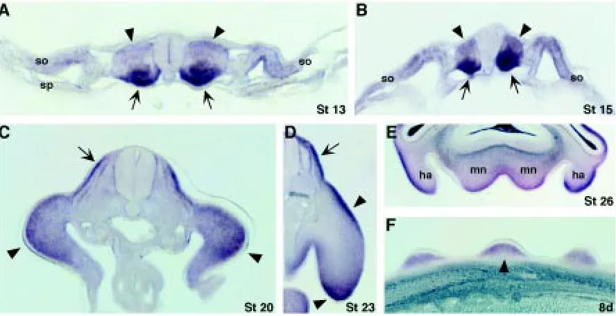
Chick Twist (cTwist) transcripts were first detected at stages 11-12 in the medial region of the somites (Fig. 2A). A very dynamic pattern of expression is seen during somite differentiation. At stages 13-15, cTwist expression is very strong in the ventral medial portion (presumptive sclerotome; Fig. 3 A,B; arrows) and weak in the dorsal portion (presumptive dermomyotome; Fig. 3 A,B; arrow-heads). At stages 20-23, cTwist expression in the somites is
Fig. 1. Alignment of the chick Twist deduced amino acid sequence with the human, mouse, Xenopus, zebrafish and Drosophila homologues
localized to the dermatome (higher levels) and sclerotome (lower levels), and excluded from the myotome (Fig. 3 C,D; arrows). cTwist transcripts could be detected in the somites until the last stage analyzed (stage 28; data not shown).
At stages 13-14, cTwist transcripts are detected in the anterior lateral plate mesoderm (Fig. 2B; arrowheads) and faintly in the posterior lateral mesoderm (Fig. 2B; arrow). A transverse section of these embryos shows that transcripts are found in the somatic component of the lateral mesoderm (somatopleura) but not in the splanchnic component (splanchnopleura; Fig. 3 A,B). At stage 16, cTwist transcripts are found all over the lateral plate mesoderm (Fig. 2C; arrowheads). cTwist expression remains in the flank mesoderm until stage 24 (Fig. 2 D,E, and data not shown). In the
developing limb, cTwist is expressed throughout the limb bud mesenchyme at stages 19-20 (Fig. 2 D,E; Fig. 3C; arrowheads). From stage 22 on, cTwist expression in the limb bud is excluded from the mesenchyme of the central and proximal regions and is detected in the anterior, distal and posterior mesenchyme (Fig. 2 F-H; Fig. 3D; arrowheads). At stages 28-30, transcripts are present at higher levels in the posterior mesenchyme (Fig. 2 H,I; arrow-heads) and in the interdigital regions (Fig. 2 H,I; arrows). The pattern of expression in these regions becomes restricted to areas in the vicinity of the developing digits at stage 30 (Fig. 2I, arrows). At 7 days of development, cTwist expression is still detected in a discrete region of the distal posterior mesenchyme (Fig. 2K; arrowhead). cTwist expression is similar in both the forelimb and hindlimb buds (data not shown).
Fig. 2.Whole mount in situ hybridization analysis of cTwist in the chick embryo. (A) At stage 12, cTwist transcripts are detected in the medial portion of the somites (arrow). (B) At stage 14, expression is found in the lateral plate mesoderm. The staining is stronger in the anterior (arrowheads) than in the posterior (arrow) LPM. (C) At stage 16, the expression of cTwist in the LPM has a broader domain spanning the limb and flank fields (arrowheads). (D) At stage 19, the expression is detected in the limb bud mesenchyme and in the flank (arrowheads). (E) At stage 20, cTwist expression remains in the flank and allover the mesenchyme of the limb buds (arrowheads). cTwist transcripts can also be detected in the branchial arches (asterisks). (F) At stage 22, expression in the limb buds is excluded from the central portion, remaining in the anterior, distal and posterior mesenchyme (arrowheads). In a stage 25 limb bud (G), cTwist expression is stronger in the anterior, distal and posterior mesenchymal regions (arrowheads). (H) At stage 28, transcripts are detected in the posterior mesenchyme (arrowhead) and in the interdigital spaces (arrow) of the limb bud. (I) cTwist expression remains in the posterior mesenchyme of the limb bud (arrowhead) and in the interdigital mesenchyme close to the differentiating digits (arrows). (J) In the developing face, transcripts are found in the head mesenchyme (arrow), in the maxillary processes (mx), mandibular processes (mn), hyoid arch (ha), third branchial arch (b3) and fourth branchial arch (b4).
710 A.T. Tavares et al.
During craniofacial development, chick cTwist is expressed initially in the branchial arches (Fig. 2E; asterisks) and later in the maxillary and mandibular processes, hyoid arch, and third and fourth branchial arches (Fig. 2J), where it is restricted to the distal mesenchyme (Fig. 3E). In addition, cTwist transcripts are found in the head mesenchyme (Fig. 2J, arrow).
cTwist expression is also found in the developing feathers. At day 7 of development, transcripts are found in the interfollicular areas (Fig. 2K; arrow). By embryonic day 8, cTwist expression moves to the mesenchyme of the feather buds (Figs. 2L, 3F arrowhead), and as the feather buds elongate, expression remains in the proximal mesenchyme (Fig. 2M; arrows).
Regulation of cTwist expression by the AER, retinoic acid and SHH
Since the expression of cTwist is detected in limb areas influ-enced by the apical ectodermal ridge and the zone of polarizing activity, we analyzed the role of several molecules that mediate their biological activities.
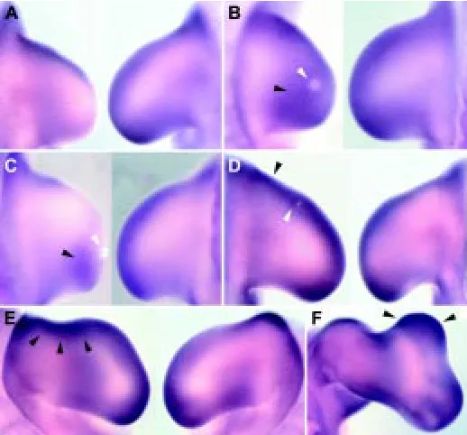
Surgical removal of the AER was performed in order to determine its implication in maintaining cTwist expression. At 6-8 hours after AER ablation, cTwist expression is downregulated and by 12 hours after removal cTwist transcripts are almost undetectable (n=12; Fig. 4A). The growth of the limb bud along the proximal-distal axis is controlled by FGFs released from the AER. To test the influence of FGFs in cTwist expression, beads soaked in FGF-4 and FGF-8 were implanted after AER removal (n=16). The expression domain of cTwist is maintained after FGF-8 treatment and is enlarged when
compared with the contralateral limb (Fig. 4B). Implantation of FGF-4 soaked beads is followed by maintenance of the cTwist expression domain, but this domain is restricted to the posterior part of the limb bud (Fig. 4C).
The expression of cTwist is also observed in the ZPA. Anterior treatments with beads incubated in RA sustain the anterior domain of cTwist (n=20; Fig. 4D), which is maintained during limb bud duplication (data not shown). To verify if cTwist is induced in response to SHH, we implanted clumps of SHH expressing cells in the anterior mesenchyme (n=14). Cells expressing SHH induce an ectopic area of cTwist in the anterior part of the limb bud (Fig. 4E), also observed in the mirror image duplication obtained 48 hours after treatment (Fig. 4F).
In order to elucidate if SHH signalling is sufficient to maintain cTwist expression, clumps of SHH expressing cells were im-planted following removal of the AER. After 12 hours, no cTwist expression is detected in the manipulated limb, suggesting that SHH signalling acts indirectly by maintaining cTwist expression (data not shown).
Discussion
During embryonic development, the expression patterns of chick Twist coincide with those reported for the mouse homologue (Wolf et al., 1991; Fuchtbauer, 1995; Gitelman, 1997; Bourgeois et al., 1998), with the obvious exceptions of MTwist expression during odontogenesis and cTwist expression in the developing feathers. In general, the pattern of cTwist expression in the developing chick
Fig. 3. Sections of chick embryos stained for cTwist by whole mount in situ hybridization. (A) Transverse section of a stage 13 chick embryo showing cTwist expression in the ventral regions of the somites (arrows) and, less intensely, in the dorsal somites (arrowheads) and somatic lateral mesoderm (so). cTwist transcripts are not found in the splancnopleura (sn). (B) At stage 15, expression remains in the ventral (arrows) and dorsal (arrowheads) somites, and becomes more intense in the somatopleura (so). (C) In a transverse section of a stage 20 embryo, cTwist expression can be seen in the mesenchyme (arrowheads) and in the somites (arrow). Transcripts are found in the dermatome (higher expression) and sclerotome but excluded from the myotome. (D)
At stage 23, expression is detected in the distal and dorsal limb mesenchyme (arrowheads) and maintained in the same regions of the somites (arrow). (E)
coincides with mesenchymal regions that are under the control of FGFs and SHH.
In the somites, cTwist is expressed first in the ventral medial portion and later in the dermatome and sclerotome compartments. Fgf-4 (Niswander and Martin, 1992), Fgf-5 (Haub and Goldfarb, 1991), Fgf-6 (deLapeyrière et al., 1993) and Fgf-8 (Vogel et al., 1996) are also expressed in the somites, and shh expression in the notochord and floor plate was shown to be required for somitic cell survival (Teillet et al., 1998, Marcelle et al., 1999).
In the branchial arches, cTwist is expressed in the mesen-chyme, whereas Fgf-3 (Mahmoon et al., 1996), Fgf-8 (Heikinheimo et al., 1994) and shh (Wall and Hogan, 1995) transcripts are detected in the ectoderm. On the other hand, cTwist and Fgf-8 are also co-expressed in the head mesenchyme (Heikinheimo et al., 1994).
During feather development, cTwist transcripts are detected initially in the interfollicular region and later in the bud mesen-chyme. Conversely, Fgf-2, Fgf-4, Fgf-18 and shh are expressed in the overlying ectodermal placodes (Nohno et al., 1995; Song et al., 1996; Ohuchi et al., 2000).
In limb buds, cTwist transcripts are initially detected throughout the limb bud mesenchyme and then become restricted to the anterior, distal and posterior mesenchyme. This expression pat-tern is very similar to that of Fgf-10 (Ohuchi et al., 1997) and partially overlaps with Fgf-18 (Ohuchi et al., 2000) Fgf-12 (cFhf-1) and Fgf-13 (cFhf-2; Munoz-Sanjuan et al., 1999) expression do-mains. Moreover, several Fgfs are expressed in the AER: Fgf-2 (Savage and Fallon, 1995), Fgf-4 (Niswander and Martin, 1992; Savage et al., 1993), Fgf-8 (Heikinheimo et al., 1994, Ohuchi et al., 1994, Crossley and Martin, 1995), and Fgf-9 (Colvin et al., 1999). The posterior domain of cTwist is also overlapped in the limb bud mesenchyme with shh expression (Echelard et al., 1993, Krauss et al., 1993, Riddle et al., 1993).
These closely tied expression domains suggest the existence of a relationship between Fgfs and cTwist. In here we present data that further reinforce this relationship. Removal of the AER results in truncated limbs (Summerbell, 1974; Rowe et al., 1982; Todt and Fallon, 1987) and downregulation of Fgf-10 and Fgf-18 expression in the mesenchyme (Ohuchi et al., 1997, Ohuchi et al., 2000), phenotypes that can be rescued by application of FGFs (Niswander et al., 1993; Fallon et al., 1994; Crossley et al., 1996; Vogel et al., 1996, Ohuchi et al., 1997, Ohuchi et al., 2000). Ablation of AER downregulates cTwist expression. The normal expression is re-stored when a bead releasing either FGF-4 or FGF-8 is implanted after removal of the AER. The maintenance of cTwist expression through FGF signalling is in agreement with several studies show-ing that Twist expression is induced by FGFs, not only in vitro (Fang et al., 2001) but also in vivo (Isaac et al., 2000).
It is also noteworthy that expression domains of cTwist and shh are closely related during development. In limb buds, the expres-sion of cTwist is also observed in the ZPA, overlapping with that of shh expression. In addition, implants of SHH expressing cells or beads soaked in RA (that are able to induce shh expression; Riddle et al., 1993) at the anterior margin of the limb bud induce cTwist expression. However, the regulation through SHH seems to be indirect since SHH expressing cells are not able to maintain cTwist expression after AER removal. The induction of cTwist by SHH in the anterior mesenchyme may be mediated by the upregulation of FGFs in the overlying AER.
Some of the possible downstream targets of Twist are the FGF receptors. In Drosophila and C. elegans, the expression of their Fgfr homologues appears to be regulated by Twist during meso-dermal patterning (Shishido et al., 1993; Harfe et al., 1998). In vertebrates, Fgfr1 and -2 expression during embryonic develop-ment coincides with that of Twist in several tissues, such as the limb bud mesenchyme (Orr-Urtreger et al., 1993; Deng et al., 1997), head mesenchyme and branchial arches (Walshe and Mason, 2000), and mesenchyme of interfollicular region and feather buds (Noji et al., 1993). In addition, and as also shown here, during chick limb development, expression of cTwist in the interdigital mesenchyme is coincident with the expression domains of several Fgfr (Noji et al., 1993, Marcelle et al., 1995).
Taken together, these results suggest that Twist may be impli-cated in the response of several mesenchymal tissues to FGF signalling, possibly as a mediator in the maintenance of Fgfr expression.
Materials and Methods
cDNA cloning
Chick cTwist full length clones were identified in the screening of a stage 20-22 chick limb bud λZAPII cDNA library with a 311 bp chick cTwist RT-PCR fragment as a probe (nucleotides 262-573; 5’ primer: CCGCAGTCCTACGAGGAGCTG;
712 A.T. Tavares et al.
3’ primer: GTGGGATGCGGACATGGACCA; from G-Twist gene sequence in Genbank with accession number Y08261). The positive clones were sequenced with an automated sequencer. The largest clone identified was 1683 bp long, included 573 bp of coding sequence and, by comparison with homologues of other species, appeared to be a full length Twist clone. The cTwist mRNA sequence has also been reported in Genbank by Isaac et al. under accession number AF093816.
Sequence comparison was performed using Clustal W (version 1.8). The SwissProt accession numbers of the protein sequences used in the alignment are: P26687 (mouse Twist), Q15672 (human Twist), P13903 (Xenopus Twist), Q9PTE3 (Zebrafish Twist), and P10627 (Drosophila Twist).
Whole-mount in situ hybridization and histology
Chicken embryos were obtained from MacIntyre Poultry (San Diego, U.S.A.) and Avipronto (Benavente, Portugal). Eggs were incubated at 38°C and staged according to Hamburger and Hamilton (Hamburger and Hamilton, 1951). Whole-mount in situ hybridization was carried out as described (Wilkinson, 1993). The riboprobe was synthesized as a digoxigenin (Boehringer) labelled probe from the whole sequence of chick cTwist (1.6kb) cloned in pSlax vector (EcoRI digestion; T7 polymerase transcrip-tion). For histology, the stained embryos were dehydrated in 30% sucrose, embedded in gelatine, frozen in an isopenthane bath, and sectioned in a cryostat.
Experimental manipulation of limb buds
Application of RA was performed with AG1-X2 ion exchange beads (BIO-RAD), and FGF-4 and FGF-8 proteins (R&D Systems) were im-planted in heparin acrylic beads (Sigma). Beads were soaked in PBS (as a control) or in a solution of the desired molecule and were implanted in the limb mesenchyme. Eggs were windowed and beads were implanted using a tungsten needle. The AER was surgically removed from the wing buds with a thin tungsten needle. In some experiments this treatment was followed by implantation of FGF soaked beads or cell clumps. Clumps of control and SHH expressing cells were grafted in the anterior mesenchyme of wing buds using a tungsten needle.
Manipulations were performed at stages 20-22 in the right wing bud, using the left one as a control. After each manipulation, the eggs were closed and reincubated during different time periods. Embryos were sacrificed, fixed in 4% paraformadehyde and processed for whole mount in situ hybridization with cTwist probe.
Preparation of beads and cell clumps
Heparin acrylic beads (Sigma) were used to apply FGF proteins (R&D Systems). Beads between 100 and 150 µm were selected, washed in PBS and then incubated for 1 hour at room temperature in the protein solution (0.5 mg/ml for each FGF). AG1-X2 ion exchange beads (BIO-RAD) between 100 and 150 µm were used for RA treatment. Beads were soaked in RA, diluted in DMSO (1 mg/ml) for 20 minutes and then washed in PBS-Phenol Red solution to remove the excess DMSO and for staining. Control beads were incubated in PBS.
QT6 control and QT6 SHH-expressing cell lines were obtained from Dr. D. Duprez. Clumps of 100 µm were selected and their preparation was performed as described in Duprez et al. (1998).
Acknowledgements
We thank J. K. Ng for comments and technical advice, L. Hooks for help preparing the manuscript, D. Duprez for providing QT6 SHH-expressing cells, Isabel Marques for her assistance with sequence analysis, I. Alves for maintaining cell cultures and Prof. J. Hurlé for useful comments. A.T.T. was supported by a fellowship from the Programa Gulbenkian de Doutoramento em Biologia e Medicina (PGDBM), Program PRAXIS XXI, Fundação Luso-Americana para o Desenvolvimento (FLAD) and Fundação Calouste Gulbenkian. J.R.L. was supported by a fellowship from the Fundação
Calouste Gulbenkian. This work was supported by grants from the Fundação para a Ciência e a Tecnologia (ref. 36192/99) and by the National Science Foundation.
References
BLOCH-ZUPAN, A., HUNTER, N., MANTHEY, A. and GIBBINS, J. (2001). R-twist gene expression during rat palatogenesis. Int. J. Dev. Biol. 45: 397-404.
BOURGEOIS, P., BOLCATO-BELLEMIN, A.L., DANSE, J.M., BLOCH-ZUPAN, A., YOSHIBA, K., STOETZEL, C. and PERRIN-SCHMITT, F. (1998). The variable expressivity and incomplete penetrance of the twist-null heterozygous mouse phenotype resemble those of human Saethre-Chotzen syndrome. Hum. Mol. Genet. 7: 945-957.
BUSHDID, P.B., BRANTLEY, D.M., YULL, F.E., BLAEUER, G.L., HOFFMAN, L.H., NISWANDER, L. and KERR, L.D. (1998). Inhibition of NF-kappaB activity results in disruption of the apical ectodermal ridge and aberrant limb morphogenesis. Nature 392: 615-618.
CHEN, Z.F. and BEHRINGER, R.R. (1995). twist is required in head mesenchyme for cranial neural tube morphogenesis. Genes Dev. 9: 686-699.
COLVIN, J.S., FELDMAN, B., NADEAU, J.H., GOLDFARB, M. and ORNITZ, D.M. (1999). Genomic organization and embryonic expression of the mouse fibroblast growth factor 9 gene. Dev. Dyn. 216: 72-88.
CROSSLEY, P.H. and Martin, G.R. (1995). The mouse Fgf8 gene encodes a family of polypeptides and is expressed in regions that direct outgrowth and patterning in the developing embryo. Development 121: 439-451.
CROSSLEY, P.H., MINOWADA, G., MACARTHUR, C.A. and MARTIN, G.R. (1996). Roles for FGF-8 in the induction, initiation and maintenance of chick limb develop-ment. Cell 84: 127-136.
deLAPEYRIERE, O., OLLENDORFF, V., PLANCHE, J., OTT, M.O., PIZETTE, S., COULIER, F. and BIRNBAUM, D. (1993). Expression of the Fgf6 gene is restricted to developing skeletal muscle in the mouse embryo. Development 118: 601-611.
DE MOERLOOZE, L. and DICKSON, C. (1997). Skeletal disorders associated with fibroblast growth factor receptor mutations. Curr. Opin. Genet. Dev. 7: 378-385.
DENG, C., BEDFORD, M., LI, C., XU, X., YANG, X., DUNMORE, J. and LEDER, P. (1997). Fibroblast growth factor receptor-1 (FGFR-1) is essential for normal neural tube and limb development. Dev. Biol. 185: 42-54.
DUPREZ, D., FOURNIER-THIBAULT, C. and LE DOUARIN, N. (1998). Sonic Hedge-hog induces proliferation of committed skeletal muscle cells in the chick limb. Development 125: 495-505.
ECHELARD, Y., EPSTEIN, D.J., ST-JACQUES, B., SHEN, L., MOHLER, J., MCMAHON, J.A. and MCMAHON, A.P. (1993). Sonic hedgehog, a member of a family of putative signalling molecules, is implicated in the regulation of CNS polarity. Cell 75: 1417-1430.
FALLON, J.F., LOPEZ, A., ROS, M.A., SAVAGE, M.P., OLWIN, B.B. and SIMANDL, B.K. (1994). FGF-2: apical ectodermal ridge growth signal for chick limb develop-ment. Science 264: 104-107.
FUCHTBAUER, E.M. (1995). Expression of M-twist during postimplantation develop-ment of the mouse. Dev. Dyn. 204: 316-322.
GRIPP, K.W., ZACKAI, E.H. and STOLLE, C.A. (2000). Mutations in the human TWIST gene. Hum. Mutat. 15: 150-155.
GITELMAN, I. (1997). Twist protein in mouse embryogenesis. Dev. Biol. 189: 205-214.
HAMBURGER, V. and HAMILTON, H.L. (1951). A series of normal stages in the development of the chick embryo. J. Morphol. 88: 49-92.
HARFE, B.D., GOMES, A.V., KENYON, C., LIU, J., KRAUSE, M. and FIRE, A. (1998). Analysis of a Caenorhabditis elegans Twist homolog identifies conserved and divergent aspects of mesodermal patterning. Genes Dev. 12: 2623-2635.
HAUB, O. and GOLDFARB, M. (1991). Expression of the fibroblast growth factor-5 gene in the mouse embryo. Development 112: 397-406.
HEIKINHEIMO, M., LAWSHE, A., SHACKLEFORD, G.M., WILSON, D.B. and MACARTHUR, C.A. (1994). Fgf-8 expression in the post-gastrulation mouse suggests roles in the development of the face, limbs and central nervous system. Mech. Dev. 48: 129-138.
ISAAC, A., COHN, M.J., ASHBY, P., ATALIOTIS, P., SPICER, D.B., COOKE, J. and TICKLE, C. (2000). FGF and genes encoding transcription factors in early limb specification. Mech. Dev. 93: 41-48.
JOHNSON, D., ISEKI, S., WILKIE, A.O. and MORRISS-KAY, G.M. (2000). Expression patterns of Twist and Fgfr1, -2 and -3 in the developing mouse coronal suture suggest a key role for twist in suture initiation and biogenesis. Mech. Dev. 91: 341-345.
KANEGAE, Y., TAVARES, A.T., IZPISUA BELMONTE, J.C. and VERMA, I. M. (1998). Role of Rel/NF-kappaB transcription factors during the outgrowth of the vertebrate limb. Nature 392: 611-614.
KRAUSS, S., CONCORDET, J.P. and INGHAM, P.W. (1993). A functionally conserved homolog of the Drosophila segment polarity gene hh is expressed in tissues with polarizing activity in zebrafish embryos. Cell 75: 1431-1444.
LAUFER, E., NELSON, C.E., JOHNSON, R.L., MORGAN, B.A. and TABIN, C. (1994). Sonic hedgehog and Fgf-4 act through a signaling cascade and feedback loop to integrate growth and patterning of the developing limb bud. Cell 79: 993-1003.
LEPTIN, M., CASAL, J., GRUNEWALD, B. and REUTER, R. (1992). Mechanisms of early Drosophila mesoderm formation. Dev. Suppl. 23-31.
LI, L., CSERJESI, P. and OLSON, E.N. (1995). Dermo-1: a novel twist-related bHLH protein expressed in the developing dermis. Dev. Biol. 172: 280-292.
MAHMOOD, R., MASON, I.J. and MORRISS-KAY, G.M. (1996). Expression of Fgf-3 in relation to hindbrain segmentation, otic pit position and pharyngeal arch morphol-ogy in normal and retinoic acid-exposed mouse embryos. Anat. Embryol. (Berl.) 194: 13-22.
MARCELLE, C., WOLF, J. and BRONNER-FRASER, M. (1995). The in vivo expression of the FGF receptor FREK mRNA in avian myoblasts suggests a role in muscle growth and differentiation. Dev. Biol. 172: 100-114.
MARCELLE, C., AHLGREN, S. and BRONNER-FRASER, M. (1999). In vivo regulation of somite differentiation and proliferation by Sonic Hedgehog. Dev. Biol. 214: 277-287.
MOON, A.M., BOULET, A.M. and CAPECCHI, M.R. (2000) Normal limb development in conditional mutants of Fgf4. Development 12: 989-996.
MUNOZ-SANJUAN, I., SIMANDL, B.K., FALLON, F., and NATHANS, J. (1999). Expression of chicken fibroblast growth factor homologous factor (FHF)-1 and of differentially spliced isoforms of FHF-2 during development and involvement of FHF-2 in chicken limb development. Development 126: 409-421.
NISWANDER, L., and MARTIN, G.R. (1992). Fgf-4 expression during gastrulation, myogenesis, limb and tooth development. Development 114: 755-768.
NISWANDER, L., TICKLE, C., VOGEL, A., BOOTH, I. and MARTIN, G. R. (1993). FGF-4 replaces the apical ectodermal ridge and directs outgrowth and patterning of the limb. Cell 75: 579-587.
NISWANDER, L., JEFFREY, S., MARTIN, G.R. and TICKLE, C. (1994). A positive feedback loop coordinates growth and patterning in the vertebrate limb. Nature 371: 609-612.
NOHNO, T., KAWAKAMI, Y., OHUCHI, H., FUJIWARA, A., YOSHIOKA, H. and NOJI, S. (1995). Involvement of the Sonic hedgehog gene in chick feather formation. Biochem. Biophys. Res. Commun. 206: 33-39.
NOJI, S., KOYAMA, E., MYOKAI, F., NOHNO, T., OHUCHI, H., NISHIKAWA, K. and TANIGUCHI, S. (1993). Differential expression of three chick FGF receptor genes, FGFR1, FGFR2 and FGFR3, in limb and feather development. Prog. Clin. Biol. Res. 383: 645-654.
OHUCHI, H., YOSHIOKA, H., TAMAKA, A., KAWAKAMI, T., NOHNO, T. and NOJI, S. (1994). Involvement of androgen-induced growth factor (FGF-8) gene in mouse embryogenesis and morphogenesis. Biochem. Biophys. Res. Commun. 204: 882-888.
OHUCHI, H., NAKAGAWA, T., YAMAMOTO, A., ARAGA, A., OHATA, T., ISHIMARU, Y., YOSHIOKA, H., KUWANA, T., NOHNO, T., YAMASAKI, M., ITOH, N. and NOJI, S. (1997). The mesenchymal factor, FGF10, initiates and maintains the outgrowth of the chick limb bud through interaction with FGF8, an apical ectodermal factor. Development 124: 2235-2244.
OHUCHI, H., KIMURA, S., WATAMOTO, M. and ITOH, N. (2000). Involvement of fibroblast growth factor (FGF)18-FGF8 signaling in specification of left-right asym-metry and brain and limb development of the chick embryo. Mech. Dev. 95: 55-66.
ORR-URTREGER, A., BEDFORD, M.T., BURAKOVA, T., ARMAN, E., ZIMMER, Y., YAYON, A., GIVOL, D. and LONAI, P. (1993). Developmental localization of the splicing alternatives of fibroblast growth factor receptor-2 (FGFR2). Dev. Biol. 158: 475-486.
RICE, D.P., ABERG, T., CHAN, Y., TANG, Z., KETTUNEN, P.J., PAKARINEN, L., MAXSON, R.E. and THESLEFF, I. (2000). Integration of FGF and TWIST in calvarial bone and suture development. Development 127: 1845-1855.
RIDDLE, R.D., JOHNSON, R. L., LAUFER, E. and TABIN, C. (1993). Sonic hedgehog mediates the polarizing activity of the ZPA. Cell 75: 1401-1416.
SAUNDERS, J.W. JR. (1948). The proximo-distal sequence of origin of the parts of the chick wing and the role of the ectoderm. J. Exp. Zool. 108: 363-403.
SAUNDERS, J.W. and GASSELING, M.T. (1968). Ectoderm-mesenchymal interac-tions in the origin of wing symmetry. In Epithelial Mesenchymal Interacinterac-tions, (ed. R. Fleischmajer and R. Billingham), Williams and Wilkins, pp. 78-97.
SAVAGE, M.P., HART, C. E., RILEY, B.B., SASSE, J., OLWIN, B.B., and FALLON, J.F. (1993). Distribution of FGF-2 suggests it has a role in chick limb bud growth. Dev. Dyn. 198: 159-170.
SAVAGE, M.P. and FALLON, F.P. (1995). FGF-2 mRNA and its antisense message are expressed in a developmentally specific manner in the chick limb bud and mesonephros. Dev. Dyn. 202: 343-353.
SHISHIDO, E., HIGASHIJIMA, S., EMORI, Y. and SAIGO, K. (1993). Two FGF-receptor homologues of Drosophila: one is expressed in mesodermal primordium in early embryos. Development 117: 751-761.
SONG, H., WANG, Y. and GOETINCK, P.F. (1996). Fibroblast growth factor 2 can replace ectodermal signaling for feather development. Proc. Natl. Acad. Sci. U.S.A. 93: 10246-10249.
SUMMERBELL, D. (1974). A quantitative analysis of the effect of excision of the AER from the chick limb-bud. J. Embryol. Exp. Morphol. 32: 651-660.
SUN, X., LEWANDOSKI, M., MEYERS, E.M., LIU, Y.H., MAXSON JR, R.E. and MARTIN, G.R. (2000). Conditional inactivation of Fgf4 reveals complexity of signalling during limb bud development. Nature Genet. 25: 83-86.
TEILLET, M., WATANABE, Y., JEFFS, P., DUPREZ, D., LAPOINTE, F. and LE DOUARIN N.M. (1998). Sonic hedgehog is required for survival of both myogenic and chondrogenic somitic lineages. Development 125: 2019-2030.
TICKLE, C., ALBERTS, B., WOLPERT, L. and LEE, J. (1982). Local application of retinoic acid to the limb bud mimics the action of the polarizing region. Nature 296: 564-566.
THISSE, B., EL MESSAL, M. and PERRIN-SCHMITT, F. (1987). The twist gene: isolation of a Drosophila zygotic gene necessary for the establishment of dorsoven-tral pattern. Nucleic. Acids. Res. 15: 3439-3453.
VOGEL, A., RODRIGUEZ, C. and IZPISUA-BELMONTE, J.C. (1996). Involvement of FGF-8 in initiation, outgrowth and patterning of the vertebrate limb. Development 122: 1737-1750.
WALL, N.A. and HOGAN, B.L. (1995). Expression of bone morphogenetic protein-4 (BMP-4), bone morphogenetic protein-7 (BMP-7), fibroblast growth factor-8 (FGF-8) and sonic hedgehog (SHH) during branchial arch development in the chick. Mech. Dev. 53: 383-392.
WALSHE, J. and MASON, I. (2000). Expression of FGFR1, FGFR2 and FGFR3 during early neural development in the chick embryo. Mech. Dev. 90: 103-110.
WANG, S.M., COLJEE, V.W., PIGNOLO, R.J., ROTENBERG, M.O., CRISTOFALO, V.J. and SIERRA, F. (1997). Cloning of the human twist gene: its expression is retained in adult mesodermally-derived tissues. Gene 187: 83-92.
WILKINSON, D.G. (1993). Whole mount in situ hybridisation of vertebrate embryos. In In situ Hybridization, (ed. Wilkinson, D.G.). University Press, Oxford, pp. 75-83.
WOLF, C., THISSE, C., STOETZEL, C., THISSE, B., GERLINGER, P. and PERRIN-SCHMITT, F. (1991). The M-twist gene of Mus is expressed in subsets of mesodermal cells and is closely related to the Xenopus X-twi and the Drosophila twist genes. Dev. Biol. 143: 363-373.
XU, X., WEINSTEIN, M., LI, C. and DENG, C. (1999). Fibroblast growth factor receptors (FGFRs) and their roles in limb development. Cell. Tissue. Res. 296: 33-43.